Introduction
The temporal bone is a complex anatomical structure that contains numerous air cells, formed as epithelium invaginates into the developing bone. Pneumatisation begins at about 24 weeks of gestation and continues until early adulthood. Its degree and distribution vary considerably between individuals [1][2].
Recent studies have shown that pneumatisation patterns correlate with the development of otologic pathology, particularly chronic otitis media, and that they directly influence surgical accessibility and complication rates in skull base surgery [3][4]. Given this clinical significance, standardised classification systems have been developed to allow objective radiological assessment and preoperative planning [5].
This paper examines the major classification systems, their radiological basis and their clinical and surgical correlations. The aims of this study were: (i) to identify and categorise pneumatisation patterns of the temporal bone on high-resolution computed tomography (CT), focusing on the petrous apex, mastoid and infralabyrinthine compartments; and (ii) to analyse the relationship between the prevalence of these patterns and factors such as age, sex, bilateral symmetry and relevant clinical, surgical and pathological diagnoses in Indian patients.
Methods
Study design: Retrospective cross-sectional observational study.
Population: One hundred patients who underwent high-resolution CT of the temporal bone for otologic indications over a six-month period were included.
Data collection: Age, sex, laterality, underlying otologic pathology and degree of pneumatisation were recorded. Pneumatisation was classified using established systems.
Classification Systems
Anatomical regions: The pneumatised portions of the temporal bone are organised into three main regions: the mastoid air cell system, the infralabyrinthine (peritympanic) air cells and the petrous apex air cells (Figures 1 and 2).
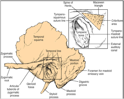
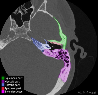
Dexian Tan classification: This system evaluates three anatomical regions separately.
The mastoid air cell system is graded I to IV:
Grade I, very hypopneumatised;
Grade II, hypopneumatised;
Grade III, normally pneumatised;
Grade IV, hyperpneumatised.
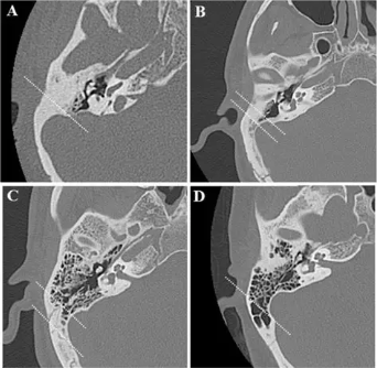
The petrous apex is classified into four groups:
Group I, completely sclerotic with no pneumatisation;
Group II, less than 50% pneumatisation medial to the labyrinth;
Group III, more than 50% pneumatisation medial to the labyrinth;
Group IV, completely pneumatised, with extensive air cell development medial to the labyrinth and carotid canal.
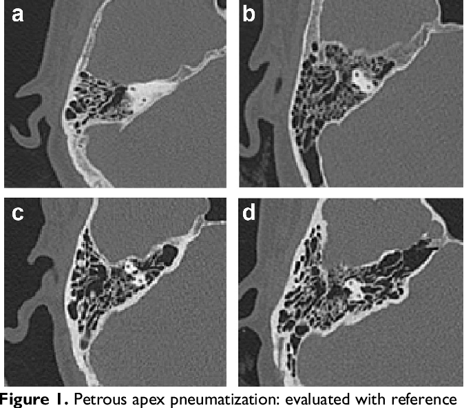
The infralabyrinthine region is
Grade I (good pneumatisation),
Grade II (moderate pneumatisation)
Grade III (hypopneumatisation).
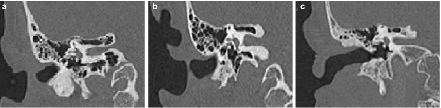
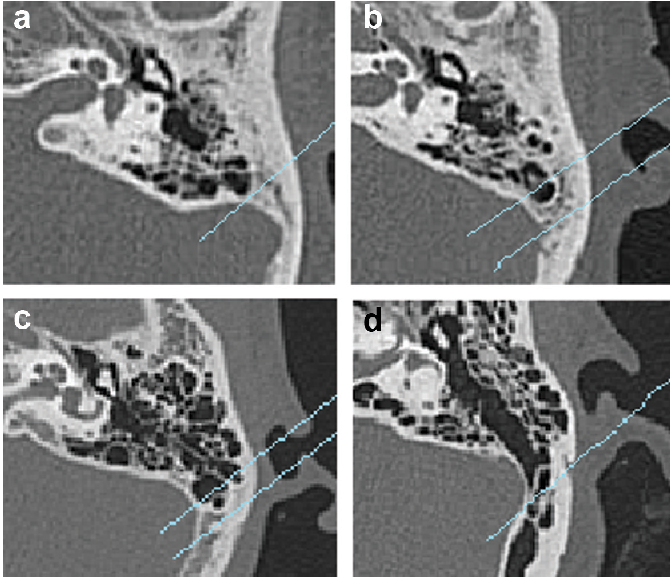
Han classification: This alternative system is based on the extent of pneumatised air cells in relation to the sigmoid sinus [5]:
Presinusoidal type (anterior pattern; hypopneumatisation),
Sinusoidal type (air cells adjacent to the sigmoid sinus; good pneumatisation)
Postsinusoidal type (air cells posterior to the sigmoid sinus; hyperpneumatisation).
Statistical analysis
Prevalence, correlations between pneumatisation and demographic, clinical and surgical factors, and bilateral symmetry were analysed Data will be entered and analysed using IBM SPSS Statistic
Results
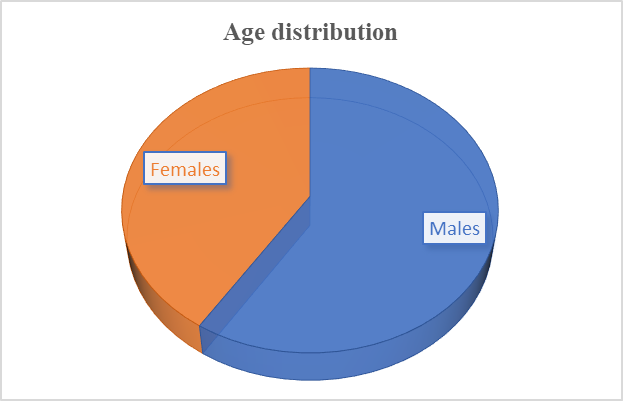
Study population: A total of 100 patients were included in the study, with a male preponderance (65%) and with mean age of 48 ± 20
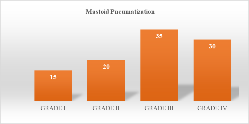
Mastoid pneumatisation: Using the Dexian Tan grading, 15 patients (15%) were Grade I, 20 (20%) Grade II, 35 (35%) Grade III and 30 (30%) Grade IV (Table 1, Figure 3,8).
| Grade | Description | N | % |
| I | Very hypopneumatised | 15 | 15 |
| II | Hypopneumatised | 20 | 20 |
| III | Normal pneumatisation | 35 | 35 |
| IV | Hyperpneumatised | 30 | 30 |
n: number of patients.
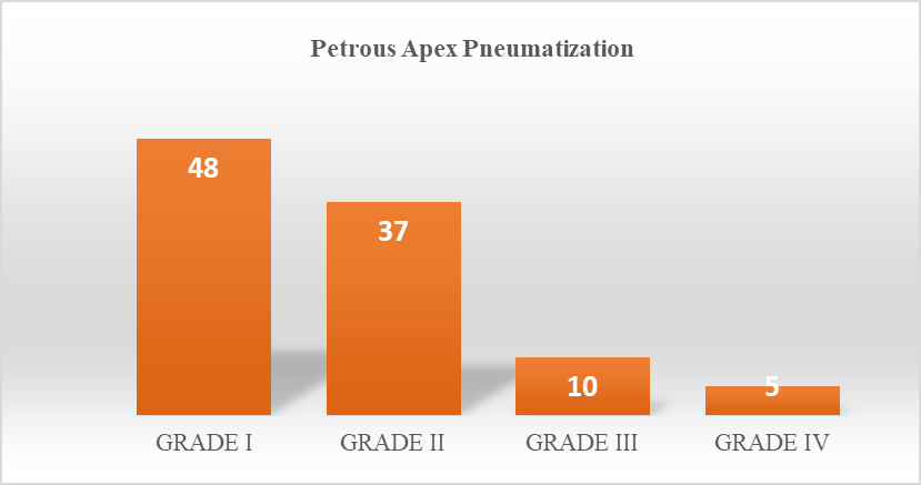
Petrous apex pneumatisation. The petrous apex was completely sclerotic (Group I) in 48 patients (48%). Group II was seen in 37 patients (37%), Group III in 10 (10%) and Group IV in 5 (5%) (Table 2, Figure 4,9).
| Group | Description | n | % |
| I | Completely sclerotic, no pneumatisation | 48 | 48 |
| II | < 50% pneumatisation medial to the labyrinth | 37 | 37 |
| III | > 50% pneumatisation medial to the labyrinth | 10 | 10 |
| IV | Completely pneumatised, extending medial to the labyrinth and carotid canal | 5 | 5 |
n: number of patients.
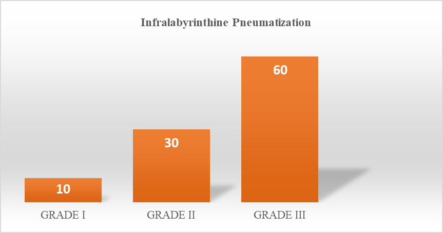
Infralabyrinthine pneumatisation: Grade I (good) pneumatisation was seen in 10 patients (10%), Grade II (moderate) in 30 (30%) and Grade III (hypopneumatisation) in 60 (60%) (Table 3, Figure 5,10).
| Grade | Description | N | % |
| I | Good pneumatisation | 10 | 10 |
| II | Moderate pneumatisation | 30 | 30 |
| III | Hypopneumatisation | 60 | 60 |
n: number of patients.
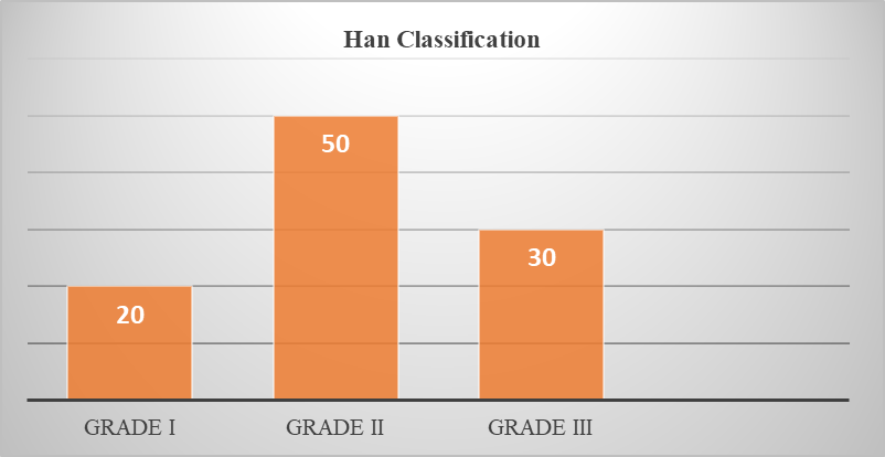
Han classification: The presinusoidal type was seen in 20 patients, the sinusoidal type in 50 and the postsinusoidal type in 30 (Table 4, Figure 6,11).
| Type | Description | n |
| Presinusoidal | Anterior pattern; hypopneumatisation | 20 |
| Sinusoidal | Air cells adjacent to the sigmoid sinus; good pneumatisation | 50 |
| Postsinusoidal | Air cells posterior to the sigmoid sinus; hyperpneumatisation | 30 |
n: number of patients.
Discussion
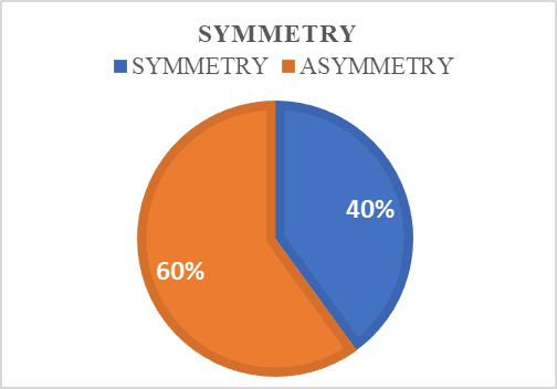
Bilateral symmetry. 40% of individuals in this series demonstrated bilateral symmetry of pneumatisation. Asymmetries that do occur can be indicative of prior disease, particularly chronic otitic disease. (Figure 12).
Age-related changes: Pneumatisation patterns tend to stabilise in adulthood. However, secondary changes can follow infection and lead to mastoid sclerosis.
Choice of classification system: The Dexian Tan system describes the three compartments of the temporal bone separately, whereas the Han system, which is based on the relation of air cells to the sigmoid sinus, is the simplest and the most widely used [5].
Chronic otitis media: Hypopneumatised (sclerotic) mastoids are significantly associated with a greater risk of squamous-type cholesteatoma and chronic suppurative disease (Figure 13). Chronic otitis media in the contralateral ear has been significantly associated with Group 2 mastoid pneumatisation [3].
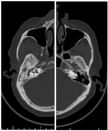
Cerebrospinal fluid leak. Cerebrospinal fluid leakage is a significant complication after retrosigmoid craniotomy for skull base lesions [4, 6]. In the study by Zaczkowski et al., patients with cerebrospinal fluid leakage had significantly higher pneumatisation volumes on both sides than those without leakage [4] (Figure 14).
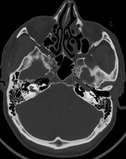
Temporal bone fractures. Temporal bone pneumatisation has been speculated to act as a shock absorber in temporal bone fractures, potentially directing fracture lines away from vital structures (Figure 15). Extensive pneumatisation may distribute traumatic forces across a larger surface area, reducing concentrated stress on critical anatomical structures.
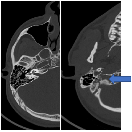
Limitations
This was a retrospective study of 100 patients collected over a six-month period, which limits generalisability.
Conclusion
In conclusion, temporal bone pneumatisation is a fundamental anatomical variable with significant implications for radiological reporting, prediction of pathophysiology and surgical planning.
The Han system is the simplest and most widely used classification.
Hypopneumatised (sclerotic) mastoids are associated with a greater risk of squamous-type cholesteatoma and chronic suppurative disease, whereas a greater degree of pneumatisation has been associated with cerebrospinal fluid leak in retrosigmoid surgery.
Surgical outcome data indicate that aeration patterns predict operative difficulty and morbidity.
Systematic assessment of pneumatisation patterns in temporal bone imaging provides valuable preoperative information for the surgical team and a basis for follow-up by clinicians, and the classification should be incorporated into radiology reports.
Declarations
Conflict of interest
The authors have no conflicts of interest to declare.
Funding
This research received no specific grant from any funding agency in the public, commercial, or not-for profit sectors.
Ethics
This retrospective study was conducted in accordance with the ethical guidelines of the institution, including adherence to the legal requirements of the study country. The study was approved by the institutional review board / research ethics committee, and the requirement for individual patient consent was waived given the retrospective, de identified nature of the analysis.
Declaration of Human Rights
The authors confirm that they accept and agree with the United Nations' Declaration of Human Rights.
Trial details
Not applicable (retrospective observational study).
Author contribution
Dr. S. Charishma: Conceptualisation, data collection, image analysis, and drafting of the manuscript.
Dr. N. L. N. Moorthy: Study supervision, critical review of imaging findings, and critical revision of the manuscript for important intellectual content.
Dr. G. Ramesh Goud: Data analysis and critical revision of the manuscript.
All authors read and approved the final manuscript.
Acknowledgements
None