Introduction
Wunderlich syndrome is defined as spontaneous renal hemorrhage in the absence of trauma and may involve the subcapsular and perirenal spaces. It is an uncommon entity. Jo et al. reported 46 episodes in 38 patients treated at a tertiary center between 2011 and 2024; flank pain was present in 44 episodes (95.7%) and gross hematuria in three (6.5%) [1]. The classic Lenk triad adds a palpable mass and hypovolemic shock to pain, although the complete triad is not consistently present. These figures describe a hospital-based series and cannot be used to estimate prevalence in the general population.
The systematic review by Ahn et al. included 102 cases from 79 publications between 2000 and 2016. Renal neoplasms accounted for 56.9% of causes; among cases of tumor origin, 74.1% were angiomyolipomas. Polyarteritis nodosa was identified in 11.8% of all cases [2]. These are proportions derived from published cases and are subject to selection bias and the availability of follow-up, rather than population-based estimates. The predominance of tumor-related causes remains relevant even when the initial computed tomography (CT) does not show a mass.
CT can identify the hematoma before its underlying cause is necessarily established; a small lesion may be obscured by the hemorrhagic content [3]. In the present patient, no renal mass was identified. Bleeding persisted on the 48-hour follow-up study, when delayed contrast leakage at the level of the renal pelvis was also described. We report the clinical, laboratory, and CT evolution up to the time of transfer for embolization.
Case presentation
A 56-year-old male patient with a history of controlled arterial hypertension presented on July 12, 2026, with severe right-sided lumbar pain and hematuria. On admission, he was hemodynamically stable, with vital signs within normal limits. He denied abdominal or lumbar trauma and recent urologic procedures. He was not receiving anticoagulants and had no known history of chronic kidney disease, renal neoplasm, or cystic kidney disease. The clinical timeline is summarized in Table 1.
| Date | Clinical course |
| July 12, 2026 | Presentation with severe right-sided lumbar pain and hematuria. Hemodynamically stable on admission. Hemoglobin 15.8 g/dL, hematocrit 46.5%, leukocytes 19,000/µL, and creatinine 1.40 mg/dL. Computed tomography urography showed a right perirenal hematoma with active contrast extravasation; no renal mass was identified and the initial collecting-system reconstructions showed no contrast leakage. |
| July 13, 2026 | Hemoglobin 10.9 g/dL, hematocrit 32.7%, leukocytes 18,000/µL, and creatinine 2.00 mg/dL. |
| July 14, 2026 | Repeat computed tomography urography at 48 hours showed enlargement of the hematoma, persistent active extravasation, and delayed contrast leakage at the level of the renal pelvis, compatible with collecting system involvement. Hemoglobin 10.0 g/dL and creatinine 1.60 mg/dL. |
| July 15, 2026 | Hemoglobin 9.0 g/dL and hematocrit 27.0%. Blood pressure decreased to 100/60 mmHg with a heart rate of 110 beats/min. The patient was transferred to Hospital Eugenio Espejo for renal embolization. |
| After transfer | The available information does not establish whether the embolization procedure was completed or what its outcome was. |
Admission hemoglobin was 15.8 g/dL and hematocrit was 46.5%. Leukocytosis of 19,000/µL was present, with 88.6% neutrophils. Creatinine was 1.40 mg/dL; prothrombin time (PT), international normalized ratio (INR), and activated partial thromboplastin time (aPTT) were reported as normal. On the unenhanced phase of CT urography, a hyperdense hematoma was observed in relation to the lower pole of the right kidney, accompanied by marked perirenal fat stranding. No masses or fat-containing components suggestive of angiomyolipoma were identified.
The focus of contrast extravasation was identified within the hemorrhagic area. In the corticomedullary phase, linear hypoattenuating nonenhancing areas and focal loss of parenchymal architecture were seen in the lower pole; asymmetric enhancement of the right kidney persisted in the nephrographic phase. Initial reconstructions of the collecting system showed no contrast leakage (Figure 1).
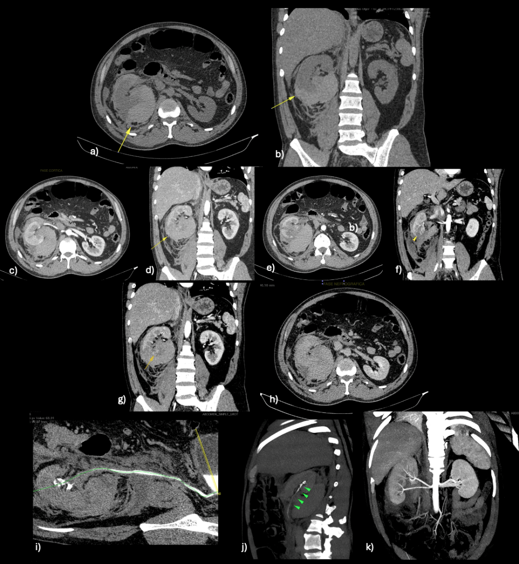
On July 14, CT urography was repeated 48 hours after the initial study. Active extravasation persisted in the corticomedullary phase. The hematoma was larger and showed heterogeneous attenuation related to the evolution of the hemorrhagic content; delayed acquisitions demonstrated contrast leakage at the level of the renal pelvis, compatible with collecting system involvement (Figure 2).
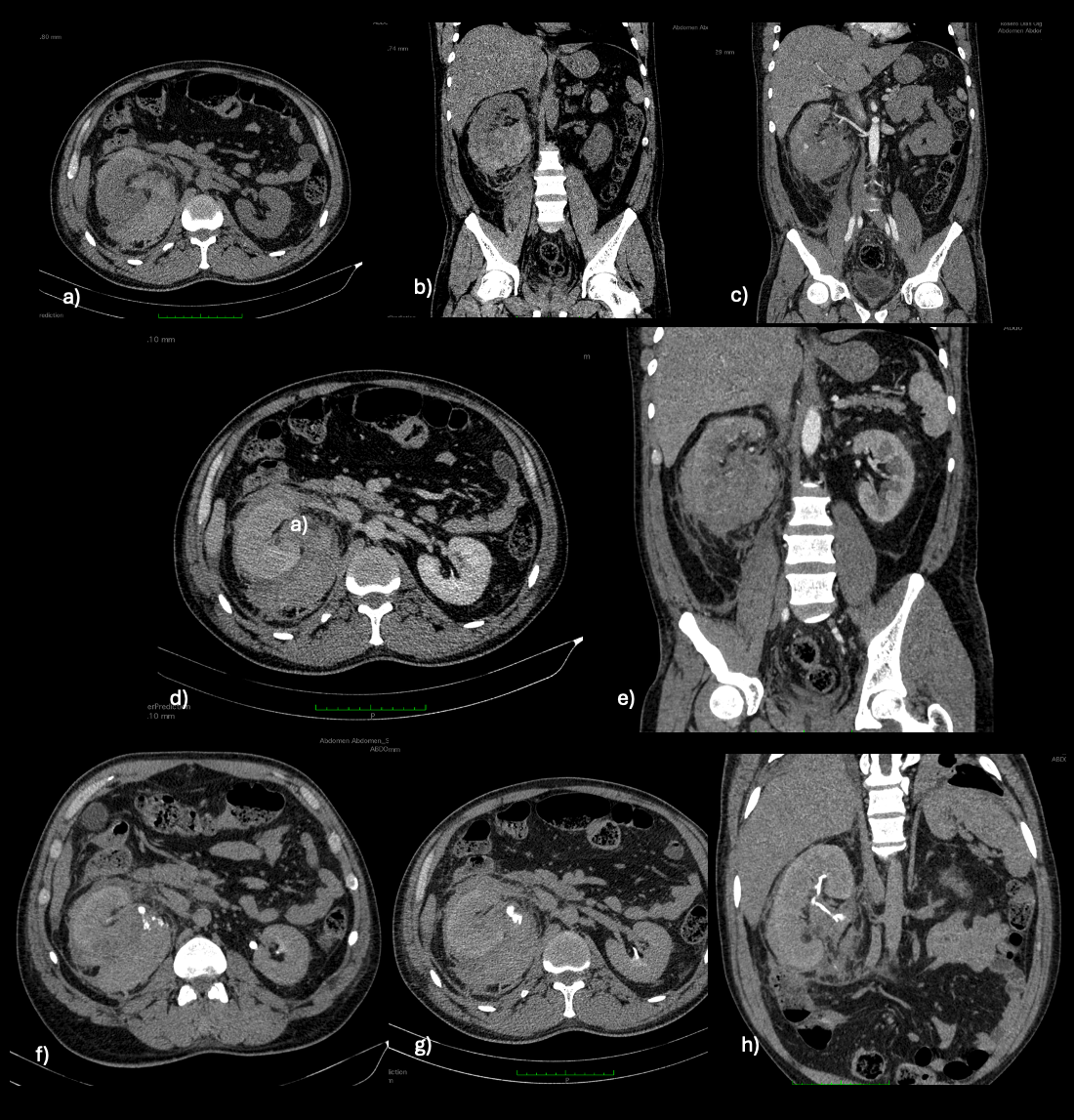
Hemoglobin decreased during the recorded period to 9.0 g/dL on July 15, with a hematocrit of 27.0% (Table 2). Creatinine peaked on July 13 at 2.00 mg/dL; subsequent values were 1.60 and 1.70 mg/dL, without returning to the admission value. Leukocytosis decreased. Clinical stability was documented during follow-up, with a progressive decrease in blood pressure to 100/60 mmHg and mild tachycardia of 110 beats/min on the last recorded day. The spontaneous perirenal hematoma supported the diagnosis of Wunderlich syndrome. Because of persistent extravasation and hematologic impact, the patient was transferred to Hospital Eugenio Espejo for renal embolization.
| Parameter | Admission July 12 | July 13 | July 14 | July 15 |
| Hemoglobin (g/dL) | 15.8 | 10.9 | 10.0 | 9.0 |
| Hematocrit (%) | 46.5 | 32.7 | 30.0 | 27.0 |
| Leukocytes (/µL) | 19 000 | 18 000 | 16 000 | 13 000 |
| Neutrophils (%) | 88.6 | 87.0 | 85.0 | 83.0 |
| Absolute neutrophils (/µL) | 16 834 | 15 660 | 13 600 | 10 790 |
| Creatinine (mg/dL) | 1.40 | 2.00 | 1.60 | 1.70 |
| PT | Normal | Normal | Normal | Normal |
| INR | Normal | Normal | Normal | Normal |
| aPTT | Normal | Normal | Normal | Normal |
PT: prothrombin time; INR: international normalized ratio; aPTT: activated partial thromboplastin time.
Discussion
Severe right-sided lumbar pain prompted the consultation despite initial hemodynamic stability. Hemorrhagic activity was documented by contrast extravasation and persisted on the July 14 follow-up study, accompanied by enlargement of the hematoma [3,4]. By that date, hemoglobin had decreased from 15.8 to 10.0 g/dL; the decline continued the following day. A decrease in blood pressure and tachycardia developed during this course. Therefore, the initial assessment alone did not reflect the degree of clinical impact that the bleeding would reach over the following days.
The absence of a visible mass poses a difficulty that has also been described in published series. In 13 of the 46 episodes analyzed by Jo et al., no distinct lesion was identified adjacent to the hematoma [1]. A small neoplasm may be obscured by blood, which supports follow-up imaging once the hematoma has evolved sufficiently to allow better assessment of the kidney [3]. In the surgical series by Tao et al., histologic examination identified one angiomyolipoma and two clear cell carcinomas in tissue obtained during surgery [5]. In this case, the cause was not established during the documented period.
Controlled arterial hypertension was the patient's only relevant medical history. By itself, it does not allow the hemorrhage to be attributed to hypertension. Coagulation times remained normal, and there was no anticoagulant use.
The collecting system showed no contrast leakage on the initial reconstructions. The finding was described on delayed acquisitions of the follow-up study, at the level of the renal pelvis, when the hematoma had already enlarged and the enhancement abnormalities persisted. It was considered compatible with collecting system involvement. Linear nonenhancing areas in the lower pole had been present since the first study; the mechanism linking both findings was not demonstrated, nor was parenchymal extension toward the pelvis. Comparison between the studies made it possible to recognize this change.
Creatinine reached 2.00 mg/dL and subsequently decreased, although the latest values remained above the admission level. The patient was referred to Hospital Eugenio Espejo for renal embolization in the setting of persistent bleeding, progressive hemoglobin loss, and changes in blood pressure and heart rate. Transarterial embolization allows targeted treatment of the hemorrhagic focus and has been used in both series and case reports of spontaneous renal hemorrhage [2,4,6]. The available information confirms the purpose of the transfer but does not establish whether the procedure was completed or what its outcome was.
Conclusion
In conclusion, the spontaneous hematoma progressed during the first 48 hours without a renal mass to explain the bleeding. Hemodynamic stability at admission coexisted with active hemorrhage and was followed by a decrease in hemoglobin, lower blood pressure, and mild tachycardia. Delayed contrast leakage compatible with collecting system involvement was identified on follow-up CT. The clinical course prompted transfer to Hospital Eugenio Espejo for renal embolization.
Declarations
Ethical Clearance
This report describes the patient's clinical care and imaging studies. The information is presented without direct personal identifiers, and no interventions performed for experimental purposes are described.
Informed consent
Written informed consent was obtained from the patient for publication of this case.
Acknowledgements
No additional acknowledgments.
Conflict of interest
The authors have no conflicts of interest to declare.
Funding/ financial support
This case report received no specific funding.