Introduction
Dengue is one of the most widespread mosquito-borne viral infections in the world and continues to place an enormous burden on health systems across the tropics and subtropics. The virus belongs to the Flaviviridae family and is spread mainly through the bites of Aedes aegypti and Aedes albopictus mosquitoes. Over the past few decades, rapid urban growth, expanding populations, a shifting climate and patchy vector control have together pushed dengue from an occasional outbreak into a firmly endemic disease in many developing countries, India among them [1][5]. Four antigenically distinct serotypes circulate, and while infection with one gives lasting immunity to that serotype, it offers only brief and partial protection against the others.
A later infection with a different serotype can actually make things worse through antibody-dependent enhancement, in which non-neutralising antibodies help the virus enter host cells and amplify the immune response. Shifts in the dominant circulating serotype have been linked to more severe outbreaks and a heavier disease burden in several regions [6][7]. Clinically, dengue spans a wide range, from a silent or mildly febrile illness to life-threatening dengue haemorrhagic fever and dengue shock syndrome. The classical illness begins abruptly with high fever, severe headache, retro-orbital pain, myalgia, arthralgia, nausea, vomiting and a rash [8][9]. Symptoms usually appear three to fourteen days after infection and settle within a week, though some patients slip into severe dengue marked by plasma leakage, bleeding, organ dysfunction and shock [10].
The central problem in severe dengue is a leaky vascular endothelium. Plasma escapes into tissue spaces and body cavities such as the pleura and abdomen, thickening the blood and producing hypotension, effusions, pulmonary oedema and, ultimately, shock. This leakage tends to start as the fever breaks and lasts one to two days, the so-called critical phase [11]. If this window is missed and fluid therapy is delayed, complications and death can follow. The World Health Organization criteria for dengue haemorrhagic fever rest on thrombocytopenia together with evidence of plasma leakage, such as a rising haematocrit, hypoalbuminaemia, pleural effusion or ascites [12]. The trouble is that these signs often show up late, after substantial leakage has already occurred, and a reliable, non-invasive early marker is still lacking, which is exactly why simple prognostic indicators matter. In endemic areas dengue also affects children heavily and carries real morbidity and mortality in that group [13].
Population studies from India have reported an annual dengue incidence of roughly 49.5 per 1,000 child-years among children with fever lasting more than three days. How severe the illness becomes depends on age, the infecting serotype, immune status and any comorbidities. Because the risk factors for severe disease are hard to pin down at presentation, many uncomplicated cases end up admitted for observation through the critical phase, straining hospitals, patients and families alike [14]. From the laboratory side, thrombocytopenia is the most consistent abnormality in dengue. Its causes are tangled, spanning marrow suppression, immune-mediated destruction, increased peripheral consumption, complement-mediated lysis and platelet apoptosis. Platelet count is a routine part of dengue care, but on its own it does not capture the full picture of platelet production, activation and consumption that shapes how the disease behaves [15].
Newer work suggests that platelet indices from automated analysers, mean platelet volume, platelet distribution width and plateletcrit, may help gauge severity. Mean platelet volume reflects average platelet size and indirectly indicates marrow activity; a high value points to the release of larger, younger platelets in response to peripheral destruction, while a low value suggests impaired production [16][17]. Platelet distribution width captures the variability in platelet size and rises during activation and morphological change [18][19]. Plateletcrit, the product of platelet count and mean platelet volume, measures the total circulating platelet mass and therefore reflects both the number and the character of platelets in a way the count alone cannot. Studies show it drops sharply in severe thrombocytopenia and tracks severity and bleeding risk more closely than platelet count [20][21]. A falling plateletcrit signals heightened consumption, while a rising one suggests recovery, making it a more rounded window on platelet behaviour during dengue [22].
Dengue does not stop at the blood. It also injures the vascular endothelium and the glomerular filtration barrier, and subtle renal involvement is common even without overt kidney failure, often surfacing as microalbuminuria, the leakage of small amounts of albumin into urine above normal but below what a dipstick detects. Microalbuminuria is a recognised early marker of endothelial dysfunction and capillary leak in diabetes, sepsis and critical illness, and both proteinuria and microalbuminuria have been documented in dengue, particularly during the acute and critical phases [23]. A raised urine albumin-to-creatinine ratio has been seen in dengue compared with other febrile illnesses, and higher urinary albumin excretion is tied to severe disease, plasma leakage and poorer outcomes [24]. Measuring microalbumin from a spot sample is a simple, non-invasive and inexpensive alternative to a 24-hour collection, which is especially valuable in acute care and resource-limited settings [25][26]. Bringing plateletcrit and urine microalbumin together, one reflecting platelet depletion and the other endothelial injury, may give a more timely read on severity than either alone. This study set out to examine how urine microalbumin and plateletcrit relate to disease severity in dengue, with the aim of sharpening early risk stratification.
Materials and Methods
This was a hospital-based observational cross-sectional study carried out in the Department of Medicine at Moti Lal Nehru Medical College and the associated Swaroop Rani Nehru Hospital, Prayagraj, over a period of one year. Approval was obtained from the Institutional Ethics Committee before the study began, and written informed consent was taken from every participant. All patients admitted with suspected dengue during the study period were considered for inclusion, and the final cohort comprised 108 patients in whom dengue was confirmed serologically.
Patients aged 15 years and above of either sex with confirmed dengue were included. Those with comorbidities likely to confound the markers of interest, namely hypertension, diabetes mellitus, chronic liver disease or chronic kidney disease, were excluded, as were patients taking ACE inhibitors or dapagliflozin, pregnant women, and anyone who declined consent. Because this was a purposive, hospital-based study, all dengue patients presenting during the period who met the criteria were enrolled consecutively rather than sampled at random.
Each patient underwent a detailed clinical assessment at admission, with a structured history covering fever pattern, arthralgia, headache, rash, vomiting, abdominal pain and any bleeding, followed by a full general and systemic examination. Vital signs, including blood pressure, pulse and temperature, were recorded, and patients were examined for hepatomegaly, splenomegaly, pleural effusion and ascites. Routine investigations included a complete blood count with platelet count and plateletcrit measured by automated haematology analyser (electronic impedance), liver function tests (SGPT and SGOT), and renal function tests (serum urea and creatinine). Dengue was confirmed using NS1 antigen and IgM antibody serology. Urine microalbumin was estimated by the pyrogallol red colorimetric method, in which urinary protein reacts with pyrogallol red and molybdate to form a coloured complex whose intensity, read spectrophotometrically, is proportional to protein concentration.
Patients were classified into mild dengue (without warning signs), moderate dengue (with warning signs) and severe dengue according to standard WHO criteria, severe dengue being defined by plasma leakage leading to shock or fluid accumulation with respiratory distress, severe bleeding, or severe organ impairment. All data were entered on a structured case record form and analysed statistically. Continuous variables are expressed as mean ± standard deviation and were compared across severity groups using one-way ANOVA with post-hoc pairwise testing; categorical variables were compared using the chi-square test. Univariate linear regression identified factors associated with severity, and a receiver operating characteristic (ROC) curve assessed the predictive value of urine microalbumin. A p-value below 0.05 was taken as statistically significant.
Results
A total of 108 confirmed dengue patients were studied. Their demographic and serological profile is summarised in Table 1. The cohort was predominantly male (72.2%), and cases were spread fairly evenly across adult age bands, with the 21–30 and 51–60 year groups each contributing the largest share (20.4%). NS1 antigen and IgM antibody were each positive in exactly half of the patients, while none tested positive for IgG, consistent with a population of largely primary, acute-phase infections.
| Number (n) | Percentage (%) | ||
| Age | ≤20 years | 16 | 14.8 |
| 21–30 years | 22 | 20.4 | |
| 31–40 years | 16 | 14.8 | |
| 41–50 years | 18 | 16.7 | |
| 51–60 years | 22 | 20.4 | |
| >60 years | 14 | 13.0 | |
| Mean±SD | 40.15 ± 15.97 | ||
| Gender | Male | 78 | 72.2 |
| Female | 30 | 27.8 | |
| Serological profile | NS1 antigen positive | 54 | 50.0 |
| IgM antibody positive | 54 | 50.0 | |
| IgG antibody positive | 0 | 0.0 |
The clinical picture is presented in Table 2. Constitutional and classical dengue features dominated: lethargy (89.8%), arthralgia (88.9%) and headache (75.9%) were the most frequent complaints. Among the signs of plasma leakage, pleural effusion was the commonest, seen in 43.5% of patients, followed by ascites and hepatomegaly, while splenomegaly was not observed at all. Bleeding manifestations such as epistaxis, melaena and haematemesis were relatively uncommon.
| Symptom / Sign | Number (n) | Percentage (%) |
| Lethargy | 97 | 89.8 |
| Joint pain (arthralgia) | 96 | 88.9 |
| Headache | 82 | 75.9 |
| Rashes | 49 | 45.4 |
| Vomiting | 40 | 37.0 |
| Retro-orbital pain | 32 | 29.6 |
| Abdominal pain | 17 | 15.7 |
| Dysgeusia | 12 | 11.1 |
| Epistaxis | 9 | 8.3 |
| Melaena | 8 | 7.4 |
| Saddle-back fever | 2 | 1.9 |
| Haematemesis | 2 | 1.9 |
| Pleural effusion | 47 | 43.5 |
| Ascites | 16 | 14.8 |
| Hepatomegaly | 12 | 11.1 |
| Splenomegaly | 0 | 0.0 |
When patients were graded by severity, half had mild dengue without warning signs, 42.6% had moderate dengue with warning signs, and 7.4% had severe dengue. Table 3 sets out the haemodynamic and haematological findings across these groups. Age did not differ significantly between the groups (p = 0.142). Blood pressure, however, fell steeply in severe dengue, with mean systolic pressure dropping to 83.5 mmHg (p < 0.001). The most striking laboratory finding was the progressive collapse of both platelet count and plateletcrit as disease worsened: plateletcrit fell from 0.18 in mild dengue to 0.06 in severe dengue (p < 0.001), mirroring the fall in platelet count.
| Mild | Moderate | Severe | p-value | |
| Systolic BP (mmHg) | 106.96 ± 7.14 | 104.04 ± 8.02 | 83.50 ± 1.77 | <0.001 |
| Diastolic BP (mmHg) | 74.22 ± 5.48 | 72.09 ± 8.24 | 56.50 ± 1.41 | <0.001 |
| Haemoglobin (g/dL) | 13.13 ± 1.49 | 13.66 ± 1.54 | 12.15 ± 0.68 | 0.018 |
| Neutrophils (%) | 66.96 ± 10.0 | 67.80 ± 8.22 | 58.34 ± 13.46 | 0.038 |
| Platelet count (×10⁵/µL) | 1.03 ± 0.22 | 0.51 ± 0.23 | 0.17 ± 0.08 | <0.001 |
| Plateletcrit (%) | 0.18 ± 0.04 | 0.11 ± 0.06 | 0.06 ± 0.02 | <0.001 |
| TLC (/µL) | 6386 ± 1616 | 6240 ± 2012 | 5755 ± 2208 | 0.654 |
BP, blood pressure; TLC, total leucocyte count. Comparisons by one-way ANOVA.
The biochemical profile, shown in Table 4, told a similar story of escalating organ involvement. Liver enzymes climbed sharply with severity, SGPT reaching 319.4 IU/L and SGOT 353.0 IU/L in severe dengue (both p < 0.001), and serum creatinine rose significantly to 1.82 mg/dL. Most notably, urine microalbumin, the marker of chief interest, was more than doubled in severe dengue at 54.91 mg/L compared with roughly 19–21 mg/L in the mild and moderate groups (p < 0.001).
| Mild | Moderate | Severe | p-value | |
| SGPT (IU/L) | 129.04 ± 78.21 | 168.17 ± 80.53 | 319.43 ± 140.96 | <0.001 |
| SGOT (IU/L) | 155.30 ± 88.34 | 190.54 ± 78.63 | 352.99 ± 143.36 | <0.001 |
| Serum urea (mg/dL) | 42.55 ± 16.29 | 39.65 ± 18.85 | 55.63 ± 24.11 | 0.073 |
| Serum creatinine (mg/dL) | 1.21 ± 0.33 | 1.24 ± 0.41 | 1.82 ± 0.57 | <0.001 |
| Urine microalbumin (mg/L) | 18.96 ± 6.31 | 20.80 ± 4.67 | 54.91 ± 12.40 | <0.001 |
Disease severity also translated directly into longer hospitalisation. Of the whole cohort, 42 patients (38.9%) were managed as outpatients, all with mild disease, while 66 required admission. Among those admitted, mean length of stay lengthened from 3.67 days in mild dengue to 9.00 days in severe dengue (p < 0.001), and every pairwise comparison between severity groups was significant. Univariate regression (Table 5) confirmed that lower platelet count and higher urine microalbumin were significantly associated with greater severity, alongside rising transaminases, older age and longer hospital stay.
| B | t | p-value | |
| Age | 0.003 | 2.18 | 0.032 |
| Platelet count at admission | −0.79 | −6.18 | <0.001 |
| Urine microalbumin | −0.014 | −3.58 | 0.001 |
| SGPT | 0.0029 | 5.16 | <0.001 |
| SGOT | 0.0027 | 4.89 | <0.001 |
| Total duration of hospital stay | 0.15 | 6.45 | <0.001 |
| Plateletcrit value | −0.22 | −0.32 | 0.753 |
| Serum creatinine | −0.03 | −0.39 | 0.696 |
B, unstandardised regression coefficient. Only selected variables shown.
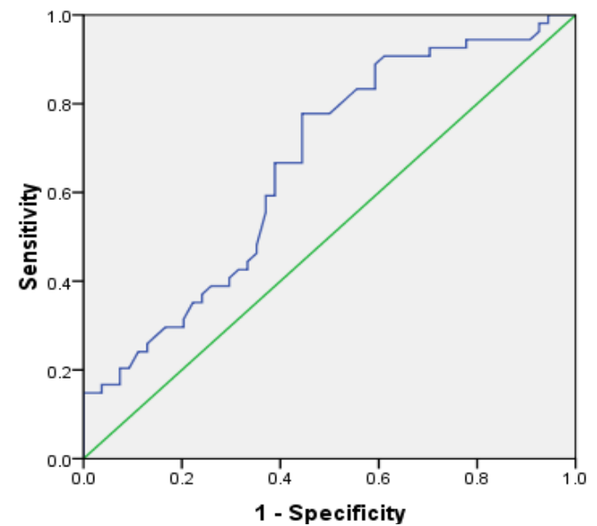
Finally, the discriminating ability of urine microalbumin for severe dengue was tested using ROC analysis (Table 6 and Fig. 1). A cut-off of 19.50 mg/L gave a sensitivity of 72.2% and a specificity of 65.6%, with an area under the curve of 0.663 (p = 0.004), indicating moderate but statistically significant predictive value for identifying severe disease.
| Cut-off (mg/L) | Sensitivity (%) | Specificity (%) | AUC | 95% CI | p-value |
| 19.50 | 72.2 | 65.6 | 0.663 | 0.56–0.77 | 0.004 |
AUC, area under the curve; CI, confidence interval.
Discussion
This study set out to see whether two simple, inexpensive laboratory markers, plateletcrit and urine microalbumin, could track the severity of dengue, and the results are clear on both counts. As disease worsened, plateletcrit fell steadily while urine microalbumin climbed, and the two together painted a consistent picture of intensifying platelet consumption and endothelial leak. My cohort was predominantly young to middle-aged and male, a pattern that echoes much of the published literature. Male preponderance in dengue is well described and is usually attributed to greater outdoor exposure and daytime Aedes biting, and my male share of 72.2% sits comfortably alongside earlier hospital-based series [16][22]. The even spread across adult age bands, without a significant age difference between severity groups, likewise fits the view that dengue affects all adult ages, with the peak varying by region and setting.
The clinical spectrum we observed, dominated by arthralgia, headache and lethargy, with pleural effusion the leading sign of leakage and splenomegaly conspicuously absent, matches the classical and warning-sign profile laid out in WHO guidance. That pleural effusion topped the list of signs is itself telling, since it reflects the capillary leak that defines moderate and severe disease [11][12]. Half of my patients had mild disease and only a small minority progressed to severe dengue, which is what one expects in a non-epidemic, hospital-based population and mirrors the severity distribution reported elsewhere.
The haemodynamic findings reinforced the pathophysiology. Blood pressure, particularly systolic pressure, dropped steeply in severe dengue while remaining stable across mild and moderate cases, consistent with the plasma leakage and circulatory compromise that characterise the critical phase [11]. Alongside this, the fall in platelet count with rising severity was one of the most robust signals in my data, in keeping with the recognised mechanisms of thrombocytopenia in dengue, which range from marrow suppression to immune-mediated destruction and increased peripheral consumption [15].
Where my study adds value is in plateletcrit. Rather than simply restating the platelet count, plateletcrit fell significantly at every step from mild to moderate to severe disease, behaving as an independent gradient that closely shadowed severity. This aligns with work showing that plateletcrit and related indices track thrombocytopenia severity and disease progression better than the count alone, and that a declining plateletcrit marks heightened consumption while a rising value heralds recovery [16][17][20][21]. Because it reflects the total circulating platelet mass, plateletcrit captures both the quantity and the character of platelets in a single, automated, no-extra-cost number, which makes it an attractive bedside marker [22].
The biochemical results extended the same theme of multi-organ involvement. Both transaminases rose sharply and significantly in severe dengue, pointing to the hepatic injury, part direct viral, part immune-mediated, that is most pronounced in the critical phase [13]. Serum creatinine also rose significantly in severe cases, consistent with the renal hypoperfusion and direct viral effects that can culminate in acute kidney injury. These findings underline that severe dengue is rarely confined to one organ system and that leak, liver and kidney injury tend to move together.
The urine microalbumin results were perhaps the most compelling. Levels were roughly comparable in mild and moderate dengue but jumped more than two-fold in severe disease, and microalbumin emerged as a significant correlate of severity on regression. On ROC analysis it offered moderate discrimination for severe dengue, with a cut-off around 19.5 mg/L. This dovetails with a substantial body of work on urinary protein markers in dengue, in which raised albumin-to-creatinine or protein-to-creatinine ratios have repeatedly been linked with plasma leakage, haemorrhagic complications and adverse outcomes, and have performed as useful predictors of severe disease [23][24]. Mechanistically, microalbuminuria is a well-established readout of generalised endothelial dysfunction, and in dengue, where widespread endothelial activation and vascular leak sit at the heart of severe disease, its rise is biologically coherent [25][26]. A spot urine test is simple, non-invasive and cheap, which makes it particularly suited to the acute setting and to resource-limited environments where early risk stratification matters most.
The clinical and economic weight of getting this right is captured by the length-of-stay data. Hospital stay lengthened markedly and significantly with severity, and severity in turn was mirrored by my two markers of interest. Identifying, early and cheaply, the patients likely to deteriorate could therefore ease pressure on beds and resources while improving outcomes. Taken as a pair, a falling plateletcrit and a rising urine microalbumin give complementary information, one on platelet dynamics and the other on endothelial integrity, that may flag impending severe dengue before the classical late signs appear.
Several limitations temper these conclusions. The sample was modest, and the severe dengue group in particular was small, which limits precision and generalisability. As a single-centre study, the findings may not transfer directly to other populations. We measured plateletcrit and microalbumin at a single time point rather than serially, so we could not chart how they evolve through the illness, and we did not fully account for every factor that can influence microalbuminuria. Larger, multi-centre studies with serial sampling are needed to confirm these associations and to establish standardised cut-offs.
Conclusion
In this cohort of adult dengue patients, plateletcrit and urine microalbumin moved in opposite directions as disease severity increased. Plateletcrit fell steadily while microalbumin rose sharply, each tracking the underlying platelet depletion and endothelial leak that drive severe dengue. Both are simple, inexpensive and widely available, and used together they provide clinicians with an early, practical signal of patients at risk of progressing to severe disease. Their routine use, ideally with serial monitoring and alongside clinical assessment, could aid timely intervention, particularly in resource-limited settings, though larger multicentre studies are warranted to validate their prognostic value and define standardised thresholds.
Declarations
Ethical Approval
Proper Institutional Ethical committee approval was taken from the institute ‘Moti Lal Nehru Medical College Prayagraj, (Ethics Committee Registration No. ECR/922/inst/UP/RR-22 issued under New Drugs and Clinical Trial Rules, 2019).
Patient Consent
The authors certify that they have obtained all appropriate patient consent involved in study.
Conflicts of interest
None
Financial support and sponsorship/Funding
Nil
Consent to Participate
All patients were informed about the study and proper consent taken after explaining the advantages and risks to the patient.
Written Consent for Publication by Patients
The subjects involved in this study, have given their consent for getting their data to be published in your journal ‘SNCCM’. We declare that this manuscript is original, has not been published before and is not currently being considered for publication elsewhere and we give consent to be published in your journal ‘SNCCM’.
Availability of Data and Material
No data taken from outside.
Contribution of Author
A.S, K.S Chose the topic, planned the execution. All evaluated the subjects and collected the data. All wrote the manuscript. K.S, A.K.C and P.G made the tables. All reviewed the manuscript.