Introduction
Pelvic ectopic kidney is a relatively uncommon congenital renal /malformation, with an estimated incidence of 1 in 2,200 to 1 in 3,000 individuals. The abnormal position is often associated with malrotation of the ipsilateral side, altered anatomy of the pelvicalyceal system and anomalous renal vasculature [1].
This altered anatomy makes the management of renal stone disease challenging in an ectopic kidney. Moreover, the pelvic kidney which is anterior to the sacrum is often surrounded by bowel loops. Consequently, a conventional fluoroscopic guided Percutaneous nephrolithotomy (PCNL) carries a significantly higher risk of injury to the adjacent bowel loops and major blood vessels [2]. While RIRS remains a feasible option in patients with stones in anomalous kidneys; a tortuous ureter, high ureteral insertion and altered intrarenal anatomy may make access and complete stone clearance difficult. SWL is also less effective due to difficulty in positioning the stone within the focal zone and impaired fragment drainage. In patients in whom these approaches are unsuccessful or unsuitable, laparoscopic-assisted stone surgery provides an alternative by allowing direct visualization of the kidney and surrounding structures [2][3].
Esghi and colleagues first reported using laparoscopically assisted percutaneous nephrolithotomy (PCNL) for a pelvic ectopic kidney in 1985. Since then, several modifications of the technique have been described, including transperitoneal and extraperitoneal approaches [3][4].
In our study, we report our experience and outcomes with laparoscopic-assisted PCNL in six patients with pelvic ectopic kidney and renal calculi.
Materials and Methods
A retrospective analysis was conducted at our tertiary care hospital after obtaining institutional and ethical committee clearance. All patients with a diagnosis of pelvic ectopic kidney with renal lithiasis, who underwent laparoscopic-assisted PCNL between 2017 and 2023 were identified and included in the study. A total of six patients were included of whom five patients were male and one was a female. 4/6 patients had previously undergone unsuccessful RIRS and 2/6 had prior failed SWL.
Inclusion Criteria
Pelvic ectopic kidney with a solitary renal calculus measuring >10 mm.
Exclusion Criteria
Stone size <10mm, non-urological contraindications to laparoscopic surgery, active urinary tract infection, pregnancy, haematological abnormalities.
Apart from a detailed clinical evaluation and routine investigations including urine culture, as per institutional protocol, all patients underwent preoperative CT urography to identify the position of the ectopic kidney, assess stone characteristics, describe pelvicalyceal anatomy and evaluate the relationship of the kidney with the surrounding structures.
Surgical technique
All procedures were performed under general anaesthesia after obtaining informed/written consent. An intravenous antibiotic was administered at induction and patient initially positioned in the dorsal lithotomy position. Under aseptic precautions, a 6-French ureteric catheter was inserted under fluoroscopic guidance and retrograde pyelography was performed to delineate the location of the ectopic kidney and the pelvicalyceal anatomy. This information was used to determine the optimal and ergonomically feasible position of the laparoscopic trocars.
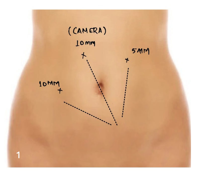
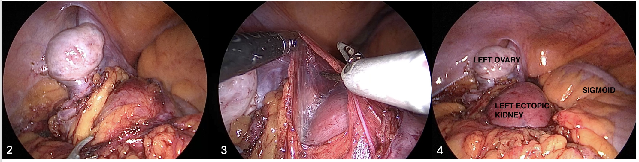
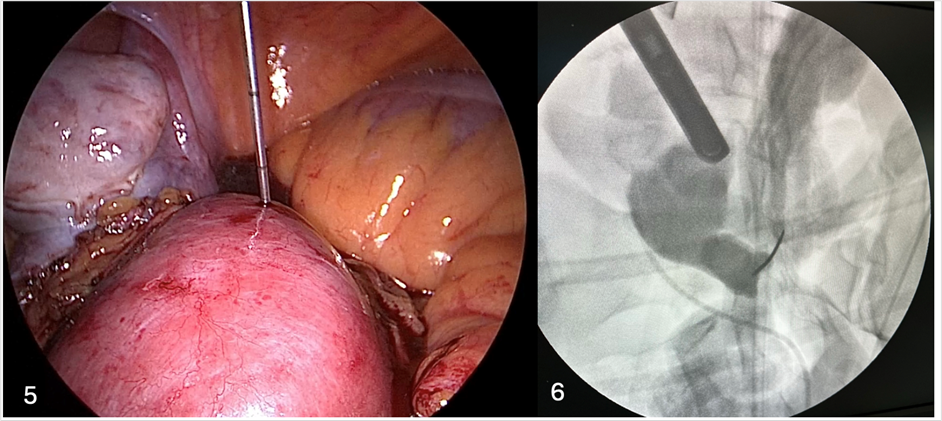
Three laparoscopic trocars were used: two 10-mm ports and one 5-mm port Standard trocar placement for all patients was configured as shown in Figure 1. After creating pneumoperitoneum and placement of the trocars, the descending and sigmoid colon were mobilized medially to enter the retroperitoneal space. Gerota's fascia was opened, and adequate dissection done till the anterior surface of the kidney was exposed and optimally visualised. (Figure 2-4) The desired calyx was punctured percutaneously under combined laparoscopic and fluoroscopic guidance. (Figure 5,6)
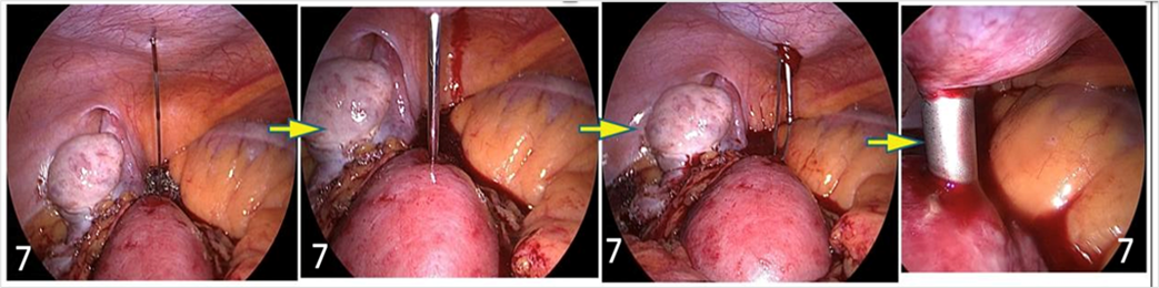
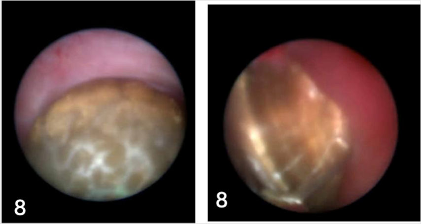
Once a successful access was obtained, the intra-abdominal pressure was reduced from 14 mm Hg to 8 mm Hg. The percutaneous tract was serially dilated up to 30 French using coaxial Alken’s metal dilators. A 30-F Amplatz sheath was placed and using 26-French rigid nephroscope, standard nephroscopic examination was performed. The stones were fragmented using a pneumatic lithotripter and the fragments were extracted (Figure 7,8).
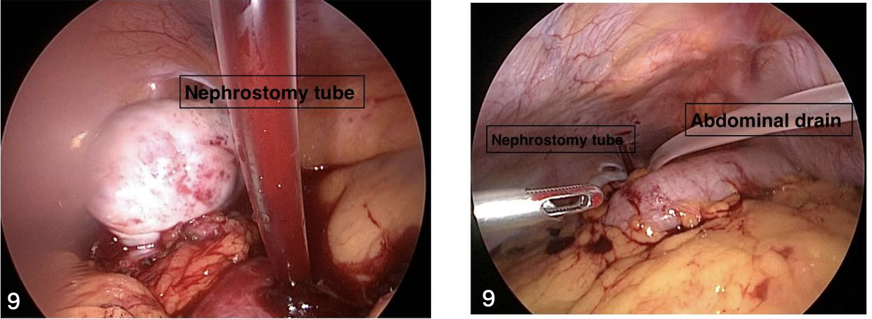
On confirming clearance of the stone both visually and under fluoroscopy, a 5/26 French DJ stent placed and a 16-French nephrostomy tube placed through the access tract. The Amplatz sheath was removed, and a 12-French abdominal drain was placed within the peritoneal cavity adjacent to the kidney (Figure 9). Pneumoperitoneum was s released and the laparoscopic port sites were closed and the drains were secured.
Nephrostomy tube was removed on post operative day 2, Intrabdominal drain was removed on day 3. All patients underwent a non-contrast CT KUB (NCCT KUB) at 1 month post procedure to determine the clearance rates. A successful stone clearance was defined by no stone or residual fragment measuring <4 mm on NCCT KUB at 1 month post procedure.
Parameters recorded including age, sex, stone size, side of stone, total operative time of procedure, complications, duration of hospitalization and stone-free status at 1 month.
Results
Six patients underwent laparoscopic-assisted PCNL during the study period. The mean age was 31.17 4.45 years, 5/6 patients were male. The mean stone size was 18 4 mm (Table 1).
All six patients had stones in the left pelvic ectopic kidney. 4/6 stones were located in the left lower calyx, 1 in the left middle calyx and 1 in the left renal pelvis. The mean operative time was 90 18.97 minutes. Mean drop in Hb was < 1gm%
The nephrostomy tube was removed on postoperative day 2 in all patients. The abdominal drain and Foley catheter were removed on postoperative day 3. The mean duration of hospitalization was 4 days.
No significant intraoperative or postoperative complications were recorded in 5 patients. One patient developed post operative ileus which was managed conservatively.
At the one-month postoperative visit, all patients underwent NCCT KUB to assess the stone clearance rate. 5/6 patients met the predefined stone-free criterion. One patient with a 5-mm residual stone in the left lower calyx was managed conservatively. The overall stone-free rate in our series was 83.3%.
| Patient | Age (years) | Sex | Stone size (mm) | Side | Stone location | Operative time (min) | Complications | Hospital stay (days) | Stone-free status at 1 month |
| 1 | 25 | M | 12 | Left | Lower calyx | 70 | None | 3 | Stone-free |
| 2 | 32 | M | 18 | Left | Middle calyx | 90 | None | 3 | Stone-free |
| 3 | 30 | M | 16 | Left | Lower calyx | 70 | None | 4 | Stone-free |
| 4 | 37 | M | 18 | Left | Lower calyx | 90 | None | 4 | Stone-free |
| 5 | 28 | F | 20 | Left | Renal pelvis | 100 | Ileus | 5 | Stone-free |
| 6 | 35 | M | 24 | Left | Lower calyx | 120 | None | 5 | 5-mm residual stone |
Discussion
Pelvic ectopic kidney is usually located in the retroperitoneum, anterior to the sacrum surrounded by bowel loops. An associated malrotation, altered calyceal orientation and anomalous renal vessels pose a significant challenge in the management of renal calculi in such kidneys. Hence, a blind puncture during PCNL is not advisable due to a higher risk of injury to the surrounding bowel and major vessels [1][2][5].
Alternative treatment modalities for the management of renal stones in ectopic pelvic kidneys include SWL, RIRS and open/laparoscopic pyelolithotomy. SWL in pelvic kidneys may be difficult to perform due to the surrounding pelvic bones that interfere with stone focusing. Associated ureteral anatomy abnormality leading to poor drainage can impair the drainage and thereby clearance of stone fragments [6].
RIRS is the preferred treatment option in many centres for ectopic kidney with stone disease. Weizer et al.,[7] has reported a 75% success rate following RIRS for renal calculi in pelvic and horseshoe kidneys. Ugurlu et al.,[8] however, reported a lower success rate of 61.5% following a single session of RIRS in 13 patients with ectopic kidneys. While the literature is still emerging, one should be aware that the access to the collecting system can be difficult, particularly, because of abnormal ureteral anatomy and the altered anatomical orientation of the renal pelvis and calyces [5].
Laparoscopic-assisted PCNL is a suitable alternative in patients when RIRS/SWL is not preferred, before considering open stone surgery. The benefit lies is the ability to directly expose and visualize the kidney and adjacent structures during puncture of the kidney limiting surrounding structure injury. Eshghi et al. first described a laparoscopic-assisted percutaneous approach to a pelvic kidney in 1985 [3]. The procedure gained further evidence when Holman and Toth reported successful transperitoneal laparoscopic-assisted PCNL in 15 patients. They modified the procedure by mobilising the bowel loops to expose the kidney laparoscopically for percutaneous access [9].
Since then, various technical modifications have been described by many [4][10]. Zafar and Lingeman [10] described a tubeless procedure with intracorporeal suturing of the nephrotomy site. Troxel et al.,[11] performed laparoscopic-assisted PCNL via extraperitoneal approach, while Soylemez et al.,[12] described both mesocolon dissection and a transmesocolic puncture technique to reduce the extent of bowel mobilization and peritoneal dissection thereby reducing postoperative ileus. The modifications establish that the management of stones in pelvic ectopic kidneys needs to be individualized according to renal position, stone burden, collecting-system anatomy and surgeon expertise [10-14].
In our study, all six patients had a solitary renal stone measuring >10 mm. The mean stone size was 18 mm, with a range of 12–24 mm. Four patients had previously failed RIRS and two had previously failed SWL. The cohort represented patients in whom conventional minimally invasive treatment had been unsuccessful.
A transperitoneal laparoscopic approach was used in all cases. The key advantage with our technique is that the renal access was obtained under combined laparoscopic and fluoroscopic guidance after bowel mobilization and exposure of the ectopic kidney ensuring direct visualization of the kidney during puncture. Simultaneous fluoroscopy used to confirm precise entry into the desired collecting system.
Another key step in our technique was the reduction of intra-abdominal pressure from 14 mmHg to 8 mmHg after renal puncture and before tract dilatation. This enabled the continuous laparoscopic monitoring during percutaneous tract dilatation.
The mean operative time was 90 minutes. Five of six patients achieved the predefined stone-free status at one month, resulting in a stone-free rate of 83.3%. The single residual stone measured 5 mm and was located in the lower calyx; this patient was managed conservatively. One patient developed post operative ileus which was managed conservatively. Our stone-free rate is comparable to the results reported in the existing literature [10-14].
The limitations of this study include small sample size of six patients, a retrospective study, single-center design and lack of a comparator group. Hence a direct comparison of outcomes with alternative procedures such as RIRS, SWL, open surgery or other PCNL approaches cannot be made.
Nevertheless, In patients with pelvic ectopic kidneys, with large stones or when conventional endourological treatments have failed, laparoscopic-assisted PCNL should be considered. This technique identifies kidney in a controlled manner, while evaluating the surrounding structures and provides a precise percutaneous access without the need for a blind puncture.
Conclusion
Laparoscopic-assisted PCNL is a safe and effective minimally invasive approach for the management of renal calculi in selected patients with pelvic ectopic kidneys. Combined laparoscopic and fluoroscopic guidance enables visualization of the kidney and surrounding structures allowing for a controlled and safe percutaneous access with minimal complications. Treatment should, however, be individualized according to stone characteristics, renal anatomy, previous treatment, available expertise and institutional resources.
Declarations
Ethics approval
Ethical approval was obtained from the Institutional Ethics Committee of Ramaiah Medical College, Bengaluru, India (Approval No.: MSRMC/EC/AP-04/188-29).
Informed Consent
Written informed consent was obtained from the participant for the publication of the clinical data.
Data Availability
The datasets used and/or analysed during the current study are available from the corresponding author on reasonable request.
Competing Interests
The authors declare that they have no competing interests.
Financial Support or Sponsorship
None
Authors' contributions
Author 1: Concept, Design, Supervision, Resources, Writing Manuscript, Analysis and/or Interpretation, Critical Review
Author 2: Design, Data Processing, Analysis and/or Interpretation, Literature Search, Critical Review
Author 3: Data Collection, Processing, Analysis and/or Interpretation, Writing Manuscript
Author 4: Data Processing, Analysis, Critical Review
Acknowledgements
Nil.