Introduction
Stroke remains one of the most prevalent and disabling neurological illnesses worldwide, ranking as the second leading cause of death after ischaemic heart disease and a leading contributor to years lived with disability [1][2]. Its burden continues to rise with increasing life expectancy, urbanisation and the growing prevalence of vascular risk factors such as hypertension, diabetes, dyslipidaemia and smoking, particularly in low- and middle-income countries.
Two broad categories are recognised. Acute ischaemic stroke (AIS), accounting for roughly 80–85% of cases, results from sudden occlusion of a cerebral artery, whereas haemorrhagic stroke (HS) follows rupture of a blood vessel and carries disproportionately higher early mortality [2][3]. Despite advances in neuroimaging, thrombolysis, mechanical thrombectomy and neurocritical care, a substantial proportion of survivors are left with lasting neurological impairment [3][4].
Clinical course after stroke is notoriously heterogeneous: patients with comparable stroke subtype and radiological findings may follow markedly different trajectories, shaped by age, sex, lesion size and location, comorbidities, baseline severity and quality of care. Validated instruments such as the National Institutes of Health Stroke Scale and the modified Rankin Scale capture neurological and functional status but do not directly reflect the physiological stress response driving secondary injury. This has prompted growing interest in blood-based biomarkers that can complement clinical and imaging assessment by providing an objective window onto the systemic stress, inflammatory and neuroendocrine changes that follow a stroke.
Acute stroke is a potent physiological stressor that activates the hypothalamic–pituitary–adrenal (HPA) axis and perturbs thyroid hormone metabolism [5][6]. Cortisol, the terminal effector of the HPA axis, mobilises glucose, sustains vascular tone and modulates immune function during acute illness, but an excessive or prolonged rise is maladaptive: higher circulating cortisol has repeatedly been linked to greater stroke severity, larger infarct volume and increased mortality [7][9], alongside hyperglycaemia, endothelial dysfunction and heightened susceptibility to infection.
Triiodothyronine (T3), the biologically active thyroid hormone derived from thyroxine (T4), is essential for neuronal survival, synaptic transmission, myelination and mitochondrial function [10][11]. A transient fall in serum T3 with preserved or low T4 and thyroid-stimulating hormone (TSH) commonly termed low-T3 syndrome or non-thyroidal illness syndrome is well described during acute illness, including stroke, and has been linked to greater severity, poorer functional recovery and higher mortality [12][13]. Experimentally, T3 limits neuroinflammation, glutamate-mediated excitotoxicity and promotes neurogenesis and brain-derived neurotrophic factor expression, suggesting a genuinely neuroprotective role rather than a mere epiphenomenon of illness severity.
Cortisol and T3 have chiefly been studied in isolation, yet the HPA and hypothalamic–pituitary–thyroid axes are closely interconnected: elevated cortisol and pro-inflammatory cytokines such as interleukin-6 can inhibit peripheral deiodinase activity, impairing conversion of T4 to T3 and precipitating low-T3 syndrome during acute stress [14][15]. This neuroendocrine crosstalk may compound neuronal injury and worsen recovery, yet head-to-head comparisons of cortisol and T3 as joint prognostic markers across both ischaemic and haemorrhagic stroke, and specifically contrasting survivors with non-survivors remain limited, particularly among South Asian populations where most available studies examine a single hormone or stroke subtype.
Given the rising burden of stroke, its increasing occurrence at a younger age, and the scarcity of rehabilitation resources in many developing health systems, there is a pressing need for simple, inexpensive and widely available predictive biomarkers. Serum cortisol and serum T3 are routinely measurable, low-cost assays that could fulfil this role. This study therefore examined the association between serum cortisol and serum T3 and clinical outcome in patients with acute ischaemic and haemorrhagic stroke, with the aim of improving early risk stratification and informing timely management.
Aims and Objectives
-
To compare serum cortisol and serum T3 as prognostic markers in acute ischaemic and haemorrhagic stroke.
-
To evaluate serum cortisol as a prognostic marker distinguishing survivors from non-survivors in acute ischaemic and haemorrhagic stroke.
-
To evaluate serum T3 as a prognostic marker distinguishing survivors from non-survivors in acute ischaemic and haemorrhagic stroke.
Materials And Methods
Study Design and Setting
This cross-sectional observational study was conducted in the Department of General Medicine of a tertiary-care teaching hospital in northern India over a one-year period (2025–2026), following approval from the Institutional Ethics Committee. Written informed consent was obtained from all participants or their attendants, and patient confidentiality was maintained throughout.
Participants
Patients aged 18 years or older with acute ischaemic or haemorrhagic stroke, confirmed by computed tomography (CT) or magnetic resonance imaging (MRI) and presenting within 72 hours of symptom onset, were eligible. Based on an expected proportion of 8% and a precision of 5% at 95% confidence, the calculated minimum sample size was 113; 120 patients were ultimately enrolled. Patients with a history of thyroid disorder or thyroid hormone replacement, Cushing's syndrome, adrenal insufficiency or other known endocrine disease, current corticosteroid therapy, severe concurrent infection, malignancy or recent major surgery were excluded, to minimise confounding of the cortisol–thyroid axis.
Clinical and Radiological Assessment
Demographic data (age, sex, body mass index), vascular risk factors (hypertension, diabetes mellitus, smoking, alcohol use) and time from symptom onset to presentation were recorded at admission. Stroke severity was graded using the National Institutes of Health Stroke Scale (NIHSS) and level of consciousness using the Glasgow Coma Scale (GCS); functional outcome at discharge was assessed with the modified Rankin Scale (mRS). All patients underwent CT or MRI to confirm stroke subtype and to quantify infarct volume (ischaemic stroke) or haematoma volume (haemorrhagic stroke).
Laboratory Investigations
Fasting venous blood samples were drawn within 24 hours of admission. Serum cortisol was measured by a competitive immunoenzymatic (chemiluminescent) assay, and serum T3, T4 and TSH by radioimmunoassay or electrochemiluminescence immunoassay; laboratory personnel were blinded to patients' clinical details to reduce assessment bias. Fasting blood sugar, glycated haemoglobin (HbA1c), a fasting lipid profile, renal function and serum electrolytes were obtained concurrently.
Outcome Assessment and Statistical Analysis
Patients were followed throughout their hospital stay and up to 30 days, with the primary outcome recorded as survived or expired. Data were entered in Microsoft Excel and analysed using SPSS. Continuous variables are expressed as mean ± standard deviation and were compared using Student's t-test; categorical variables are expressed as number and percentage and were compared using the chi-square test. A two-tailed p-value < 0.05 was considered statistically significant.
Results
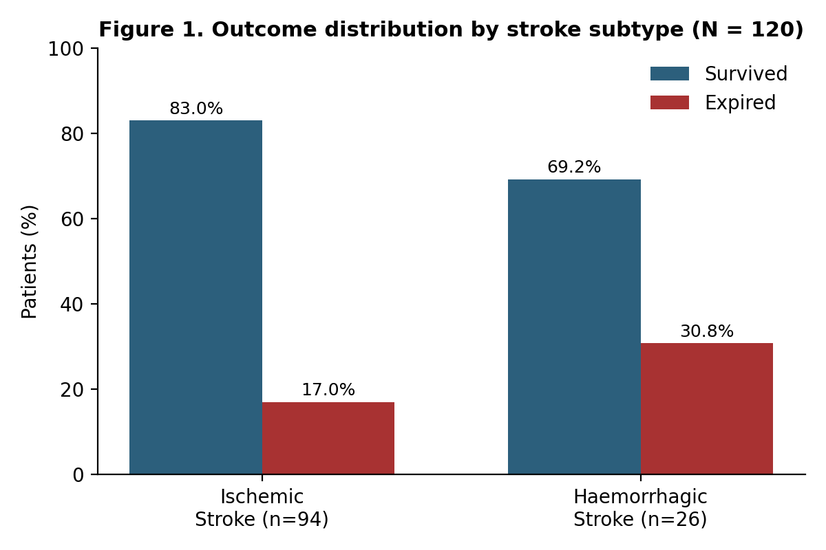
Of 120 patients admitted with acute stroke, 94 (78.3%) had ischaemic stroke and 26 (21.7%) had haemorrhagic stroke. In-hospital mortality was substantially higher in the haemorrhagic subgroup: 8 of 26 patients (30.8%) died, compared with 16 of 94 (17.0%) with ischaemic stroke (Figure 1).
Demographic and anthropometric comparisons showed no significant difference in age or body mass index between survivors and non-survivors with ischaemic stroke. Among haemorrhagic stroke patients, however, survivors were significantly older than non-survivors (68.67 ± 8.61 vs 57.25 ± 13.30 years; p = 0.015), a reversal probably reflecting the fact that younger haemorrhagic-stroke patients in this cohort tended to present with larger, more rapidly fatal bleeds rather than any protective effect of youth. Male sex was significantly associated with mortality in ischaemic stroke (75.0% of non-survivors were male versus 38.5% of survivors; p = 0.016) but not in haemorrhagic stroke (p = 1.00). Hypertension, diabetes, smoking and alcohol use showed no statistically significant association with outcome in either stroke subtype (all p > 0.05).
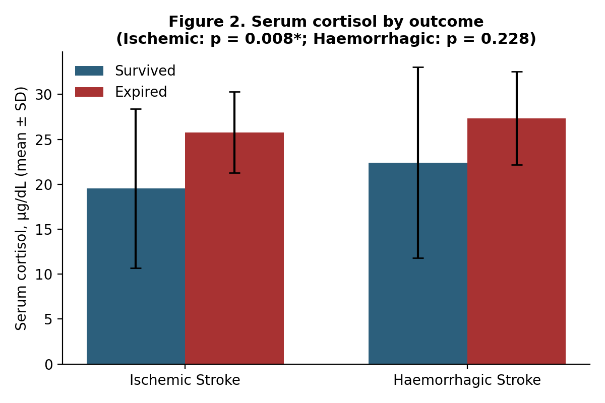
Serum cortisol was significantly higher among patients who died of ischaemic stroke (25.77 ± 4.51 µg/dL) than among survivors (19.54 ± 8.86 µg/dL; t = -2.732, p = 0.008). A similar direction of effect was seen in haemorrhagic stroke 27.35 ± 5.19 µg/dL in non-survivors versus 22.42 ± 10.64 µg/dL in survivors but this difference did not reach statistical significance (t = -1.238, p = 0.228), likely reflecting the smaller haemorrhagic subgroup (Figure 2).
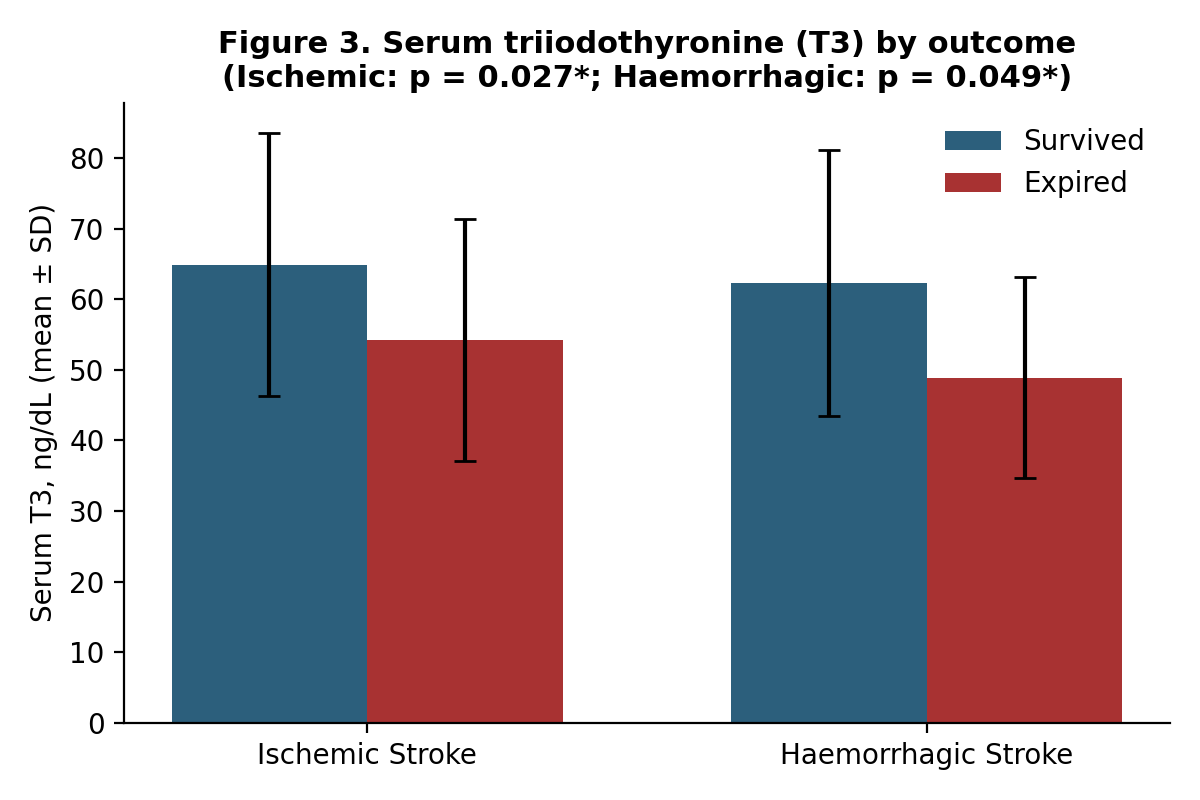
Serum T3 showed the inverse pattern and reached significance in both subtypes: levels were lower in non-survivors than survivors in ischaemic stroke (54.20 ± 17.15 vs 64.94 ± 18.70 ng/dL; t = 2.24, p = 0.027) and in haemorrhagic stroke (48.90 ± 14.22 vs 62.36 ± 18.90 ng/dL; t = 2.05, p = 0.049) (Figure 3). Neither T4 nor TSH differed significantly between survivors and non-survivors in either subgroup (all p > 0.05), consistent with a selective fall in the biologically active hormone rather than a global thyroid-axis derangement.
Clinical severity scores (GCS, NIHSS at admission and at 72 hours, and mRS at discharge) were each worse among non-survivors in both stroke subtypes, consistent with the biochemical findings. Glycaemic parameters showed a mixed picture: HbA1c was paradoxically higher among ischaemic-stroke survivors (7.07 ± 0.92% vs 6.38 ± 1.11%; p = 0.009), while fasting blood sugar was significantly higher among non-survivors with haemorrhagic stroke (181.00 ± 27.31 vs 143.33 ± 30.80 mg/dL; p = 0.007). Lipid profile, renal function and serum electrolytes did not differ significantly by outcome in either subgroup.
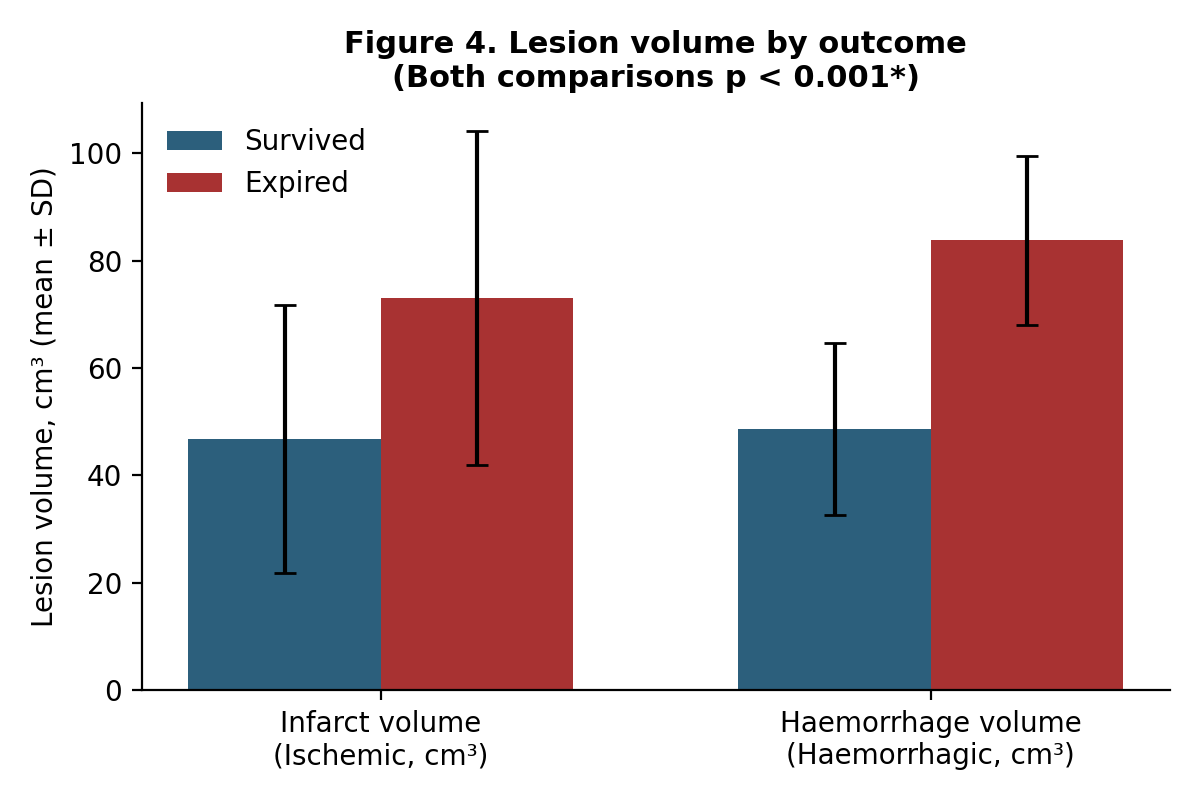
Radiological lesion size closely tracked mortality. Mean infarct volume was significantly larger among non-survivors with ischaemic stroke (73.05 ± 31.05 cm³) than among survivors (46.74 ± 24.91 cm³; t = -3.69, p < 0.001), and mean haematoma volume was similarly larger among non-survivors with haemorrhagic stroke (83.80 ± 15.71 cm³) than among survivors (48.56 ± 16.03 cm³; t = -5.21, p < 0.001) (Figure 4).
Finally, time from symptom onset to hospital presentation was significantly longer among non-survivors with haemorrhagic stroke (38.50 ± 9.09 hours) than among survivors (19.33 ± 13.01 hours; p < 0.001); a similar but non-significant trend was observed in ischaemic stroke (32.50 ± 10.32 vs 26.49 ± 12.23 hours; p = 0.070). Hospital stay itself did not differ significantly by outcome in either subgroup.
Discussion
In this cohort of 120 patients with acute stroke, haemorrhagic stroke carried nearly double the in-hospital mortality of ischaemic stroke (30.8% vs 17.0%), a pattern consistent with previously published series in which haemorrhagic stroke, though less common, disproportionately drives early case-fatality [20]. The higher mortality among males with ischaemic stroke in our cohort mirrors observations elsewhere and may relate to a greater prevalence of large-vessel atherosclerotic disease and vascular risk factor burden in men, although comorbidities such as hypertension, diabetes, smoking and alcohol use did not independently predict mortality in either subgroup in our data suggesting that acute biochemical and radiological status may be more informative than chronic risk-factor burden in the immediate prognosis of an evolving stroke.
The central finding of this study is the divergent but complementary behaviour of serum cortisol and serum T3 across outcome groups. Elevated cortisol at admission was strongly associated with mortality in ischaemic stroke, in keeping with a substantial body of literature linking hypercortisolaemia to greater stroke severity and higher mortality [8][9][16]. Cortisol release after stroke reflects activation of the HPA axis as part of the acute stress response; while a modest, transient rise may be adaptive, sustained or exaggerated elevation is associated with hyperglycaemia, endothelial dysfunction, immunosuppression and aggravation of secondary neuronal injury [9], plausibly explaining the excess mortality seen in our expired ischaemic-stroke patients. Comparable correlations between cortisol and stroke severity have been reported across a range of hospital-based cohorts, including recent Indian and South Asian series [20][27][28][31], reinforcing the generalisability of cortisol as an accessible prognostic marker.
In haemorrhagic stroke, cortisol was also higher among non-survivors, but the difference did not reach statistical significance, most plausibly reflecting the smaller haemorrhagic subgroup (n = 26) and consequently reduced statistical power rather than a genuine absence of association. Larger haemorrhagic-stroke cohorts would be needed to clarify whether this trend represents a true, if more modest, effect or simply insufficient sample size.
Serum T3 showed a consistent and statistically significant inverse relationship with mortality in both stroke subtypes a pattern that, unlike cortisol, held even in the smaller haemorrhagic-stroke group. This is compatible with the low-T3 or non-thyroidal illness syndrome long described in critically ill and stroke patients, in which serum T3 falls while T4 and TSH remain relatively preserved, reflecting reduced peripheral conversion of T4 to T3 under conditions of severe systemic stress [12][17]. Several groups have similarly reported lower T3 in patients with more severe strokes and poorer functional outcomes [18,21-23], and meta-analytic data support thyroid hormone status as a genuine, reproducible prognostic signal in acute cerebrovascular disease [21]. That T4 and TSH were unaffected in our cohort argues against primary thyroid pathology and instead supports a stress-induced, centrally mediated suppression of T3 synthesis mechanistically linked to the same HPA-axis activation responsible for the cortisol rise [14][19], since elevated cortisol and inflammatory cytokines can themselves inhibit deiodinase activity and impair T4-to-T3 conversion.
Taken together, the concurrent rise in cortisol and fall in T3 observed here support the concept of an integrated neuroendocrine stress signature accompanying poor outcome in acute stroke, rather than two independent, unrelated biomarkers. This is biologically plausible given the well-documented crosstalk between the hypothalamic–pituitary–adrenal and hypothalamic–pituitary–thyroid axes [6][14], and is consistent with experimental evidence that T3 itself is neuroprotective–limiting neuroinflammation, glutamate excitotoxicity and promoting neurotrophic support [11][13] – such that its depletion may not merely mark severity but actively contribute to worse recovery.
Beyond the hormonal findings, lesion volume emerged as the strongest single correlate of mortality in both stroke subtypes, with infarct and haematoma volumes roughly 1.5 to 1.7 times larger among non-survivors. This is unsurprising given the direct relationship between lesion burden and both primary neurological deficit and the risk of secondary complications such as raised intracranial pressure, herniation and malignant oedema, and reinforces that biochemical markers should be interpreted alongside, rather than instead of, radiological assessment.
Delayed hospital presentation was strongly and independently associated with mortality in haemorrhagic stroke, an association not reaching significance in ischaemic stroke in our cohort. Similar findings have been reported in other South Asian series, where longer onset-to-assessment intervals correlated with elevated cortisol and worse severity scores [20][26][28], consistent with cumulative physiological decompensation during untreated illness. The particularly strong delay–mortality relationship we observed in haemorrhagic stroke is clinically intuitive: haematoma expansion is most rapid in the first few hours after onset, and blood pressure control, anticoagulation reversal and surgical evaluation are all time-critical interventions. This underscores the public-health importance of community stroke-awareness initiatives aimed at reducing pre-hospital delay, particularly for haemorrhagic presentations.
Clinically, the practical appeal of serum cortisol and T3 lies in their availability, low cost and rapid turnaround compared with advanced neuroimaging or genomic biomarkers, making them realistic adjuncts to clinical and radiological assessment in resource-constrained settings [9][17]. Measured together at admission, alongside NIHSS, GCS and lesion volume on initial imaging, these two hormones could help flag patients at highest risk of early mortality who may warrant closer monitoring or more aggressive supportive care, although prospective validation in larger, multicentre cohorts would be required before any such biomarker-guided pathway could be recommended for routine practice.
Limitations
This study has several limitations. The relatively small sample size, particularly in the haemorrhagic stroke subgroup (n = 26), limits statistical power and may explain the non-significant cortisol association in that group. The single-centre, cross-sectional observational design precludes causal inference and limits generalisability. Cortisol and thyroid hormones were measured only once, at admission, without serial sampling to characterise the trajectory of neuroendocrine change over the course of illness. Finally, potential confounders such as circadian variation in cortisol secretion and unmeasured acute-phase inflammatory markers were not directly controlled for.
Conclusion
In this cohort, haemorrhagic stroke carried substantially higher in-hospital mortality than ischaemic stroke. Elevated serum cortisol and depressed serum T3 at admission were each associated with poor outcome cortisol most clearly in ischaemic stroke, and T3 consistently across both stroke subtypes alongside larger infarct or haemorrhage volume and, in haemorrhagic stroke, delayed hospital presentation. These findings support a combined neuroendocrine and radiological signature of prognosis in acute stroke. Given their low cost and wide availability, serum cortisol and T3 merit further evaluation as adjunctive, early risk-stratification tools that could help identify patients at highest risk of mortality and guide timelier, more targeted management, particularly in resource-limited settings.
Declaration
Ethics approval
Proper Institutional Ethical committee approval was taken from the institute ‘Moti Lal Nehru Medical College Prayagraj, (Ethics Committee Registration No. ECR/922/inst/UP/RR-22 issued under New Drugs and Clinical Trial Rules, 2019)
Consent to Participate
All patients were informed about the study and proper consent taken after explaining the advantages and risks to the patient.
Written Consent for publication
We declare that this manuscript is original, has not been published before and is not currently being considered for publication elsewhere and we give consent to be published in your journal.
Data Availability
Data transparency is maintained, and available on corresponding author upon responsible request.
Generative AI Declaration
Not used
Conflicts of interest
None
Financial support and sponsorship/Funding
Nil
Authors' contributions
M.K.M, A.S Chose topic and Planned execution. All the authors did the investigative part. All collected data. A.S, A.K.C, M.K.M, P.G wrote the main manuscript. A.S, P.G. A.K.C and J.S prepared tables and figures and contributed in discussion. A.S, A.K.C, J. S contributed in preparing Discussion and Formatted References. All authors reviewed the manuscript. All authors contributed equally.