Introduction
Soft tissue sarcomas are an uncommon and heterogeneous group of malignant tumours of mesenchymal origin, with a reported incidence of approximately 4-8 per 100,000 in Western populations; their tendency toward local infiltration and delayed diagnosis contributes disproportionately to cancer-related morbidity [1]. The 2020 World Health Organization classification divides soft tissue sarcomas into more than a dozen broad categories comprising over 70 histological subtypes, each with distinct morphological, immunohistochemical, and molecular features [1,2].
Magnetic resonance imaging (MRI) is the imaging modality of choice for the local staging of soft tissue tumours because of its superior soft tissue contrast, multiplanar capability, and ability to characterise signal behaviour, enhancement pattern, and involvement of adjacent neurovascular and osseous structures [3,4]. While no single MRI feature is pathognomonic for malignancy, certain signal and enhancement patterns, when interpreted together with patient age, tumour location, and growth pattern, considerably narrow the differential diagnosis and help direct biopsy and surgical planning [5,6].
This study was undertaken to evaluate the spectrum of MRI features of histologically confirmed malignant soft tissue tumours encountered at our institution and to correlate these imaging findings with histopathology, with the aims of (a) describing the key MRI characteristics of malignant soft tissue tumours, (b) assessing staging features including neurovascular and osseous involvement, (c) correlating MRI findings with histopathological subtypes, and (d) identifying distinctive imaging features across this 25-case series.
Materials and Methods
This was a retrospective observational study of 25 patients with histologically proven malignant soft tissue tumours evaluated in the Department of Radiology. All patients underwent MRI as the primary imaging modality prior to biopsy or surgical excision, and the histopathological diagnosis obtained subsequently was used as the reference standard for confirmation of the imaging diagnosis.
MRI examinations were performed on a 1.5-T MRI system (Siemens Healthineers, Erlangen, Germany) using dedicated surface coils. Standard multiplanar T1-weighted, T2-weighted, short tau inversion recovery (STIR), diffusion-weighted, and post-contrast fat-suppressed T1-weighted sequences were acquired [7]. Intravenous gadolinium-based contrast material (0.1 mmol/kg) was administered when indicated.
Results
Of the 25 histologically confirmed malignant soft tissue tumours evaluated, soft tissue sarcoma not otherwise specified (NOS) was the most frequently encountered diagnosis (4 cases), followed by synovial sarcoma, Kaposi sarcoma, clear cell sarcoma, and rhabdomyosarcoma (3 cases each). Spindle cell sarcoma, neurofibroma, kaposiform haemangioendothelioma, and nerve sheath tumour accounted for 2 cases each, while metastatic deposits were identified in 1 case. The distribution of histological diagnoses is shown in Table 1 and Figure 1.
| Diagnosis | Number of cases |
| Soft tissue sarcoma (NOS) | 4 |
| Synovial sarcoma | 3 |
| Kaposi sarcoma | 3 |
| Spindle cell sarcoma | 2 |
| Clear cell sarcoma | 3 |
| Neurofibroma | 2 |
| Rhabdomyosarcoma | 3 |
| Kaposiform haemangioendothelioma | 2 |
| Nerve sheath tumour | 2 |
| Metastatic deposits | 1 |
| Total | 25 |
Table footnote: NOS, not otherwise specified. Percentages of the total cohort (n = 25) are reported in the text and in Figure 1.
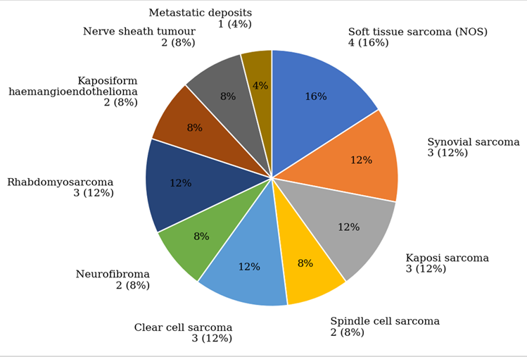
On qualitative review of the MRI findings across the cohort, heterogeneous T2 hyperintensity was the most common feature, present in 85% of cases. Necrosis or haemorrhage was identified in 70% of lesions, most frequently in soft tissue sarcoma, spindle cell sarcoma, and rhabdomyosarcoma. Restricted diffusion was noted in 80% of high-grade tumours. Heterogeneous post-contrast enhancement was seen in 75% of cases, whereas Kaposi sarcoma characteristically showed predominantly homogeneous enhancement. Neurovascular involvement was identified in 6 cases and osseous infiltration in 3 cases, both indicating locally advanced disease at presentation. Synovial sarcoma demonstrated the characteristic triple-signal pattern in 2 of 3 cases, and clear cell sarcoma showed T1 hyperintensity in all 3 of its cases, reflecting its melanocytic differentiation.
Discussion
The overall imaging–histopathology concordance in this series was high, particularly for synovial sarcoma, nerve sheath tumour, and clear cell sarcoma, in keeping with previously reported diagnostic performance of MRI for soft tissue sarcoma subtyping [5,6]. The individual case findings are summarised below.
Synovial sarcoma
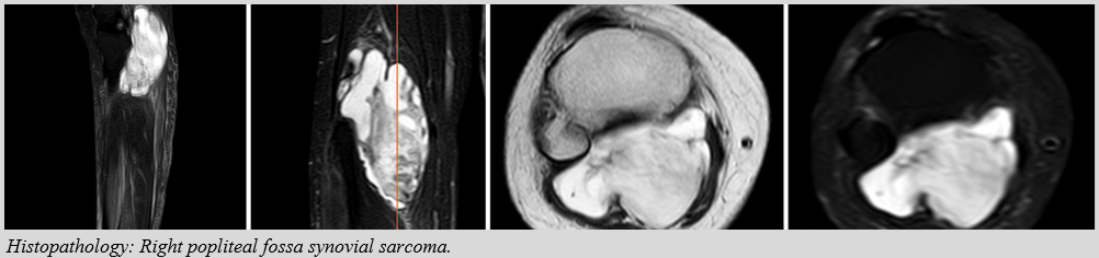
A 28-year-old male presented with progressive swelling and pain in the posterior knee. MRI demonstrated a large lobulated soft-tissue mass in the right popliteal fossa and proximal leg with mixed T2/STIR signal, restricted diffusion, and heterogeneous enhancement. The mass involved the posterior compartment muscles, more prominently medially, and compressed and encased the popliteal vessels without definite loss of flow void, findings consistent with previously described appearances of synovial sarcoma, including the characteristic triple-signal pattern [8,9].
Rhabdomyosarcoma
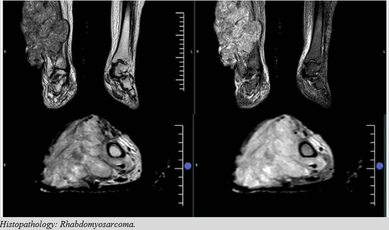
A 58-year-old female presented with rapidly progressive, painful swelling of the right leg. MRI showed a lobulated, infiltrative soft-tissue mass arising from muscle, hypointense on T1 and markedly hyperintense on T2/STIR with internal myxoid and necrotic areas. The lesion showed heterogeneous enhancement, restricted diffusion, ill-defined margins with intermuscular spread, neurovascular displacement and encasement, surrounding oedema, and probable regional lymphadenopathy, in keeping with previously described rhabdomyosarcoma imaging characteristics [10].
Nerve sheath tumour
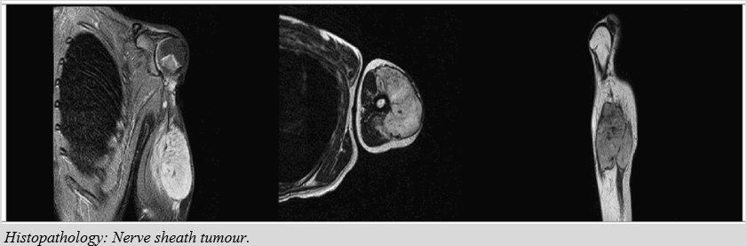
A 52-year-old female presented with a gradually increasing left arm mass and associated discomfort. MRI of the proximal arm showed a well-defined, ovoid lesion within or adjacent to the biceps muscle compartment, T1 hypointense and T2/STIR hyperintense with thin internal septations and smooth margins, without surrounding muscle oedema or bone involvement; the neurovascular bundle was preserved.
Spindle cell sarcoma
A 45-year-old female presented with a painless, slowly enlarging swelling in the anterolateral forearm. MRI showed a well-defined, hyperintense soft-tissue lesion in the anterolateral forearm with a preserved muscle interface and a few internal flow voids, suggesting a neurogenic origin. The underlying bones were normal, the elbow joint showed minimal physiological effusion, neurovascular bundles were preserved, and no deep extension or bony involvement was seen.
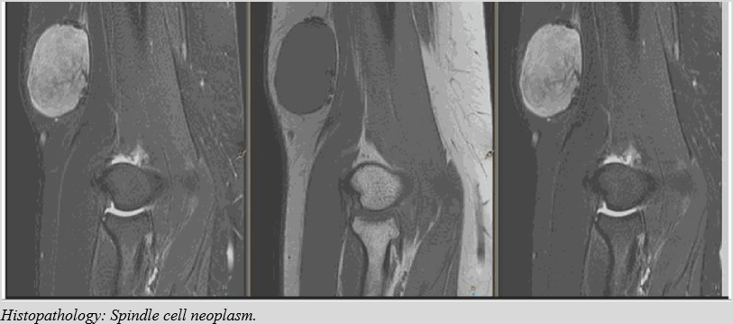
Soft tissue sarcoma
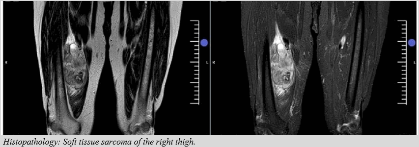
A 77-year-old male presented with a firm, enlarging medial thigh swelling. MRI showed a large, well-defined mass in the medial right thigh, involving the vastus intermedius and vastus medialis, with low T1 and heterogeneous high T2/proton-density signal, and heterogeneous post-contrast enhancement. No underlying bone involvement was seen.
Clear cell sarcoma
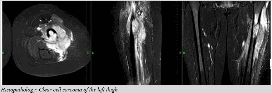
A 32-year-old female presented with a slowly enlarging, painful mass in the upper thigh. Imaging of the left proximal femur showed an infiltrative soft-tissue mass with periosteal and medullary extension, involving adjacent thigh muscles but sparing the major neurovascular bundles, with surrounding oedema and moderate post-contrast enhancement — findings suggestive of a malignant lesion, later confirmed as clear cell sarcoma. Reflecting its melanocytic differentiation, this tumour, like the other two cases in the series, showed T1 hyperintensity, consistent with previously reported imaging features of clear cell sarcoma [11].
Kaposiform haemangioendothelioma
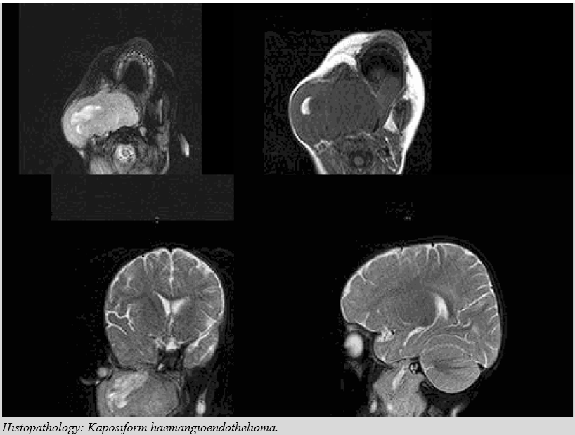
A 10-year-old female presented with facial swelling and gradually increasing jaw fullness. MRI showed a large, ill-defined, infiltrative mass in the right masticator space extending into the infratemporal and submandibular spaces, with T2 hyperintensity and cystic/serpiginous areas, T1 dark-to-intermediate signal, and no major flow voids - features suggestive of an infiltrative vascular tumour and consistent with previously described imaging of kaposiform haemangioendothelioma [12].
Kaposi sarcoma
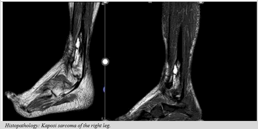
A 48-year-old male with a history of chronic lymphoedema presented with leg pain and swelling. MRI showed a fairly well-defined, T2/STIR-hyperintense and T1 iso- to hypointense, lobulated expansile lesion involving the distal third of the right fibula at the metadiaphyseal region, causing mild medullary expansion and patchy anterior cortical destruction. In keeping with the pattern observed across all three cases of Kaposi sarcoma in this series, enhancement was predominantly homogeneous, in contrast to the heterogeneous pattern seen in most other tumour subtypes, consistent with reported Kaposi sarcoma imaging patterns [13].
Neurofibroma
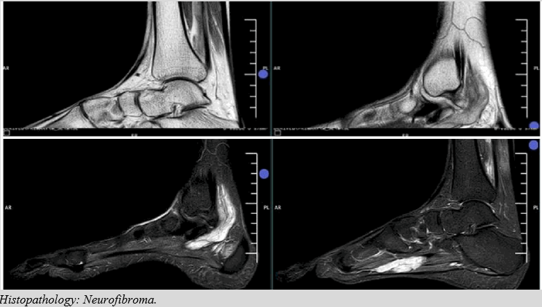
A 26-year-old male presented with progressively worsening ankle pain and tingling along the plantar aspect of the foot. A lobulated, beaded lesion, T2/STIR hyperintense and showing restricted diffusion, was noted along the neurovascular bundle close to the posterior tibial vessels, extending from the ankle to the plantar aspect of the foot and dividing at the level of the midfoot — a distribution and morphology characteristic of a peripheral nerve sheath origin.
Metastatic deposits
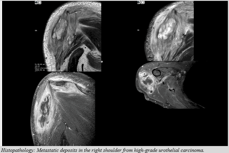
A patient with known carcinoma of the urinary bladder, with nodal and skeletal metastases following chemotherapy and immunotherapy, presented with swelling in the right shoulder. MRI showed T1-hypointense marrow lesions in the proximal humeral shaft and scapula with STIR hyperintensity and a hair-on-end periosteal reaction, in keeping with metastatic deposits. Lobulated, T1-low/T2-high intramuscular collections were also noted in the deltoid with surrounding intra- and intermuscular oedema, favoured to represent post-radiation change.
Across the series, benign nerve sheath tumours (schwannoma/ neurofibroma) demonstrated well-defined margins, a target sign, and homogeneous or mildly heterogeneous enhancement, distinguishing them from malignant peripheral nerve sheath tumours (MPNST), which characteristically showed loss of the target sign and ill-defined margins, correlating with malignant transformation [14,15]. These findings are consistent with recent literature describing MRI features that differentiate benign from malignant soft-tissue tumours, including margin definition, degree of T2 heterogeneity, and the presence of necrosis, as reproducible discriminators of grade [16-18]. Standardised whole-body staging protocols proposed by the European Society of Musculoskeletal Radiology emphasise systematic assessment of compartmental involvement and neurovascular proximity, both of which were key discriminating features in this series, particularly in cases with locally advanced disease such as the synovial sarcoma and clear cell sarcoma cases described above [19,20].
Limitations
Limitations of this study include its retrospective, single-centre design and the relatively small sample size, which precludes formal statistical analysis of diagnostic accuracy; larger multicentre cohorts, ideally incorporating radiomic and multiparametric analysis, would help validate the imaging–histopathological associations identified here [2,21-24].
Conclusion
In conclusion, a total of 25 malignant soft tissue tumours were evaluated on MRI and confirmed by histopathology. Heterogeneous T2 hyperintensity, necrosis or haemorrhage, restricted diffusion, and heterogeneous enhancement were the most frequent imaging findings. MRI demonstrated strong concordance with histopathology and remains a valuable modality for tumour characterisation, local staging, and pre-operative planning.
Key Messages
-
MRI reliably differentiates histological subtypes of malignant soft tissue tumours based on characteristic signal, enhancement, and diffusion patterns.
-
Heterogeneous T2 hyperintensity, necrosis/haemorrhage, and restricted diffusion are frequent features of high-grade malignant tumours in this cohort.
-
Synovial sarcoma and clear cell sarcoma demonstrate distinctive imaging signatures (triple-signal pattern and T1 hyperintensity, respectively) that aid diagnosis.
-
MRI accurately delineates neurovascular and osseous involvement, supporting its central role in pre-treatment staging and surgical planning.
Declarations
Conflict of interest
The authors have no conflicts of interest to declare.
Funding
This research received no specific grant from any funding agency in the public, commercial, or not-for-profit sectors.
Ethical Approval
This retrospective study was conducted in accordance with the ethical guidelines of the institution, including adherence to the legal requirements of the study country. The study was approved by the institutional review board / research ethics committee, and the requirement for individual patient consent was waived given the retrospective, de-identified nature of the analysis.
Author Contribution
Sai Keerthana Sarabudla: Conceptualisation, data collection, image analysis, and drafting of the manuscript.
N. L. N. Moorthy: Study supervision, critical review of imaging findings, and critical revision of the manuscript for important intellectual content.
Padmaja Singireddy: Data analysis, histopathological correlation, and critical revision of the manuscript. All authors read and approved the final manuscript.
Declaration of Human Rights
The authors confirm that they accept and agree with the United Nations' Declaration of Human Rights.