Introduction
Since its introduction in 1995, laparoscopic living donor nephrectomy has become the preferred approach for living kidney donation due to reduced postoperative pain, shorter faster recovery, improved cosmetic outcomes, and excellent graft function [1]. The left kidney is generally preferred for donation owing to its longer renal vein, which facilitates donor hilar dissection and recipient venous anastomosis [2,3].
Procedural success relies on meticulous preoperative assessment of renal vascular anatomy, experience and thorough knowledge of anatomical variations. Although anatomical variations of the renal vessels are common, a retro aortic left renal vein (RLRV) is encountered in approximately 2–9% of living kidney donors. In this variant, the left renal vein courses posterior to the aorta before draining into the inferior vena cava. This altered hilar anatomy may increase the technical complexity of vascular dissection, particularly during identification of the renal artery and adrenal vein [4,5].
Preoperative contrast-enhanced CT angiography with multiplanar reconstruction is the standard imaging modality for identifying renovascular anomalies and planning donor nephrectomy [6]. However, beyond preoperative identification of the anomaly, reproducible intraoperative anatomical landmarks that facilitate safe hilar dissection may further improve donor and recipient outcomes.
During laparoscopic donor nephrectomy in donors with an RLRV, we consistently observed a distinct triangular anatomical space bounded by the aorta, left renal artery, RLRV, and renal hilum with the left adrenal vein as the principal content of this space. Its consistent appearance may serve as an intraoperative landmark that could be clinically useful. We propose this anatomical relationship as the Ramaiah Triangle.
Patients and Observation
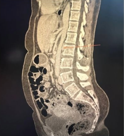
A retrospective review was performed of all laparoscopic donor nephrectomies carried out between January 2013 and December 2023 at our institution. Among 300 living kidney donors, 20 donors (6.5%) were identified as having a RLRV on preoperative CT angiography. Preoperative vascular evaluation of the anatomical relationship between the aorta, renal artery, renal vein, and adrenal vein was performed using contrast-enhanced CT angiography and correlated with intraoperative findings (Figure 1).
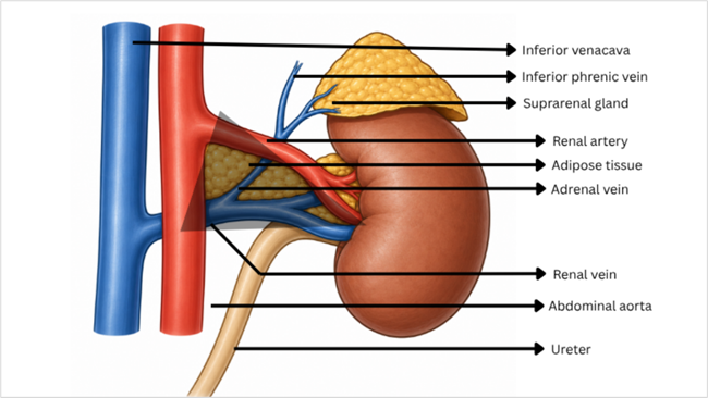
In all 20 cases, a consistent triangular anatomical space was identified during hilar dissection that contained the adrenal vein and perivascular adipose tissue. Additionally, the left inferior phrenic vein was observed in a few instances.
This triangle that we have named the Ramaiah Triangle is bounded superiorly by the left renal artery, inferiorly by the RLRV, medially by the abdominal aorta, with the renal hilum at its apex.
In donors with an RLRV, the renal vein arises from the renal hilum and courses obliquely posterior to the abdominal aorta before draining into the inferior vena cava. Throughout its hilar course, the left renal artery lies superior to the RLRV, thereby defining the superior and inferior boundaries of the Ramaiah Triangle. The left adrenal vein consistently drained into the RLRV, while the left inferior phrenic vein joined the adrenal vein or the renal vein in a proportion of cases (Figure 2). Shows a schematic representation of the triangle described.
The Ramaiah Triangle may be identified following exposure of the renal hilum. Once the left renal artery is identified superiorly and the RLRV inferiorly, the triangular space bounded medially by the abdominal aorta and laterally by the renal hilum can be visualised. The left adrenal vein consistently encountered within this space is then identified and divided to allow for safe hilar dissection (Figure 3).
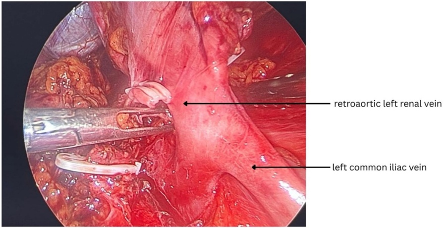
RLRV anomalies are traditionally classified into four types based on their course and site of venous drainage. Type I is the most common variant, in which the retro aortic vein drains into the inferior vena cava (IVC) at its normal orthotopic position. Type II drains into the IVC at a lower (L4–L5) level after receiving the gonadal and ascending lumbar veins, Type III represents the circumaortic left renal vein with both preaortic and retro aortic components, and Type IV is the rarest variant, in which the retro aortic vein courses caudally to drain into the left common iliac vein (Table 1). lists the incidence of the variants of RLRV described in the literature [7].
Recognition of the Ramaiah Triangle may assist the surgeon in identifying the left adrenal vein, maintaining orientation in the presence of altered retroaortic venous anatomy, and guiding meticulous hilar dissection. This reproducible anatomical landmark may facilitate safe vascular dissection around the renal hilum while preserving adjacent vascular structures.
Discussion
The RLRV is a well-recognized venous variant believed to result from persistence of the dorsal limb of the embryonic renal venous collar with regression of the ventral limb. It has been reported in approximately 2–9% of living kidney donors and is of particular importance during donor nephrectomy because it alters the normal hilar vascular relationships [8,9].
Although RLRV is no longer considered a contraindication to laparoscopic donor nephrectomy, altered venous anatomy may increase the technical complexity of hilar dissection [10,11]. Careful preoperative CT angiography allows accurate identification of these vascular variations and assists operative planning. However, successful donor nephrectomy ultimately depends on reliable intraoperative recognition of anatomical relationships.
Several anatomical landmarks have previously been described to facilitate laparoscopic donor nephrectomy, including the Danger Triangle (or the Golden Triangle), the Triangle of Marcille in retroperitoneal surgery help surgeons maintain orientation and identify critical structures during dissection [3,12].
The Ramaiah Triangle is not proposed as a new anatomical structure, but rather as a reproducible surgical landmark identified during laparoscopic donor nephrectomy in donors with a retroaortic left renal vein. Similar to other established anatomical landmarks used in surgery, it is intended to serve as an intraoperative reference point that may help maintain orientation during hilar dissection and facilitate identification of the left adrenal vein in the presence of altered venous anatomy.
In our experience, the Ramaiah Triangle was consistently identified in all 20 donors with a retroaortic left renal vein over the study period. Although the present study does not establish whether recognition of this landmark improves operative outcomes, we believe that its consistent anatomical relationship may form a useful adjunct during hilar dissection and may be of practical value.
The present study has several limitations. It is a retrospective, single-centre study with a relatively small sample size. The effect of recognising the Ramaiah Triangle on operative time, blood loss, vascular injury, or other surgical outcomes was not evaluated, and the clinical utility of this landmark has yet to be established. Larger prospective studies are required to validate these findings and to evaluate the clinical utility of the landmark in laparoscopic donor nephrectomy.
Conclusion
The Ramaiah Triangle represents a reproducible anatomical landmark observed during laparoscopic donor nephrectomy in donors with retroaortic left renal veins. Recognition of this triangular space may facilitate identification of the adrenal vein and assist surgeons during hilar dissection thereby optimising procedural quality. Additional studies are required to validate its applicability in larger patient populations.
Declarations
Acknowledgements
The authors would like to express their sincere gratitude to all the faculty and staff of the Department of Urology for their support and to the living kidney donors whose contribution made this study possible.
Conflict of interest
The authors declare that they have no conflicts of interest
Funding/ financial support
No external funding or financial support was received for this study
Contributors
Dr. Venkata Krishna Reddy Pendyala, MS Ramaiah Medical college; Dr. Amit Patil, MS Ramaiah Medical college; Dr. Venkata Mahesh Chalumuru, MS Ramaiah Medical college; Dr. Tarun Dilip Javali – Professor, MS Ramaiah Medical college Ramaiah Medical college
Ethical Clearance
Ethical approval was obtained from the institutional ethics committee
References
-
Turk IA, Deger S, Davis JW, Ingolf A, Loening SA, Jordan GH, et al. Laparoscopic live donor right nephrectomy: A new technique with preservation of vascular length. J Urol. 2002; 167:630-3. doi:10.1016/S0022-5347(01)69100-6.
-
Gupta N, Raina P, Kumar A. Laparoscopic donor nephrectomy. J Minim Access Surg. 2005; 1:155-64. doi:10.4103/0972-9941.19262.
-
Kanani F, Kozin M, Ben Avraham Y, Avitan E, Gurevich M, Nesher E, et al. Laparoscopic living donor nephrectomy: Learning curve analysis through 1446 cases and outcomes from 200 consecutive mastery-phase procedures-How I do it. J Clin Med. 2026;15(4):1363. doi:10.3390/jcm15041363.
-
Valenzuela Fuenzalida JJ, Vera-Tapia K, Urzúa-Márquez C, Yáñez-Castillo J, Trujillo-Riveros M, Koscina Z, et al. Anatomical variants of the renal veins and their relationship with morpho functional alterations of the kidney: A systematic review and meta-analysis of prevalence. J Clin Med. 2024;13(13):3689. doi:10.3390/jcm13133689.
-
Tatarano S, Enokida H, Yamada Y, Nishimura H, Yoshino H, Ishihara T, et al. Anatomical variations of the left renal vein during laparoscopic donor nephrectomy. Transplant Proc. 2019; 51:1311-3. doi: 10.1016/j.transproceed.2019.01.132.
-
Casadiego Duran JT, Ferreira Arquez H. Anatomical study of retro-aortic left renal vein. J Chem Pharm Res. 2016; 8:1011-8.
-
Alharbi Y, Jan MM. Anatomical study of the rare left renal vein anomaly. Artery Res. 2025; 31:15. doi:10.1007/s44200-025-00082-y.
-
Shreevastava AK, Das RS, Mishra A. Protean drainage patterns of the left renal vein: A cadaveric and retrospective clinical study on the surgical implications and technical feasibility. Cureus. 2024; 16:e63037. doi:10.7759/cureus.63037.
-
Hostiuc S, Rusu MC, Negoi I, Dorobanțu B, Grigoriu M. Anatomical variants of renal veins: A meta-analysis of prevalence. Sci Rep. 2019; 9:10802. doi:10.1038/s41598-019-47280-8.
-
Mang J, Hennig L, Biernath N, Liefeldt L, Bichmann A, Ralla B, et al. Is a retroaortic vein a risk factor in laparoscopic living donor nephrectomy? Urol Int. 2020; 104:641-5. doi:10.1159/000507642.
-
Lin CH, Steinberg AP, Ramani AP, Abreu SC, Desai MM, Kaouk J, et al. Laparoscopic live donor nephrectomy in the presence of circumaortic or retroaortic left renal vein. J Urol. 2004; 171:44-6. doi:10.1097/01.ju.0000099895.62724.04.
-
Lee P, Francis KE, Solomon MJ, Ramsey-Stewart G, Austin KKS, Koh C. Triangle of Marcille: The anatomical gateway to lateral pelvic exenteration. ANZ J Surg. 2017; 87:582-6. doi:10.1111/ans.13872.