Introduction
Primary cardiac tumors are rare (<0.1-0.2% at autopsy), with approximately 75% being benign; nearly half of these benign lesions are myxomas. Myxomas most commonly originate in the left atrium (LA), typically from the interatrial septum (IAS) near the fossa ovalis, and may be pedunculated or sessile [1]. The clinical manifestations of atrial myxomas are typically divided into three categories, such as obstructive symptoms (dyspnea, syncope), embolic phenomena (TIA/stroke, limb ischemia), and constitutional findings (fever, weight loss), reflecting tumor size, mobility, and friability [2]. Large LA myxomas may intermittently obstruct the mitral valve, producing auscultatory features reminiscent of mitral stenosis (MS), including a low-pitched diastolic murmur and the characteristic early diastolic “tumor plop” [2].
Echocardiography is the cornerstone of diagnosis with high sensitivity, while preoperative coronary angiography (CAG) is often considered in adults to exclude concomitant coronary artery disease and occasionally to visualize tumor vascularity [3][4].
This present study reports a surgically treated case of a large sessile LA myxoma causing severe functional mitral valve obstruction and integrates current evidence to guide clinicians through diagnostic and therapeutic decision-making.
Case Description
Patient Information
A 51-year-old male with pre-diabetes presented with progressive exertional dyspnea (New York Heart Association class II) and episodic nocturnal dry cough for four months. There was no history of syncope, strokes, transient ischemic attacks, peripheral embolization, or prior cardiac surgery. The patient was a non-smoker and non-alcoholic. Family history was non-contributory.
Clinical findings
On examination, the patient was afebrile and hemodynamically stable with systolic blood pressure 110-115 mmHg, diastolic blood pressure 68-70 mmHg, and heart rate 70-80/min. Cardiovascular examination revealed normal S1 and S2, an additional S3, a mid-diastolic crescendo-decrescendo murmur at the apex, and an early diastolic “tumor plop.” Respiratory examination demonstrated normal air entry bilaterally [2].
Diagnostic assessment
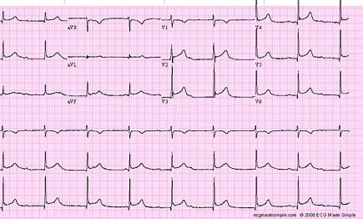
Electrocardiography (ECG): Incomplete right bundle branch block (RBBB) was noted, while the ECG patterns in myxoma are heterogeneous and non-specific [Figure 1] [2].
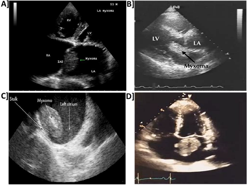
Echocardiography: The transthoracic echocardiography (TTE) demonstrated a large left atrium (LA) mass (6.3 × 3.6 cm) attached to the IAS, prolapsing into the mitral orifice, with mildly dilated LA and preserved left ventricle (LV) systolic function (ejection fraction (EF) ~60%). Echocardiography (TTE/ transesophageal echocardiography (TEE)) exhibits high sensitivity (≈90-97%) for cardiac myxoma detection [Figure 2] [3].
Coronary angiography (CAG): Radial-access selective angiography revealed normal coronaries. The CAG is commonly performed in adults >40 years to rule out occult CAD and, in select cases, to define tumor vascularity [4].
Histopathology
Gross examination identified a gelatinous, lobulated mass. Microscopic evaluation revealed lepidic (stellate/polygonal) cells arranged in cords and nests within a myxoid stroma, with hemorrhage and hemosiderin deposits, a features diagnostic of myxoma [5].
Therapeutic intervention
Given the tumor’s size, sessile attachment, and obstructive physiology, urgent surgical excision was recommended. The operative approach comprised median sternotomy, aorto-bicaval cannulation, cardioplegic arrest, transseptal/right atrial exposure extended into the LA roof, wide excision of the mass including a cuff of septal tissue, meticulous clearance of the LA, pulmonary veins, and LV cavity for fragments, and interatrial septal reconstruction using autologous pericardium [6].
Follow-up and outcomes
The patient was extubated on the day of surgery, remained in sinus rhythm without sustained pacing requirements, and was discharged on postoperative day five. At early clinic follow-up, he reported resolution of dyspnea with stable vitals and a healthy wound. Patient was advised with periodic echocardiographic surveillance.
Differential Diagnosis
In a patient with exertional dyspnea and a diastolic murmur, the differential diagnosis includes: rheumatic/degenerative mitral stenosis (MS), left atrial thrombus, papillary fibroelastoma, atrial lipoma, and other cardiac tumors (e.g., angiosarcoma). The ECG aids differentiation by demonstrating mass attachment to the IAS, mobility, and tissue characteristics. Magnetic resonance imaging can further delineate tissue features when thrombus or non-myxomatous tumor is suspected [2][3][4].
Discussion
Myxomas typically arise from multipotent mesenchymal/ endocardial cells near the fossa ovalis, and their clinical behavior depends on size, mobility, and site. The “tumor plop” is a classic, though infrequent, auscultatory sign linked to mass motion across the mitral orifice. Comparative reports confirm that tumor plop is present in 15-20% of cases and can be confused with opening snaps of rheumatic mitral stenosis [1][2][7].
The ECG remains the first-line modality, TTE defines size, attachment, and dynamics, and TEE enhances visualization of stalk origin and mobility. Sensitivity approaches 90-97%. Computed tomography (CT) and MRI add tissue characterization and can help distinguish myxoma from thrombus or other masses. Functional mitral stenosis due to LA myxoma has been described in multiple case series, often misdiagnosed initially as rheumatic MS or restrictive cardiomyopathy [3][4][8].
Preoperative coronary evaluation (invasive CAG or coronary CT angiography) may be considered in middle-aged/older adults to exclude coronary artery disease (CAD) and, occasionally, to identify tumor neovascularization ("tumor blush"), which can influence intraoperative strategy [4].
The operative goal is complete en bloc excision with adequate margins around the stalk to minimize recurrence. Sessile, broad-based attachments frequently require interatrial septal patch reconstruction; careful clearance of the LA, pulmonary veins, and LV cavity reduces the risk of residual fragments. Myocardial protection and meticulous de-airing are essential to support uneventful weaning from bypass [6].
Surgical excision is curative in most sporadic cases, with excellent early and long-term outcomes. Series demonstrate low recurrence in sporadic myxomas and survival comparable to age-matched populations after complete resection. Late complications may include postoperative atrial arrhythmias; scheduled echocardiographic surveillance is prudent. Comparative literature shows that patients with functional MS due to myxoma experience rapid symptomatic improvement post-excision, unlike rheumatic MS cases requiring valve intervention [6][8][9][10].
This case underscores that functional mitral obstruction due to an LA mass can mimic primary valvular disease. High clinical suspicion, prompt echocardiography, and definitive resection prevent embolic and obstructive complications. Clinicians should be aware of mimickers such as LA thrombus and fibroelastoma, which differ in management [2][3][8].
Conclusion
A large sessile left atrial myxoma presented with functional mitral stenosis and was successfully treated by complete excision with septal patch reconstruction. The ECG facilitated rapid diagnosis, preoperative coronary evaluation refined risk assessment, results in meticulous surgery yielded excellent recovery. Clinicians should maintain vigilance for myxoma in patients with unexplained diastolic murmurs and dyspnea, as timely intervention offers outstanding outcomes.
Patient perspective
The patient reported early relief of breathlessness and appreciated structured discharge guidance, including pulmonary hygiene and wound care. He resumed light activity as advised at the first follow-up.
Declaration
Acknowledgements
None
Conflict of interest
Chetan Gharat is employee of Lupin Digital Health, Mumbai. All other authors have no conflict of interest to declare.
Funding/ financial support
No external funding was received.
Contributors
AG and CG conceived the study. CG and AG managed the patient and collected clinical data. AG performed the literature review. CG drafted the manuscript. AG and CG critically revised the manuscript. All authors approved the final version and take responsibility for the integrity of the work.
Ethical Clearance
This study utilizes de-identified records. The clinical consent for surgery was obtained from the patient. Case reporting complied with institutional policies. The study adheres to the CARE (CAse REport) guidelines for structure and transparency and adhered to institutional ethical standards for de-identified presentation. The narrative is supported by peer-reviewed literature and incorporates AMA/Vancouver references.
Trial details
None