Introduction
Chest drainage is an essential therapeutic procedure in the management of selected pleural emergencies. It allows evacuation of air, blood, pus, or other pathological pleural collections and may restore lung expansion and improve respiratory mechanics. In emergency settings, timely drainage can be critical in patients with tension pneumothorax, traumatic hemothorax, hemopneumothorax, or pleural infection associated with significant respiratory or hemodynamic compromise. Current international and multidisciplinary guidelines emphasize that the indication, technique, drain size, and urgency of chest drainage should be adapted to the underlying pleural condition and the patient's clinical status [1-4].
Pleural conditions requiring drainage represent an important component of respiratory and trauma-related morbidity. Clinicians frequently encounter pleural effusions in practice, which can complicate pneumonia and other infectious conditions [5]. Their presence at the initial emergency department evaluation has also been associated with hospital admission, prolonged hospital stay, and worse clinical outcomes among patients with pneumonia [6]. Chest tube thoracostomy, however, carries risks. Complications related to tube placement have been reported in both traumatic and non-traumatic settings, including tube malposition or dislodgement, iatrogenic pneumothorax, obstruction, and other procedure-related adverse events [7].
In sub-Saharan Africa, chest drainage remains an important component of emergency management for thoracic trauma and other acute pleural conditionsPre-hospital intercostal chest drainage has also been considered a potential component of emergency trauma care in resource-constrained settings, highlighting the importance of timely access to chest drainage when clinically indicated [8]. In a study conducted at the National Hospital Abuja, Nigeria, thoracic trauma was associated with substantial morbidity, and chest tube insertion was among the initial management strategies used for injured patients [9]. Tube thoracostomy itself may be associated with iatrogenic complications, emphasizing the importance of appropriate patient selection and procedural technique [10]. Similarly, a prospective case series from Kenya described patients managed with tube thoracostomy for indications including hemopneumothorax, empyema thoracis, and pneumothorax, with tube-related complications occurring in a substantial proportion of patients [11]. These findings illustrate both the importance of chest drainage in emergency thoracic care and the potential for procedure-related morbidity.
In the Democratic Republic of the Congo (DRC), published evidence on patients requiring pleural drainage remains limited and has primarily focused on pleural infection. At the University Clinics of Kinshasa, Subayi et al. reported a large series of 416 patients with purulent pleuritis and described pleural drainage as an important component of surgical management [12]. More recently, Filaka et al. retrospectively evaluated 60 patients with pleural empyema at the University Clinics of Kinshasa, of whom 51 (85%) underwent pleural drainage. Overall, in-hospital mortality was 30%, with no statistically significant difference between patients treated with pleural drainage and those undergoing decortication [13]. However, these studies primarily addressed pleural infection and did not examine the broader spectrum of acute pleural emergencies or the timing of drainage in routine emergency practice.
Reports from other low- and middle-income African settings also illustrate the heterogeneity of indications and practices surrounding tube thoracostomy. In Cameroon, tube thoracostomy has been reported for both traumatic and non-traumatic free pleural effusions, with procedure-related morbidity observed in a resource-constrained setting [14]. Similarly, studies of traumatic chest injury in Ethiopia have documented the use of chest tube drainage as part of the management of thoracic trauma and have highlighted the clinical burden associated with these injuries [15]. Current international recommendations for spontaneous pneumothorax further emphasize that management should be individualized according to the clinical presentation, underlying condition, and available treatment options rather than based on a single drainage strategy for all patients [16].
In resource-constrained emergency care settings, access to appropriate imaging, trained personnel, equipment, and standardized procedural pathways may influence the diagnosis, safety, and timeliness of pleural drainage. Thoracic ultrasound can provide rapid bedside assessment of pleural and thoracic abnormalities and may offer diagnostic advantages over conventional chest radiography for selected pulmonary and pleural conditions [17]. Computed tomography can provide more detailed characterization of thoracic injuries and may improve the detection of pneumothorax and hemothorax in selected trauma patients, particularly when clinically appropriate [18]. In trauma settings, point-of-care approaches such as extended focused assessment with sonography for trauma (E-FAST) may further support the early identification of thoracic injuries in the emergency department [19][20]. Nevertheless, the availability and use of these imaging modalities may vary substantially according to the resources, organization, and expertise of individual healthcare facilities.
Despite the clinical importance of prompt and appropriate pleural drainage, data describing real-world drainage practices, delays between hospital admission and chest tube placement, and hospital outcomes among adults with pleural emergencies in the DRC remain scarce. The present study therefore aimed to describe the clinical characteristics, diagnostic and chest drainage practices, timing of drainage, and hospital outcomes among adults with pleural emergencies requiring chest drainage in Kinshasa. Particular attention was given to the interval between hospital admission and chest tube placement, using 6 hours as a predefined operational descriptive threshold rather than as a universally validated definition of delayed drainage.
Methods
Study design and setting
This was a prospective, multicenter, descriptive observational study conducted in Kinshasa, Democratic Republic of the Congo (DRC), over an 8-month period from November 2025 to June 2026. The study was conducted in seven healthcare facilities in Kinshasa: University Hospital Center Renaissance, Kinshasa Medical Center, Biamba Marie Mutombo Hospital, Sino-Congolese Friendship Hospital, Central Military Hospital, Alliance Hospital Center (Gombe), and the University Clinics of Kinshasa. Participants were recruited from emergency departments, resuscitation units, and intensive care units where chest drainage procedures were performed. These facilities were selected because chest drainage was routinely performed in their emergency and resuscitation services.
Study population
The study population consisted of patients aged 18 years or older who were managed in the emergency department or resuscitation area for a pleural emergency requiring chest drainage as part of the initial management.
For the purposes of this study, a pleural emergency was defined as an acute pleural condition in which urgent drainage was required because failure to evacuate the pleural collection promptly could compromise the patient's vital prognosis.
Sampling and participant selection
An exhaustive consecutive sampling strategy was used. All patients presenting with an acute pleural condition potentially requiring chest drainage during the study period were screened for eligibility. Patients who met the predefined inclusion criteria and underwent chest drainage were eligible for inclusion in the final analysis.
Patients were included if they were aged 18 years or older, had an acute pleural condition meeting the predefined criteria for a pleural emergency, had the diagnosis confirmed by thoracic imaging, underwent chest drainage as part of the initial management, and provided informed consent themselves or through their designated representative when applicable.
We excluded patients younger than 18 years, those who did not undergo chest drainage, those who refused to participate, and those with insufficient data for the main study variables.
Data collection
Data were collected using standardized data collection forms. After scientific review of the research protocol by the Department of Anesthesiology and Critical Care, the amended protocol was submitted to the Ethics Committee of the School of Public Health for approval. Administrative authorization was subsequently obtained from the relevant authorities of the participating healthcare facilities.
Clinical data were recorded in real time at admission when applicable, while additional information was extracted from the patients' medical records. Patients were followed daily during hospitalization until discharge or death. Before data collection, the patient or their representative provided informed consent.
Variables and measurements
The variables collected included sociodemographic, clinical, diagnostic, procedural, and hospital outcome characteristics.
Sociodemographic variables included age, sex, weight, height, body mass index, occupation, and admission source, categorized as home, intrahospital transfer, or extrahospital transfer.
Clinical variables included presenting complaints, comorbidities, vital signs before and after chest tube placement, signs of respiratory and hemodynamic instability at admission, and pain intensity according to the scale used.
Diagnostic variables included thoracic imaging findings, particularly the type and extent of the pleural collection.
Procedural variables included the professional qualification of the operator, the drainage technique, the use of ultrasound guidance, chest tube size, insertion site, and type of drainage equipment, including the use of a Heimlich valve.
The operators involved in chest tube placement and monitoring included anesthesiologists, surgeons, residents in anesthesiology and critical care or surgery, emergency physicians, trained general practitioners, nurses, and radiologists.
Admission characteristics included the initial admission unit, categorized as emergency department, resuscitation unit, intensive care unit, or other.
Hospital outcome variables included the time between hospital admission and chest tube placement, post-drainage complications, length of hospital stay, and clinical outcomes, including survival, death, readmission, or complications.
Chest drainage procedure
Chest drainage was performed using either the Seldinger technique or the trocar technique, according to the clinical situation and the operator's practice. The Seldinger technique consisted of puncture, guidewire insertion, dilation, and catheter placement.
The available drainage devices included pigtail catheters measuring 8–16 French and surgical chest tubes measuring 20–24 French. Drainage systems included underwater-seal systems and two- or three-chamber systems. A Heimlich valve was also available, particularly for pneumothorax management.
The insertion approach was either anterior or lateral. Ultrasound guidance was used when available in the participating healthcare facility. Patient monitoring was performed using available monitoring equipment, including vital-sign monitors, blood pressure devices, and pulse oximeters.
Timing of chest drainage
The chest drainage interval was defined as the time elapsed between hospital admission and actual chest tube placement.
Because there is no universally validated time threshold applicable to all indications for chest drainage, the admission-to-drainage interval was initially considered as a quantitative variable. For descriptive purposes, the interval was categorized into four groups: less than 3 hours, 3–6 hours, 6–12 hours, and 12 hours or more.
For selected descriptive comparisons, the interval was also dichotomized into ≤6 hours and >6 hours. The 6-hour threshold was considered an operational descriptive threshold and was not intended to represent an internationally validated definition of delayed chest drainage.
Data management and statistical analysis
After data collection, a quality-control process was performed to assess data completeness, accuracy, and reliability. Questionnaires and medical records were manually reviewed, and data were entered, cleaned, and coded using Microsoft Excel 2016. Statistical analyses were performed using IBM SPSS Statistics version 27. Data were presented using tables and figures.
Continuous variables were summarized using the mean and standard deviation for approximately normally distributed data, and the median and interquartile range for non-normally distributed data. Categorical variables were summarized using absolute frequencies and percentages.
Given the descriptive nature of the present study, the analysis primarily focused on detailing the epidemiological and clinical characteristics of the study population, including indications for chest drainage, drainage techniques, timing of drainage, complications, length of hospital stay, and clinical outcomes.
Where descriptive comparisons between groups were performed, Student's t test or the Mann–Whitney U test was used for quantitative variables, as appropriate, while the chi-square test or Fisher's exact test was used for qualitative variables. A two-sided p value <0.05 was considered statistically significant.
No multivariable analysis was performed for the present descriptive article. Logistic regression analyses originally included in the thesis to identify factors associated with a drainage interval of >6 hours or post-drainage complications were not retained in this article, in accordance with the descriptive objective and cross-sectional design adopted for the publication.
Ethical considerations
The study protocol was approved by the Ethics Committee of the School of Public Health of the University of Kinshasa under approval number ESP/CE/92/2026. Ethical and confidentiality requirements were observed throughout the study, in accordance with the principles of the Declaration of Helsinki. Informed consent was obtained from patients or their representatives before participation.
Results
Study population
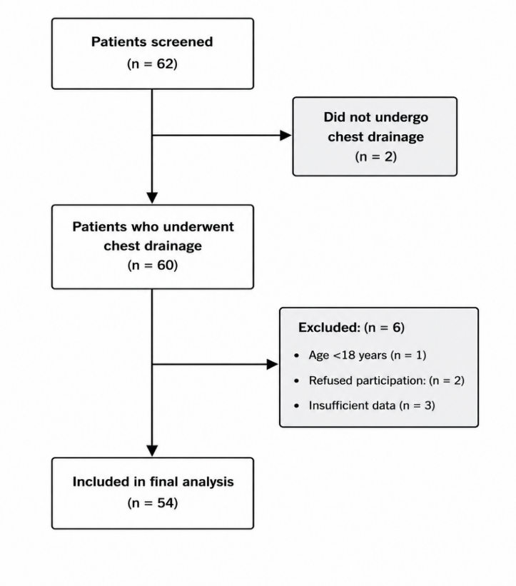
During the 8-month study period, 62 patients with an acute pleural condition potentially requiring chest drainage were screened. Two patients did not undergo chest drainage and were therefore excluded from the study population. Of the 60 patients who underwent chest drainage, six were excluded: one was younger than 18 years, two refused to participate, and three had insufficient data for the main study variables. A total of 54 patients were included in the final analysis (Figure 1).
Sociodemographic and clinical characteristics
The 54 patients included in the analysis had a mean age of 36.1 ± 15.6 years and a median age of 35 years (IQR, 25–45). Most patients were male (77.8%). Most of them (83.3%) came through the emergency room, while 7.4% went to a resuscitation unit and 7.4% went to an intensive care unit. Most patients came directly from home (66.7%), followed by intrahospital transfers (20.4%) and extrahospital transfers (13.0%). Private healthcare financing was reported for 48 patients (88.9%), while 6 patients (11.1%) had insurance coverage. Alcohol use was reported in 50.0% of patients and smoking in 27.8%. Tuberculosis was the most frequently reported comorbidity (14.8%), followed by hypertension (9.2%) and asthma (1.9%). Admission vital signs and other clinical characteristics are presented in Table 1.
| Characteristic | Value |
| Age, years (Mean ± SD) | 36.1 ± 15.6 |
| Sex, n (%) | |
| Male | 42 (77.8) |
| Female | 12 (22.2) |
| Admission unit, n (%) | |
| Emergency department | 45 (83.3) |
| Resuscitation unit | 4 (7.4) |
| Intensive care unit | 4 (7.4) |
| Other | 1 (1.9) |
| Admission source, n (%) | |
| Home | 36 (66.7) |
| Intrahospital transfer | 11 (20.4) |
| Extrahospital transfer | 7 (13.0) |
| Healthcare financing, n (%) | |
| private payment | 48 (89) |
| Insurance | 6 (11) |
| Anthropometric and physiological parameters, mean ± SD | |
| Weight, kg | 66.4 ± 16.4 |
| Height, cm | 181.5 ± 17.4 |
| Systolic blood pressure, mmHg | 113.6 ± 23.5 |
| Diastolic blood pressure, mmHg | 67.4 ± 16.5 |
| Heart rate, beats/min | 107.1 ± 19.2 |
| Respiratory rate, breaths/min | 31.4 ± 7.3 |
| Temperature, °C | 36.7 ± 0.7 |
| SpO₂, % | 89.4 ± 7.9 |
| Glasgow Coma Scale | 14.9 ± 0.4 |
| Pain score, 0–10 | 4.3 ± 1.9 |
| Body mass index category, n (%) | |
| Underweight | 7 (12.9) |
| Normal weight | 35 (64.8) |
| Overweight | 8 (14.8) |
| Obesity | 4 (7.4) |
| Lifestyle factors and comorbidities, n (%) | |
| Alcohol use | 27 (50.0) |
| Smoking | 15 (27.8) |
| Hypertension | 5 (9.2) |
| Tuberculosis | 8 (14.8) |
| Asthma | 1 (1.9) |
Abbreviations : SD, standard deviation; SpO₂, peripheral oxygen saturation.
Clinical presentation and pleural diagnoses
Chest pain was the most frequent presenting manifestation (68.5%), followed by respiratory distress (51.9%), tachypnea (48.1%), tachycardia (40.7%), and oxygen desaturation (33.3%). Signs of shock were reported in 13.0% of patients. Traumatic and non-traumatic etiologies accounted for 51.9% and 48.1% of cases, respectively. Hemothorax was the most frequent pleural diagnosis (38.9%), followed by parapneumonic pleural effusion (25.9%) and hemopneumothorax (16.7%). Chilothorax and pneumothorax accounted for 7.4% and 3.7% of cases, respectively, while other pleural conditions accounted for 7.4% (Table 2).
| Characteristic | n | % |
| Clinical manifestations | ||
| Chest pain | 37 | 68.5 |
| Respiratory distress | 28 | 51.9 |
| Tachypnea | 26 | 48.1 |
| Tachycardia | 22 | 40.7 |
| Oxygen desaturation | 18 | 33.3 |
| Low blood pressure | 3 | 5.6 |
| High blood pressure | 4 | 7.4 |
| Bradycardia | 3 | 5.6 |
| Hypothermia | 3 | 5.6 |
| Signs of shock | 7 | 13.0 |
| Etiology | ||
| Traumatic | 28 | 52 |
| Non-traumatic | 26 | 48 |
| Main pleural diagnoses | ||
| Hemothorax | 21 | 38.9 |
| Parapneumonic pleural effusion | 14 | 25.9 |
| Hemopneumothorax | 9 | 16.7 |
| Chilothorax | 4 | 7.4 |
| Pneumothorax | 2 | 3.7 |
| Other pleural conditions | 4 | 7.4 |
Note: Patients could have more than one clinical manifestation; therefore, percentages do not sum to 100%.
Diagnostic characteristics and indications for drainage
Chest radiography alone was the predominant diagnostic imaging modality, used in 88.9% of patients. Ultrasound alone was used in 5.6%, while combined or other imaging modalities were uncommon. Respiratory distress was the most frequently documented indication for drainage (57.4%), followed by oxygen desaturation (55.6%). The estimated size of the pleural collection was moderate in 40.7% of patients and large in 22.2%, while the size was not reported in 33.3%. A protocol for managing chest drainage was reported to be available in 48.1% of cases (Table 3).
| Characteristic | n | % |
| Imaging modality | ||
| Chest radiography alone | 48 | 88.9 |
| Chest radiography + ultrasound | 1 | 1.9 |
| CT scan | 1 | 1.9 |
| CT scan + MRI | 1 | 1.9 |
| Ultrasound alone | 3 | 5.6 |
| Indications for drainage | ||
| Respiratory distress | 31 | 57.4 |
| Oxygen desaturation | 30 | 55.6 |
| Agitation | 12 | 22.2 |
| Signs of shock | 8 | 14.8 |
| Chest pain | 2 | 3.7 |
| Estimated size of pleural collection | ||
| Small | 2 | 3.7 |
| Moderate | 22 | 40.7 |
| Large | 12 | 22.2 |
| Not reported | 18 | 33.3 |
| Availability of a chest drainage management protocol | ||
| No | 28 | 51.9 |
| Yes | 26 | 48.1 |
Note: Patients could have more than one indication for chest drainage; therefore, percentages do not sum to 100%. Abbreviations: CT, computed tomography; MRI, magnetic resonance imaging.
Chest drainage procedures and technical characteristics
The trocar technique was the most frequently used drainage technique (35/54, 64.8%), while the Seldinger technique was used in 19/54 procedures (35.2%). Trained general practitioners performed most chest drainages (37/54, 68.5%), followed by residents (18.5%), surgeons (9.3%), and emergency physicians (3.7%). The Monod trocar was the most frequently used drainage device (40/54, 74.1%). Most drains were 22 Fr (23/54, 42.6%), followed by 20 Fr drains (17/54, 31.5%). Blind insertion was used in 52/54 procedures (96.3%), whereas ultrasound guidance was used in 2/54 procedures (3.7%). A single-bottle collection system was used in 42/54 cases (77.8%), and suction was used in 13/54 cases (24.1%). Most procedures were performed in the operating room (38/54, 70.4%). Procedural characteristics are presented in Table 4.
| Characteristic | n | % |
| Drainage technique | ||
| Trocar technique | 35 | 64.8 |
| Seldinger technique | 19 | 35.2 |
| Drainage operator | ||
| Surgeon | 5 | 9.3 |
| Resident | 10 | 18.5 |
| Trained general practitioner | 37 | 68.5 |
| Emergency physician | 2 | 3.7 |
| Insertion guidance | ||
| Ultrasound-guided | 2 | 3.7 |
| Blind | 52 | 96.3 |
| Drain type | ||
| Joly drain | 8 | 14.8 |
| Pigtail catheter | 4 | 7.4 |
| Monod trocar | 40 | 74.1 |
| Other | 2 | 3.7 |
| Drain size | ||
| 22 Fr | 23 | 42.6 |
| 20 Fr | 17 | 31.5 |
| Other sizes | 14 | 25.9 |
| Collection system | ||
| One-bottle system | 42 | 77.8 |
| Two-bottle system | 2 | 3.7 |
| Three-bottle system | 10 | 18.5 |
| Suction | ||
| No | 41 | 75.9 |
| Yes | 13 | 24.1 |
| Place of drainage | ||
| Operating room | 38 | 70.4 |
| Resuscitation unit | 1 | 1.9 |
| Patient's bedside | 15 | 27.8 |
Abbreviation : Fr, French gauge.
Timing of diagnostic and drainage procedures
Thoracic imaging was performed within 3 hours of admission in 21 patients (38.9%), between 3 and 6 hours in 12 (22.2%), between 6 and 12 hours in 4 (7.4%), and after 12 hours in 17 (31.5%). Chest tube placement occurred within 3 hours of admission in 11 patients (20.4%), between 3 and 6 hours in 20 (37.0%), between 6 and 12 hours in 6 (11.1%), and after 12 hours in 17 (31.5%). From radiological diagnosis to chest drainage, the interval was less than 3 hours in 15 patients (27.8%), 3–6 hours in 14 (25.9%), 6–12 hours in 6 (11.1%), and more than 12 hours in 19 (35.2%). Overall, 31 patients (57.4%) underwent chest drainage within 6 hours of admission, whereas 23 (42.6%) underwent drainage after 6 hours (Table 5).
| Time interval | n (%) |
| Admission to thoracic imaging | |
| <3 hours | 21 (38.9) |
| 3-6 hours | 12 (22.2) |
| 6_12 hours | 4 (7.4) |
| >12 hours | 17 (31.5) |
| Admission to chest tube placement | |
| < 3 hours | 11 (20.4) |
| 3-6 hours | 20 (37.0) |
| 6-12 hours | 6 (11.1) |
| >12 hours | 17 (31.5) |
| Radiological diagnosis to chest drainage | |
| <3 hours | 15 (27.8) |
| 3-6 hours | 14 (25.9) |
| 6-12 hours | 6 (11.1) |
| >12 hours | 19 (35.2) |
Clinical and radiological outcomes after chest drainage
Following chest drainage, clinical improvement was documented among patients presenting with the recorded symptoms. Among patients with chest pain (n=37), 32 (86.5%) experienced complete resolution, 4 (10.8%) showed improvement, and 1 (2.7%) remained unchanged. Among patients with dyspnea (n=26), 15 (57.7%) experienced complete resolution, 8 (30.8%) improved, and 3 (11.5%) remained unchanged. Among patients with respiratory distress (n=28), 15 (53.6%) experienced complete resolution, 8 (28.6%) improved, and 5 (17.8%) remained unchangedFive (71.4%) of the seven patients with signs of shock experienced complete resolution, while one (14.3%) improved and one (14.3%) remained unchanged. Regarding radiological outcomes, 18 patients (33.3%) had complete radiological resolution, 33 (61.1%) had minimal residual pleural collection, 2 (3.7%) had moderate residual collection, and 1 (1.9%) had a persistent or important pleural collection (Table 6).
| Outcome | Improved, n (%) | Resolved, n (%) | Unchanged, n (%) | Radiological outcome | n (%) |
| Chest pain (n=37) | 4 (10.8) | 32 (86.5) | 1 (2.7) | Resolved | 18 (33.3) |
| Dyspnea (n=26) | 8 (30.8) | 15 (57.7) | 3 (11.5) | Minimal residual collection | 33 (61.1) |
| Respiratory distress (n=28) | 8 (28.6) | 15 (53.6) | 5 (17.8) | Moderate residual collection | 2 (3.7) |
| Signs of shock (n=7) | 1 (14.3) | 5 (71.4) | 1(14.3) | Persistent/important collection | 1 (1.9) |
Hospital outcomes and length of stay
At the end of hospitalization, 48 patients (88.9%) were alive and 6 (11.1%) had died. Regarding the length of hospital stay, 26 patients (48.1%) were hospitalized for ≤7 days, whereas 28 (51.9%) had a hospital stay of >7 days (Table 7).
| Outcome | n | % |
| Vital status at hospital discharge | ||
| Alive | 48 | 89 |
| Deceased | 6 | 11 |
| Length of hospital stay | ||
| ≤7 days | 26 | 48 |
| >7 days | 28 | 52 |
| Length-of-stay distribution (days) |
Discussion
Principal findings
This prospective multicenter observational study describes real-world chest drainage practices, diagnostic pathways, timing of drainage, and hospital outcomes among adults with pleural emergencies in seven healthcare facilities in Kinshasa. The study population was predominantly young and male, with traumatic and non-traumatic etiologies accounting for 52.0% and 48.0% of cases, respectively. Hemothorax was the most frequent pleural diagnosis (38.9%), followed by parapneumonic pleural effusion (25.9%) and hemopneumothorax (16.7%). Chest radiography alone was the predominant imaging modality (88.9%), whereas ultrasound-guided drainage was used in only 3.7% of procedures. Drainage timing varied substantially: 57.4% of patients underwent chest tube placement within 6 hours of admission, while 42.6% underwent drainage after 6 hours. Most patients with recorded symptoms experienced improvement or complete resolution after drainage, although 6 patients (11.1%) died during hospitalization and 28 (51.9%) remained hospitalized for more than 7 days.
Spectrum of pleural emergencies
The predominance of hemothorax in this cohort highlights the importance of traumatic pleural disease among adults requiring emergency chest drainage in Kinshasa. Nevertheless, almost half of the cases were non-traumatic, indicating that emergency chest drainage in this setting encompasses a broader spectrum of conditions than trauma alone. Parapneumonic pleural effusion and hemopneumothorax were also frequent diagnoses, while pneumothorax alone represented a smaller proportion of cases.
This distribution differs from some reports from other African settings, where traumatic chest injury, pneumothorax, and hemopneumothorax constitute important indications for tube thoracostomy [11][14][15]. Differences in case mix may reflect variations in trauma epidemiology, referral pathways, hospital organization, availability of diagnostic imaging, and local indications for drainage. Importantly, the present study was not designed to compare the epidemiology of pleural emergencies between countries or healthcare systems.
The local literature from the Democratic Republic of the Congo has largely focused on pleural infection. Earlier experience from the University Clinics of Kinshasa described a large series of patients with purulent pleuritis in whom surgical treatment included pleural drainage [12]. More recently, a retrospective cohort from the same institution reported substantial morbidity and mortality among patients with pleural empyema [13]. The present study complements these reports by including both traumatic and non-traumatic pleural emergencies and by specifically documenting the procedural and temporal aspects of chest drainage.
Diagnostic and procedural practices
Chest radiography alone was used in 88.9% of patients, whereas ultrasound alone or combined with radiography was used in only a small proportion of cases. This pattern suggests that conventional radiography remains the principal imaging resource for emergency pleural assessment in the participating facilities. Computed tomography was used in only one patient and combined CT and MRI in another.
The limited use of thoracic ultrasound is noteworthy because it can provide rapid bedside assessment of pleural fluid and thoracic abnormalities and facilitate image-guided pleural procedures. Current pleural procedure recommendations emphasize the role of thoracic ultrasound for pleural fluid procedures and recommend image guidance when appropriate to improve procedural safety [1]. Evidence comparing lung ultrasonography with chest radiography also supports the diagnostic value of ultrasound for selected thoracic conditions [17]. In trauma, ultrasound-based approaches may contribute to early identification of thoracic injuries, particularly when incorporated into structured emergency assessment pathways [19][20]. However, the present study cannot determine whether greater use of ultrasound would have reduced diagnostic or procedural complications, because imaging modalities were not compared prospectively.
The procedural characteristics also demonstrate substantial reliance on conventional techniques. Trocar insertion was used in 64.8% of procedures, while 35.2% used the Seldinger technique. The majority of insertions were performed without ultrasound guidance (96.3%), and relatively large drains were commonly used, with 22-Fr drains accounting for 42.6% and 20-Fr drains for 31.5% of procedures. The Monod trocar was the predominant drainage device (74.1%).
Contemporary recommendations emphasize adapting drain size and insertion technique to the clinical indication, making these findings relevant [1, 4]. Smaller-bore drains are appropriate for many pleural conditions, particularly pleural infection, whereas larger drains may be considered in selected unstable trauma patients or in patients with substantial air leaks [1][4]. The high proportion of trocar-based and blind procedures observed in this cohort should therefore be interpreted in the context of local equipment availability, operator training, and the heterogeneous indications for drainage. This descriptive study alone should not be taken as evidence that one technique was better than another.
Another notable finding was that trained general practitioners performed 68.5% of chest drainages. This reflects the important role of non-specialist physicians in providing emergency procedural care in the participating facilities. At the same time, it underscores the potential value of standardized training and competency-based procedural pathways for chest drain insertion. Existing guidance emphasizes appropriate procedural training, preparation, monitoring, and safety measures for pleural procedures [1].
Timing of imaging and chest drainage
The timing data provide one of the main contributions of this study. Thoracic imaging was obtained within 3 hours of admission in 38.9% of patients, whereas 31.5% underwent imaging more than 12 hours after admission. Similarly, chest tube placement occurred within 3 hours in 20.4% of patients, between 3 and 6 hours in 37.0%, and more than 12 hours after admission in 31.5%.
When admission-to-drainage time was dichotomized using the predefined operational threshold of 6 hours, 31 patients (57.4%) underwent drainage within 6 hours and 23 (42.6%) underwent drainage after 6 hours. The same variability was observed after radiological diagnosis: 27.8% underwent drainage within 3 hours, 25.9% between 3 and 6 hours, 11.1% between 6 and 12 hours, and 35.2% more than 12 hours after radiological diagnosis.
These findings indicate considerable heterogeneity in the diagnostic-to-procedural pathway. However, the 6-hour threshold used in this study was an operational descriptive threshold rather than a universally accepted definition of delayed chest drainage. The clinical significance of a given interval depends on the underlying diagnosis and the patient's physiological condition. For example, patients with severe respiratory or hemodynamic compromise may require immediate intervention, whereas the appropriate timing may differ for other pleural conditions. Current recommendations similarly emphasize that emergency pleural procedures should be guided by the clinical indication and urgency rather than by a single universal time threshold [1][4].
The observed delays may reflect several factors, including the time required to obtain imaging, availability of equipment, transfer between clinical units, availability of trained personnel, procedural organization, and the patient's clinical condition. Because these factors were not systematically evaluated in the present descriptive analysis, their relative contribution to drainage timing cannot be established.
Clinical and radiological outcomes
Clinical outcomes after drainage were generally favorable among patients with documented presenting symptoms. Among the 37 patients presenting with chest pain, 86.5% experienced complete resolution, and a further 10.8% improved. Similarly, among patients with dyspnea, 57.7% experienced complete resolution and 30.8% improved. Respiratory distress resolved completely in 53.6% and improved in an additional 28.6% of affected patients. Among the seven patients with signs of shock, five (71.4%) experienced complete resolution and one (14.3%) improved.
These findings are consistent with the physiological rationale for pleural drainage: evacuation of air or fluid can improve lung expansion and respiratory mechanics and, in appropriate clinical situations, contribute to restoration of physiological stability. However, these observations do not prove treatment efficacy because there was no control group and symptoms may have been affected by concurrent medical or surgical treatment. Radiological follow-up showed complete resolution in 18 patients (33.3%), minimal residual collection in 33 (61.1%), moderate residual collection in 2 (3.7%), and persistent or important collection in 1 (1.9%). Thus, most patients had either complete radiological resolution or only a minimal residual collection after drainage. The small number of patients with substantial residual disease precludes meaningful analysis of factors associated with incomplete radiological resolution.
Hospital mortality and length of stay
In-hospital mortality was 11.1% (6/54), while 48 patients (88.9%) were alive at discharge. More than half of the cohort, 28 patients (51.9%), had a hospital stay exceeding 7 days. These outcomes reflect the clinical severity and heterogeneity of the population, which included traumatic hemothorax, hemopneumothorax, parapneumonic pleural disease, and other acute pleural conditions.
The mortality observed in this cohort should not be attributed specifically to chest drainage practices or drainage timing. Patients requiring emergency pleural drainage may have substantial underlying trauma, infection, respiratory compromise, or hemodynamic instability, all of which can influence prognosis. Moreover, the present study was not designed or powered to identify independent predictors of mortality or prolonged hospitalization.
The relatively frequent prolonged hospital stay may also reflect factors beyond the pleural procedure itself, including the underlying disease, associated injuries or infections, complications, need for additional procedures, and local hospital organization. These factors were not analyzed in sufficient detail to establish their individual contribution.
Implications for emergency pleural care in Kinshasa
Taken together, the findings suggest several areas for potential improvement in emergency pleural care. First, the predominance of chest radiography and the very limited use of ultrasound-guided procedures indicate an opportunity to strengthen access to thoracic ultrasound and competency-based training in point-of-care pleural assessment and image-guided procedures. Second, the substantial variation in the interval between admission, imaging, and drainage suggests that standardized clinical pathways could help clarify indications, prioritize patients according to physiological severity, and reduce avoidable procedural delays.
Third, the predominance of trained general practitioners among drainage operators highlights the importance of ensuring that physicians who regularly perform chest drainage have standardized training in patient selection, insertion technique, drain size selection, procedural safety, post-procedure monitoring, and recognition of complications. Contemporary pleural procedure guidance emphasizes appropriate equipment, monitoring, aseptic technique, procedural documentation, and post-insertion assessment [1].
These observations should be considered as quality-improvement implications rather than evidence that a specific intervention would necessarily improve outcomes. Prospective implementation studies would be needed to determine whether standardized pathways, expanded ultrasound availability, or structured procedural training can reduce delays and complications in this setting.
Strengths and limitations
The principal strength of this study is its prospective multicenter design, involving seven healthcare facilities and allowing real-time collection of information on clinical presentation, imaging, drainage procedures, timing, and hospital outcomes. The use of consecutive recruitment also reduced the risk of selective enrollment among patients who underwent drainage during the study period.
Several limitations should nevertheless be acknowledged. First, the sample size was relatively small, limiting the precision of estimates and preventing robust subgroup analyses. Second, the participating facilities were selected because chest drainage was routinely performed in their emergency and resuscitation services; consequently, the findings may not be generalizable to all healthcare facilities in Kinshasa or the DRC. Third, the cohort included heterogeneous pleural emergencies, and the appropriate timing and drainage strategy may differ substantially between traumatic, infectious, and other pleural conditions.
Fourth, several procedural characteristics, including the use of ultrasound guidance, were dependent on local availability and operator practice, and the study was not designed to assess comparative effectiveness or procedural safety between techniques. Fifth, symptom outcomes were recorded only among patients who presented with the corresponding symptoms, resulting in different denominators across clinical outcomes. Finally, the descriptive observational design and absence of multivariable analysis preclude causal inference regarding the relationships between drainage timing, procedural characteristics, mortality, complications, or length of hospital stay.
Conclusion
This prospective multicenter study demonstrates that adults requiring emergency chest drainage in Kinshasa present with a heterogeneous spectrum of traumatic and non-traumatic pleural conditions, with hemothorax being the most frequent diagnosis. Chest radiography was the predominant imaging modality, while ultrasound-guided drainage was rarely used. Trocar-based drainage and procedures performed by trained general practitioners were common.
Although most patients experienced clinical improvement or resolution of their presenting symptoms after drainage, substantial variability was observed in the timing of imaging and chest tube placement. More than two-fifths of patients underwent drainage more than 6 hours after admission, while in-hospital mortality was 11.1% and more than half of patients remained hospitalized for more than 7 days.
The findings support the need for further evaluation of emergency pleural care pathways in Kinshasa, particularly regarding access to thoracic ultrasound, standardized procedural training, and organization of timely indication-specific drainage. Larger multicenter analytical studies are needed to determine which patient, organizational, and procedural factors are associated with delayed drainage, complications, prolonged hospitalization, and mortality.
Declarations
Acknowledgements
The authors would like to sincerely thank the administration and healthcare staff of the seven participating healthcare facilities in Kinshasa for their support and collaboration throughout the study: University Hospital Center Renaissance, Kinshasa Medical Center, Biamba Marie Mutombo Hospital, Sino-Congolese Friendship Hospital, Central Military Hospital, Alliance Hospital Center (Gombe), and the University Clinics of Kinshasa.
Authors' Contributions
NNL conceived and designed the study, coordinated data collection, and contributed to data analysis and manuscript preparation. AMB, WM, GM, JN, PM, MKS, JK, GMB, JNA, SSN, ASM, PDY, JNS, MB, and BB contributed to participant recruitment, data collection, interpretation of the findings, and critical revision of the manuscript. All authors reviewed and approved the final version of the manuscript and agree to be accountable for all aspects of the work.
Conflict of Interest
The authors declare that they have no conflict of interest.
Consent to Participate
Written informed consent was obtained from all participants before enrollment in the study.
Ethical Approval
The study was conducted in accordance with the principles of the Declaration of Helsinki and was approved by the appropriate institutional ethics committee.
Data Availability
The datasets generated and analyzed during the current study are available from the corresponding author on reasonable request, subject to applicable ethical and institutional requirements.
Funding
No specific funding was received for this work.
Use of Artificial Intelligence Tools
The authors affirm that they did not use any artificial intelligence tools in preparing, analyzing, or writing this manuscript.