Introduction
Preterm birth, defined as delivery before 37 completed weeks of gestation, remains a major global public health challenge. The global estimates from the WHO in 2023 demonstrated preterm births of 13.4 million babies in 2023, accounting for 9.9% of births globally. There are significant long-term respiratory, neurological, developmental and psychosocial morbidity and mortality associated with preterm birth, especially in low- and middle-income countries, and it is a major cause of death in children under 5 years of age [1][2].
Spontaneous preterm labour is a multifactorial syndrome involving various pathophysiological mechanisms such as inflammation, cervical remodelling, decidual activation, overdistention of the uterine cavity and dysfunction of the fetal membranes. Although obstetric treatment has improved over the years, predicting spontaneous preterm birth remains difficult as many women with threatened preterm labour eventually go on to full-term delivery, while others deliver preterm without any apparent risk factors. Identification of women at higher risk allows timely evidence-based interventions to be implemented, such as antenatal corticosteroids, magnesium sulphate for fetal neuroprotection, progesterone therapy, cervical cerclage and referral to facilities with appropriate neonatal care, which will improve maternal and neonatal outcomes [3].
Transvaginal ultrasonographic measurement of cervical length is the most useful biophysical measurement available for predicting the risk of spontaneous preterm delivery. A short cervix reflects premature cervical remodeling and is always related to an increased chance of spontaneous preterm delivery. Cervical length, however, has limited positive predictive value, particularly in women at low risk, whereas the Bishop score, although widely used for cervical assessment, is subject to inter-observer variability and demonstrates modest predictive performance in women with threatened preterm labour. Thus, using biochemical markers along with clinical and sonographic evaluation could help to enhance risk stratification and prediction of spontaneous preterm birth [3][4].
In pregnancy, a glycoprotein hormone called β-human chorionic gonadotropin (β-hCG) is produced mainly by the syncytiotrophoblastic cells and is found in cervicovaginal secretions; it is a promising biochemical marker for predicting spontaneous preterm birth. Previous studies have shown a significant increase in cervicovaginal β-hCG levels in women who subsequently delivered preterm as compared to those who delivered at term. Moreover, raised cervicovaginal β-hCG, either alone or in combination with transvaginal cervical length measurement, has been shown to improve the prediction of spontaneous preterm delivery in asymptomatic women at high risk of preterm birth and women presenting with symptoms of threatened preterm labour [5][7].
Therefore, the present prospective observational study aimed to evaluate cervicovaginal β-hCG as a biochemical predictor of preterm labour and determine its correlation with cervical length measured by transvaginal sonography and the Bishop score among pregnant women between 24 and 34 weeks of gestation.
Methods
Study design and setting
The present prospective observational study was carried out in the Department of Obstetrics and Gynecology at Upper India Sugar Exchange Maternity Hospital, G.S.V.M. Medical College, Kanpur, India, for a period of two years. This study included pregnant women visiting the antenatal outpatient or emergency obstetric facilities during 24 to 34 weeks of pregnancy. The study was approved by the Institutional Ethics Committee of G.S.V.M. Medical College, and written informed consent was obtained from all participants before enrolment.
Study participants
A total of 150 pregnant women with singleton pregnancies between 24 and 34 weeks of gestation were recruited. Women attending antenatal care during the study period were eligible for inclusion. Women with ruptured membranes, active preterm labor, multiple pregnancy, congenital abnormalities of the uterus, congenital abnormalities of the fetus, prior history of cervical cerclage, antepartum hemorrhage, and preterm delivery due to medical reasons were excluded.
Sample size
The sample size was estimated using the standard formula for estimating a single population proportion. An anticipated prevalence of 90% and an absolute precision of 5% were assumed during study planning, yielding a minimum sample size of 144 participants. To account for potential attrition and ensure adequate statistical precision, the final sample size was rounded to 150 participants.
Data collection
Data such as demographic data, obstetric data, maternal age, parity, socioeconomic status, gestational age, history of obstetric procedures, and medical history were collected through a pre-designed case record form. Confirmation of gestational age was done with first-trimester ultrasonography wherever available. All subjects received antenatal examination including blood pressure, body weight, Body mass index (BMI), and fetal heart rate.
Cervicovaginal β-hCG estimation
Cervicovaginal samples were obtained via a sterile cotton swab before digital vaginal examination and transvaginal ultrasonography. Swabbing was performed for 30 seconds consecutively on the endocervical canal and posterior vaginal fornix, after which the swab was put into a sterile tube with 1mL of normal saline. The sample was mixed for about one minute before being immediately transported to the laboratory. The sample was then subjected to centrifugation at 3500 x g for five minutes before testing for β-human chorionic gonadotropin (β-hCG) concentration using chemiluminescent immunoassay, and results were expressed as mIU/mL.
Transvaginal ultrasonographic assessment
Transvaginal ultrasonography was performed using a high-frequency transvaginal ultrasound probe with the patient placed in the dorsal lithotomy position. Cervical length was determined by measuring the linear distance between the internal and external os of the cervix after visualizing the endocervical canal without putting excessive pressure on the probe.
Bishop score assessment
Following ultrasonographic examination, a digital vaginal examination was performed by the attending obstetrician to determine the Bishop score based on cervical dilatation, effacement, consistency, position, and fetal station.
Follow-up and outcome assessment
The individuals underwent standard antenatal care up to the point of delivery. The main outcome measure was pre-term delivery before 37 weeks of gestational age. Subjects who delivered pre-term were termed cases, while those who delivered at term were used as controls for comparison.
Statistical analysis
Data entry was entered into Microsoft Excel, and analysis was done using SPSS version 27.0 (IBM Corp., Armonk, NY, USA). The continuous data were presented as mean ± standard deviation or median (interquartile range), while categorical data were shown as frequency and percentage. Independent Student’s t-test or Mann-Whitney U test was used to compare means of continuous data in two groups, whereas categorical data were compared using Chi-square test or Fisher’s exact test. Optimal cut-off point for cervicovaginal β-hCG level, cervical length, and Bishop score was obtained from the receiver operating characteristic (ROC) curve. Independent risk factors for spontaneous preterm birth were found using multivariable logistic regression analysis. P<0.05 was considered statistically significant.
Results
Table 1 presents the baseline characteristics and maternal risk factors among women in the study and control groups. The mean maternal age was comparable between the study and control groups (26.90 ± 3.75 vs. 27.37 ± 4.42 years), with most participants belonging to the 26–30-year age group. Socioeconomic status, gravidity, and history of abortion were similarly distributed between the two groups (p > 0.05). A previous history of preterm birth was significantly more frequent in the study group than in the control group (31.0% vs. 13.0%; p=0.019). Gestational age at recruitment also differed significantly between the groups, with a greater proportion of women in the study group recruited between 28 and 31^+6 weeks of gestation (p=0.029). Among the maternal risk factors evaluated, a history of Pelvic tract infection (PTI) (16.7% vs. 2.8%; p=0.007) and pelvic inflammatory disease (7.1% vs. 0.0%; p=0.031) were significantly more common in the study group, whereas hypertension, diabetes mellitus, poor oral hygiene, and tobacco use did not differ significantly between the groups (p>0.05).
| Variable | Category | Study Group (n=42) | Control Group (n=108) | OR (95% CI) | p-value |
| Age group (years) | 18–25 | 14 (33.3) | 29 (26.9) | — | 0.650 |
| 26–30 | 24 (57.1) | 64 (59.3) | — | ||
| >30 | 4 (9.5) | 15 (13.9) | — | ||
| Mean ± SD | 26.90 ± 3.75 | 27.37 ± 4.42 | — | ||
| Socioeconomic status | Lower | 3 (7.1) | 9 (8.3) | — | 0.768 |
| Lower middle | 23 (54.8) | 46 (42.6) | — | ||
| Upper middle | 16 (38.1) | 53 (49.1) | — | ||
| Gravida | G1 | 17 (40.5) | 24 (22.2) | — | 0.069 |
| G2–G4 | 24 (57.1) | 78 (72.2) | — | ||
| >G4 | 1 (2.4) | 6 (5.6) | — | ||
| History of abortion | Present | 13 (31.0) | 37 (34.3) | — | 0.690 |
| Absent | 29 (69.0) | 71 (65.7) | — | ||
| Previous preterm birth | Yes | 13 (31.0) | 14 (13.0) | 3.01 | 0.019 |
| No | 29 (69.0) | 94 (87.0) | — | ||
| Gestational age at recruitment | 24–27+6 weeks | 19 (45.2) | 59 (54.6) | — | 0.029 |
| 28–31+6 weeks | 22 (52.4) | 35 (32.4) | — | ||
| 32–34 weeks | 1 (2.4) | 14 (13.0) | — | ||
| Mean ± SD | 27.82 ± 1.92 | 28.25 ± 2.46 | — | ||
| Hypertension | Present | 3 (7.1) | 11 (10.2) | 0.68 | 0.793 |
| Diabetes mellitus | Present | 0 (0.0) | 6 (5.6) | — | 0.274 |
| Poor oral hygiene | Present | 9 (21.4) | 11 (10.2) | 2.4 | 0.121 |
| Tobacco use | Present | 5 (11.9) | 3 (2.8) | 4.73 | 0.067 |
| History of PTI | Present | 7 (16.7) | 3 (2.8) | 7 | 0.007 |
| Pelvic inflammatory disease | Present | 3 (7.1) | 0 (0.0) | — | 0.031 |
CI: Confidence interval; G: Gravida; OR: Odds ratio; PTI: Pelvic tract infection; SD: Standard deviation.
Table 2 compares cervicovaginal β-hCG levels and cervical length between the study and control groups. Women in the study group had significantly higher cervicovaginal β-hCG levels than those in the control group (mean ± SD: 32.77 ± 15.64 vs. 9.32 ± 14.31 mIU/mL; p<0.001). Conversely, the mean cervical length was significantly shorter in the study group compared with the control group (2.56 ± 0.32 vs. 3.54 ± 0.41 cm; p<0.001). Median values and distribution measures similarly demonstrated higher β-hCG levels and shorter cervical lengths among women in the study group.
| Parameter | Statistic | Study Group (n=42) | Control Group (n=108) | p-value |
| Cervicovaginal β-hCG (mIU/mL) | Mean ± SD | 32.77 ± 15.64 | 9.32 ± 14.31 | <0.001 |
| Median | 34.69 | 4.17 | ||
| Range | 2.92–62.12 | 1.12–73.17 | ||
| 95% CI of Mean | 27.90–37.65 | 6.59–12.06 | ||
| Cervical Length (cm) | Mean ± SD | 2.56 ± 0.32 | 3.54 ± 0.41 | <0.001 |
| Median | 2.5 | 3.55 | ||
| Range | 2.0–3.0 | 2.50–4.50 | ||
| Interquartile Range (IQR) | 2.30–2.90 | 3.30–3.89 |
β-hCG: Beta-human chorionic gonadotropin; CI: Confidence interval; IQR: Interquartile range; mIU/mL: Milli-international units per milliliter; SD: Standard deviation.
Table 3 presents the multivariable logistic regression analysis of factors associated with preterm delivery. Hypertension, history of PTI, pelvic inflammatory disease, higher cervicovaginal β-hCG levels, higher Bishop score, and shorter cervical length remained independently associated with preterm delivery after adjustment for potential confounders (p<0.05). In contrast, diabetes mellitus, poor oral hygiene, tobacco use, and thyroid-stimulating hormone levels were not independently associated with preterm delivery in the adjusted model (p>0.05).
| Variable | Adjusted Odds Ratio (AOR) | 95% Confidence Interval | p-value |
| Hypertension | 2.58 | 1.09–6.11 | 0.031 |
| Diabetes mellitus | 1.83 | 0.81–4.11 | 0.144 |
| Poor oral hygiene | 1.65 | 0.78–3.47 | 0.186 |
| Tobacco use | 2.05 | 0.99–4.24 | 0.053 |
| History of PTI | 2.29 | 1.07–4.91 | 0.033 |
| Pelvic inflammatory disease | 2.14 | 1.01–4.55 | 0.047 |
| Cervicovaginal β-hCG (per mIU/mL increase) | 1.07 | 1.03–1.11 | <0.001 |
| Cervical length (per cm increase) | 0.39 | 0.22–0.68 | <0.001 |
| Bishop score | 1.33 | 1.09–1.63 | 0.005 |
| Thyroid stimulating hormone (µIU/mL) | 1.18 | 0.94–1.48 | 0.152 |
AOR: Adjusted odds ratio; β-hCG: Beta-human chorionic gonadotropin; CI: Confidence interval; cm: Centimeter; mIU/mL: Milli-international units per milliliter; PTI: Pelvic tract infection; µIU/mL: Micro-international units per milliliter.
Table 4 shows the correlation of cervicovaginal β-hCG with cervical length and Bishop score in the study, control, and overall populations. Cervicovaginal β-hCG demonstrated a negative correlation with cervical length in both the study group (r = −0.335) and control group (r = −0.220), with a stronger negative correlation observed in the overall analysis (r = −0.571). In contrast, β-hCG showed a positive correlation with Bishop score in the study group (r = 0.421) and in the overall population (r = 0.408), whereas a weak negative correlation was observed in the control group (r = −0.202).
| Group | β-hCG vs Cervical Length (r) | β-hCG vs Bishop Score (r) |
| Study Group (Preterm Birth) | −0.335 | 0.421 |
| Control Group (Term Birth) | −0.220 | −0.202 |
| Overall | −0.571 | 0.408 |
β-hCG: Beta-human chorionic gonadotropin; r: Pearson correlation coefficient.
Cervical length demonstrated the highest diagnostic accuracy among the individual predictors, with an Area under the curve (AUC) of 0.979 and an optimal cut-off of ≤2.9 cm, followed by Bishop score (AUC=0.920) and cervicovaginal β-hCG (AUC=0.900). The combined prediction model yielded an AUC of 0.960 with an optimal probability cut-off of 0.26, achieving 95% sensitivity, 95% specificity, 89% positive predictive value, 98% negative predictive value, and an overall accuracy of 95% (Table 5 and Figure 1.).
| Predictor | AUC | Optimal Cut-off | Sensitivity (%) | Specificity (%) | PPV (%) | NPV (%) | Accuracy (%) |
| Cervicovaginal β-hCG | 0.900 | 16.61 mIU/mL | 83.3 | 88.9 | 74.5 | 93.2 | 84 |
| Cervical Length | 0.979 | ≤2.9 cm | 93.5 | 84.8 | 97.1 | 97.1 | 97.1 |
| Bishop Score | 0.920 | ≥3 | 83.3 | 87 | 71.4 | 93.1 | 86 |
| Combined Prediction Model | 0.960 | 0.26 | 95 | 95 | 89 | 98 | 95 |
AUC: Area under the curve; β-hCG: Beta-human chorionic gonadotropin; cm: Centimeter; mIU/mL: Milli-international units per milliliter; NPV: Negative predictive value; PPV: Positive predictive value.
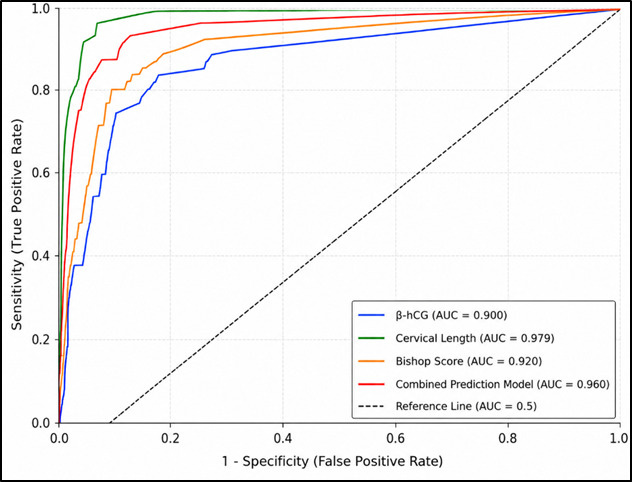
Table 6 summarizes the neonatal outcomes among the study and control groups. Neonates in the study group had a significantly lower mean birth weight than those in the control group (2.27 ± 0.46 vs. 2.86 ± 0.66 kg; p<0.001). NICU admission was significantly more frequent among neonates in the study group (47.6% vs. 22.2%; p=0.004), with a relative risk of 2.14 and an odds ratio of 3.18.
| Outcome | Study Group (n = 42) | Control Group (n = 108) | Effect Estimate | p-value |
| Birth weight (kg), Mean ± SD | 2.27 ± 0.46 | 2.86 ± 0.66 | Mean Difference = −0.59 kg | <0.001 |
| Median birth weight (kg) | 2.35 | 3 | — | — |
| Birth weight range (kg) | 0.9–2.8 | 0.9–3.7 | — | — |
| NICU admission, n (%) | 20 (47.6) | 24 (22.2) | RR = 2.14 (95% CI: 1.33–3.44) OR = 3.18 (95% CI: 1.49–6.78) | 0.004 |
CI: Confidence interval; NICU: Neonatal intensive care unit; OR: Odds ratio; RR: Relative risk; SD: Standard deviation.
Discussion
The current prospective observational study aimed to investigate the role of cervicovaginal β-hCG as a biochemical marker of spontaneous preterm delivery in pregnant women at 24-34 weeks gestation and its association with transvaginal cervical length and Bishop score. Cervicovaginal β-hCG levels were significantly higher in women who went on to deliver preterm than in those who delivered at term, and β-hCG showed a significant inverse correlation with cervical length and a positive correlation with Bishop score. Cervical length had the highest individual predictive accuracy. A combination of the three parameters gave an even better diagnostic performance. Furthermore, pelvic inflammatory disease (PID), PTI, hypertension in the mother, and previous preterm birth were independently associated with preterm delivery.
The observed increase in cervicovaginal β-hCG levels in women with preterm delivery is consistent with previous studies. In women with preterm delivery, significantly higher cervicovaginal β-hCG levels were reported by Güvenal et al., and its predictive value was shown in asymptomatic high-risk pregnancies by Adhikari et al. [5][6]. Likewise, Tripathi et al. found the mean level of β-hCG to be 27.8 ± 14.4 mIU/mL among women who gave preterm delivery, whereas 9.5 ± 14.4 mIU/mL was observed in women who delivered at term, which is very similar to the current results [8]. Cervicovaginal β-hCG is primarily produced by syncytiotrophoblastic cells and can be measured in amniotic fluid and cervicovaginal secretions throughout pregnancy [9]. Significant elevations may be due to disruption of the chorion-decidual interface, a mechanism similar to that proposed for cervicovaginal fetal fibronectin [5]. But the mechanism remains poorly understood, and no causality can be inferred from this observational study.
Receiver operating characteristic analysis showed that cervicovaginal β-hCG, at a cut-off of 16.61 mIU/mL, achieved an AUC of 0.900 with 83.3% sensitivity and 88.9% specificity. Similarly, Adhikari et al. demonstrated that cervicovaginal β-hCG predicted preterm delivery in asymptomatic high-risk women, and that the performance of the combined markers was superior to either marker alone [6]. In the present study, β-hCG demonstrated discriminatory performance comparable to cervicovaginal fetal fibronectin, which has been shown to have a good discriminatory ability in symptomatic women in a large systematic review [10]. There is, however, much greater validation of fetal fibronectin, and direct comparative studies are required to confirm the relative performance of each biomarker.
Transvaginal cervical length showed the highest AUC value of 0.979 and was predictive with a cut-off value of ≤2.9 cm. This finding is consistent with a systematic review and a recent meta-analysis demonstrating cervical length as a robust predictor of spontaneous preterm birth, which showed that the length of the cervix was a strong predictor of SPTB, and with the result of a recent meta-analysis of 447,864 pregnancies, which also demonstrated that universal cervical length screening is beneficial [11][12]. The good diagnostic performance observed in the present study may reflect the characteristics of this tertiary care antenatal cohort and the distribution of cervical length in the study population. The cut-off value of ≤2.9 cm was slightly higher than the standard of 25 mm, which may be due to population-specific characteristics of this tertiary care cohort.
More importantly, the combination of cervicovaginal β-hCG, cervical length, and Bishop score in a logistic regression model had an AUC of 0.960, which was higher than the individual parameters, with a 95% sensitivity and 95% specificity. This is in line with the principle, previously proposed by Adhikari et al. and Bagga et al., that biochemical and biophysical predictors complement each other in predicting preterm delivery [6][7]. The inclusion of Bishop score may enhance the applicability of the model in low-resource settings, where digital cervical examination remains widely practiced despite its known interobserver variability [3].
A previous history of preterm birth was more common among women who subsequently delivered preterm and was associated with a subsequent preterm delivery in multivariable analysis. In a population-based cohort study of more than 3.5 million births in Brazil, women with a history of preterm delivery had an adjusted odds ratio for recurrent preterm delivery of 2.58, which is similar to the risk observed in the current study. This association is thought to reflect persistent genetic, environmental, and uterine factors that may recur across pregnancies [13].
The independent association between PID and preterm delivery is biologically plausible. A national Taiwanese study of women found that PID was significantly associated with an increase in the cumulative incidence of preterm labour [14]. Genital tract infections stimulate inflammatory mechanisms that lead to uterine contractions, cervical ripening, and preterm premature rupture of membranes due to the production of prostaglandins and the impairment of progesterone-mediated processes [15]. Independent associations of both PID and PTI even after adjustments for β-hCG and cervical length indicate that infectious pathways are associated with PTB through pathways that are not entirely explained by these biomarkers.
Maternal hypertension was also independently linked to preterm delivery, which is similar to what was found in a population-based cohort in Spain, where the adjusted odds ratio for preterm delivery was 3.09 for women with chronic hypertension [16]. Since spontaneous and indicated preterm births were not separately analysed, the mechanism of this association could not be determined.
As expected, neonates in the preterm group were of lower birth weight and over twice as likely to be admitted to the NICU [1]. With the majority of the world's premature births taking place in LMICs, including India, accurate antenatal prediction of preterm birth is crucial to ensure early intervention, including the use of antenatal corticosteroids, neuroprotective magnesium sulphate and referral to centers with access to a neonatal intensive care unit (NICU) [1][3].
There are also several methodological strengths. A prospective design was used with standardised collection and laboratory measurement of samples (by the chemiluminescent immunoassay method), thereby minimising detection bias. Known confounders were adjusted for using multivariable logistic regression, and the parameters used to build the combination model were clinically accessible and could be used without specific equipment other than a transvaginal probe and a common immunoassay platform. The study is also one of the few from the Indian subcontinent to combine the cervicovaginal β-hCG, cervical length, and Bishop score in the same cohort, as it provides insight from a high-burden region where the burden of preterm birth remains high [1].
Several limitations must be considered. The sample size of 150 is small, and the confidence intervals of diagnostic estimates are, therefore, large. The single-centre design may bias the external validity of the results to other obstetric groups who have varying baseline risk factors and distributions of cervical length. Only one cervicovaginal β-HCG was measured per subject and the time course of this marker during gestation in relation to the time of delivery has not been characterised. Due to the non-uniform use of first trimester ultrasonography to confirm the gestational age, potential misclassification was introduced. The associations specific to the spontaneous preterm parturition pathway were not analysed separately as this could have diluted associations for spontaneous and indicated preterm deliveries. However, as in all observational studies, the possibility of residual confounding due to unobserved factors cannot be ruled out.
More research is needed to assess cervicovaginal β-hCG in larger multicentre prospective cohorts, preferably with serial measurements at specific gestational intervals for optimal timing and thresholds. Comparisons with other markers such as fetal fibronectin, placental alpha-microglobulin-1, and cervical elastography in the same population would help determine the incremental predictive value of β-hCG in current practice. It would be helpful if studies used separate models for spontaneous and indicated preterm birth and stratified the results by gestational age groups to enhance the mechanistic understanding. Future studies should evaluate the feasibility of incorporating cervicovaginal β-hCG into antenatal triage protocols to enhance clinical decision-making and improve neonatal outcomes in resource-limited settings.
Conclusion
In conclusion, the present study demonstrated that elevated cervicovaginal β-hCG levels were significantly associated with spontaneous preterm delivery and showed a significant inverse correlation with cervical length and a positive correlation with Bishop score among pregnant women. Although transvaginal cervical length remained the strongest individual predictor, The combined model demonstrated excellent diagnostic performance, with high sensitivity and specificity. Additionally, previous preterm birth, pelvic inflammatory disease, pelvic tract infections, and maternal hypertension were identified as independent risk factors for preterm delivery. These findings suggest that integrating biochemical, sonographic, and clinical parameters may enhance risk stratification for spontaneous preterm birth. However, given the single-centre design and relatively small sample size, larger multicentre prospective studies are required to validate these findings, establish optimal β-hCG cut-off values, and determine the clinical utility of incorporating cervicovaginal β-hCG into routine antenatal risk assessment.
Declarations
Authors’ Contributions
All authors contributed substantially to the conception of the study, data acquisition, analysis, drafting, and critical revision of the manuscript. All authors have read and approved the final version of the manuscript.
Acknowledgements
The authors would like to thank all participants who took part in this study and the staff of the Department of Obstetrics and Gynaecology of the participating institution for their support.
Funding/ financial support
This research received no specific grant from any funding agency in the public, commercial, or not-for-profit sectors.
Conflict of interest declaration
The authors declare that there are no conflicts of interest related to this study.
Contributors
Sugandha Patel, Department of Obstetrics and Gynecology, Ganesh Shankar Vidyarthi Memorial Medical College, Swaroop Nagar, Kanpur, Uttar Pradesh, India
Divya Dwivedi, Associate Professor, Department of Obstetrics and Gynecology, Ganesh Shankar Vidyarthi Memorial Medical College, Swaroop Nagar, Kanpur, Uttar Pradesh, India
Shaily Aggarwal, Professor, Department of Obstetrics and Gynecology, Ganesh Shankar Vidyarthi Memorial Medical College, Swaroop Nagar, Kanpur, Uttar Pradesh, India
Neena Gupta, Professor, Department of Obstetrics and Gynecology, Ganesh Shankar Vidyarthi Memorial Medical College, Swaroop Nagar, Kanpur, Uttar Pradesh, India
Renu Gupta, Professor, Department of Obstetrics and Gynecology, Ganesh Shankar Vidyarthi Memorial Medical College, Swaroop Nagar, Kanpur, Uttar Pradesh, India
Garima Gupta, Associate Professor, Department of Obstetrics and Gynecology, Ganesh Shankar Vidyarthi Memorial Medical College, Swaroop Nagar, Kanpur, Uttar Pradesh, India
Pratima Verma, Associate Professor, Department of Obstetrics and Gynecology, Ganesh Shankar Vidyarthi Memorial Medical College, Swaroop Nagar, Kanpur, Uttar Pradesh, India
Alka Nagar, Associate Professor, Department of Anatomy, Ganesh Shankar Vidyarthi Memorial Medical College, Swaroop Nagar, Kanpur, Uttar Pradesh, India
Ethical Clearance
The study was approved by the Institutional Ethics Committee of G.S.V.M. Medical College, and written informed consent was obtained from all participants before enrolment.
Trial Details
This study was a prospective observational study and was not registered as a clinical trial.