Introduction
An accessory spleen, or splenule, is a congenital nodule of normal splenic tissue that develops separately from the main organ as a result of incomplete fusion of splenic anlage during embryogenesis. It is a common finding, reported in approximately 10–30 per cent of the population, and is most often located near the splenic hilum, although ectopic sites within the left upper abdomen are recognised. Splenules are usually asymptomatic and are detected incidentally on cross-sectional imaging performed for unrelated indications.
What is not generally appreciated outside radiology practice is that, like the native spleen, an accessory spleen depends on an intact vascular pedicle and can therefore undergo torsion or vascular occlusion, resulting in infarction. When this occurs, it can present as acute left upper quadrant pain that clinically mimics other causes of an acute abdomen, and it is easily overlooked or mischaracterised on imaging if the possibility is not specifically considered. Concurrent infarction of the native spleen and an accessory spleen, as in the case reported here, is even less commonly described.
We report this case to illustrate the CECT features that allow confident, non-invasive diagnosis of combined splenic and splenunculus infarction, and to highlight why recognising this entity helps avoid unnecessary surgical intervention in patients who can be safely managed conservatively.
Patient Information and Clinical Findings
A 67-year-old woman presented to the emergency department with left upper abdominal pain of one day’s duration, associated with fever and chest pain. She reported a productive cough of one week’s duration preceding presentation. Her medical history was notable for type 2 diabetes mellitus and coronary artery disease, with a prior percutaneous transluminal coronary angioplasty.
On examination she was febrile. Chest auscultation was clear, and abdominal examination revealed mild left hypochondriac tenderness without guarding or rebound. Pulse oximetry showed desaturation (SpO₂ 86% on room air), which improved to 96% on 4 L/min supplemental oxygen. Laboratory investigations showed microcytic hypochromic anaemia, neutrophilia and moderate thrombocytopenia.
Timeline
| Time point | Clinical / diagnostic event |
| Onset of productive cough | |
| Day 0 | Onset of left upper quadrant pain and fever; presentation to Emergency Department |
| Day 0 | Clinical examination; desaturation noted (SpO₂ 86% on room air); supplemental O₂ started |
| Day 0 | Laboratory work-up: microcytic hypochromic anaemia, neutrophilia, moderate thrombocytopenia |
| Day 0 | CECT abdomen and chest performed, splenic infarct and early splenunculus infarct identified; bilateral ground-glass opacities with right middle lobe consolidation |
| Day 0–Day 2 | Subcutaneous low-molecular-weight heparin (enoxaparin/Clexane) 0.4 mg OD for 2 days |
| Day 0–Day 3 | IV fluids, supplemental O₂, and IV cefoperazone-sulbactam (Magmex Forte) 3 g BD for 3 days |
| Discharge | Discharged on a short course of aspirin (Ecosprin) and antiplatelet therapy |
| 1 month | Outpatient review, complete clinical improvement |
Diagnostic Assessment
Imaging methods
Contrast-enhanced computed tomography (CECT) of the abdomen and chest was performed on a Philips MX 16-slice CT scanner (16-slice configuration), for evaluation of acute left upper quadrant pain, fever and respiratory symptoms. Multiphase post-contrast imaging of the abdomen was obtained, including arterial, portal venous and delayed venous phases, to characterise splenic and juxtasplenic lesion enhancement.
Imaging findings
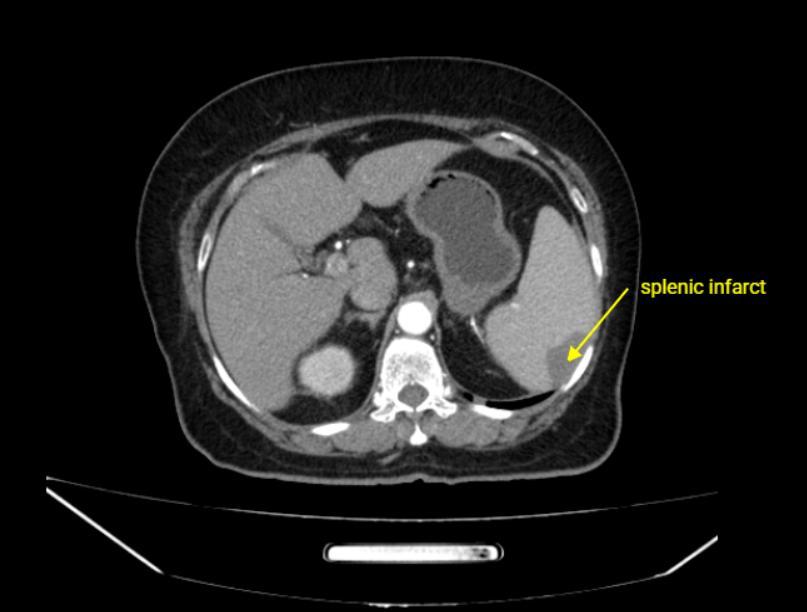
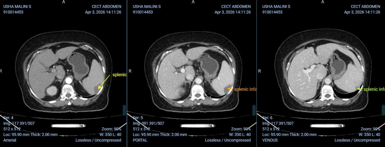
CECT of the abdomen showed borderline splenomegaly. In the anteromedial aspect of the spleen there was a small, peripheral, wedge-shaped area of hypodensity that failed to enhance on all post-contrast phases (arterial, portal venous and delayed venous), a pattern diagnostic of splenic infarction (Figure 2).
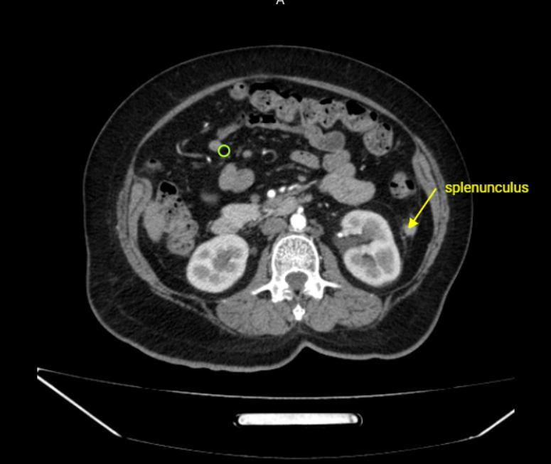
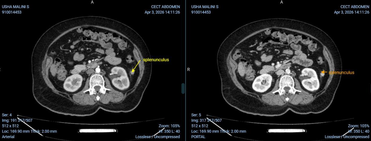
Adjacent to the lower pole of the spleen was a separate, well-defined nodule measuring approximately 12 × 11 mm, showing attenuation similar to splenic parenchyma with surrounding mesenteric fat stranding (Figure 1, Figure 3). Review of prior imaging showed that this nodule had previously enhanced in a manner similar to the native spleen, confirming its identity as an accessory spleen (splenunculus); the new perifocal fat stranding and altered attenuation on the current study were interpreted as early infarction of this splenule (Figure 3).
CT of the chest showed bilateral diffuse ground-glass opacities with foci of consolidation in the right middle lobe and mediastinal lymphadenopathy (Figure 4), findings attributed to the patient’s antecedent respiratory illness rather than to the splenic process.
Differential diagnosis
Given the acute left upper quadrant pain with fever in a patient with vascular risk factors, the differential diagnosis considered on presentation included splenic abscess, splenic infarction secondary to embolic or thrombotic disease, splenic rupture or haematoma, and a primary or metastatic splenic mass. The juxtasplenic nodule itself raised the differential of a lymph node, an exophytic splenic mass, or an accessory spleen. The non-enhancing wedge-shaped splenic lesion and the similarity of the juxtasplenic nodule’s baseline attenuation to splenic parenchyma, together with the absence of a discrete mass or rim enhancement suggestive of abscess, favoured infarction of both the native spleen and an accessory spleen over the other differential considerations.
Diagnostic reference standard
The diagnosis in this case rested on the characteristic multiphase CECT appearance, non-enhancement of the splenic lesion across arterial, portal venous and delayed venous phases, correlated with comparison to prior imaging demonstrating that the juxtasplenic nodule had previously enhanced in a manner similar to native splenic tissue, thereby confirming it as an accessory spleen rather than a lymph node or other mass. No biopsy, aspiration or surgical exploration was performed, as the patient was managed conservatively and the imaging findings were considered diagnostic; longitudinal clinical improvement at one-month follow-up provided additional supportive evidence. Histopathological confirmation was therefore not obtained, and this should be recognised as a limitation of the diagnostic certainty in this report (see Limitations).
Therapeutic Intervention
The patient was managed conservatively with intravenous fluids, intravenous antibiotics and supplemental oxygen for hypoxia. She received intravenous cefoperazone-sulbactam (Magmex Forte) 3 g twice daily for three days, together with subcutaneous low-molecular-weight heparin (enoxaparin, Clexane) 0.4 mg once daily for two days. She was discharged on a short course of aspirin (Ecosprin) and antiplatelet medication.
Outcome and Follow-up
The patient was reviewed one month after discharge and had complete clinical improvement. No interval imaging follow-up of the splenic and splenunculus findings is described in the available record.
Discussion
An accessory spleen, also known as a splenule, is a congenital focus of normal splenic tissue that is anatomically separate from the main spleen. Accessory spleens are relatively common and are often discovered incidentally during abdominal imaging. They are typically found near the splenic hilum, although they may occur in other locations within the left upper abdomen. In most individuals, splenules remain asymptomatic throughout life and require no treatment.
Rarely, a splenule may undergo torsion around its vascular pedicle or develop vascular occlusion, leading to ischaemia and eventual infarction. Splenic infarction occurs when the arterial blood supply is compromised, resulting in tissue necrosis. Predisposing factors include hypercoagulable states, haematological disorders such as sickle cell disease, malignancy, trauma, and other conditions that affect splenic perfusion.
Patients commonly present with acute or intermittent left upper quadrant abdominal pain, which may radiate to the left shoulder. Associated symptoms can include fever, nausea, and tenderness over the upper abdomen. In some cases, the clinical presentation can mimic other causes of an acute abdomen, making diagnosis challenging.
Contrast-enhanced computed tomography is the imaging modality of choice for evaluation. A normal splenule typically demonstrates imaging characteristics and enhancement patterns similar to those of the native spleen. When infarction occurs, the lesion appears as a hypoattenuating mass, usually located adjacent to the spleen, with surrounding inflammatory fat stranding. Comparison with previous imaging may reveal that a previously normally enhancing splenule has become hypoenhancing or non-enhancing, supporting the diagnosis of ischaemia and infarction.
In the present case, CECT demonstrated a well-defined hypoattenuating nodule situated anterior and inferior to the spleen, associated with mild inflammatory changes in the adjacent mesenteric fat. Review of prior imaging showed enhancement characteristics similar to the spleen, confirming its nature as an accessory spleen. Recognition of infarcted splenules is important because they can mimic other abdominal masses or surgical emergencies. Accurate diagnosis on imaging helps avoid unnecessary surgical intervention and allows appropriate conservative management in selected patients. Most uncomplicated cases respond well to symptomatic treatment, including analgesics and clinical observation, with favourable outcomes, consistent with the course observed in this patient.
Limitations
This report describes a single patient, and conclusions cannot be generalised beyond this observation. The diagnosis of splenunculus infarction was based on CECT appearance and comparison with prior imaging rather than histopathological confirmation, as the patient was managed conservatively without biopsy or surgery; the possibility that the juxtasplenic finding represents a closely related but distinct entity cannot be entirely excluded on imaging alone. Follow-up was limited to clinical review at one month without confirmatory follow-up imaging of the splenic lesions, so the study cannot describe the radiological resolution trajectory of either infarct.
Learning Points
-
Infarction of an accessory spleen (splenunculus) is a rare but recognisable cause of acute left upper quadrant pain and can occur concurrently with infarction of the native spleen.
-
On CECT, a normal splenule enhances similarly to native splenic parenchyma; loss of enhancement with perifocal fat stranding, especially when compared with prior imaging, is the key diagnostic sign of splenunculus infarction.
-
Recognising this entity on imaging can prevent an infarcted splenule from being mistaken for another abdominal mass or surgical emergency, avoiding unnecessary intervention.
-
Uncomplicated splenic and splenunculus infarction can be managed conservatively with supportive care and antiplatelet therapy, with favourable short-term outcomes.
Declarations
Ethics approval and consent to participate
waiver of consent
Conflicts of interests
NIL
Funding Statement
NIL
Author contributions (CRediT)
Ruma Chakraborty: Conceptualization, investigation, data curation, writing – original draft, visualization, and writing – review and editing.
J. Senthil Kumar: Supervision, conceptualization, investigation, and writing – review and editing.
Venit Rose: Clinical investigation, data curation, and writing – review and editing.
Venu S: Clinical investigation, interpretation, and writing – review and editing.
Narmadhar A R: Investigation, data curation, radiological interpretation, and writing – review and editing.
All authors: Reviewed and approved the final manuscript and agreed to be accountable for all aspects of the work.
Acknowledgements
NIL