Introduction
Over the decades magnetic resonance imaging (MRI) diagnostic services despite the ferromagnetic challenges have continued to rise due to the non-association with ionizing radiation and consequent wide safety margin. The production of superior anatomical imaging of water-dense body tissues (such as brain, spinal cord, abdominal viscera, joints and ligaments), based on a mechanism involving Hydrogen proton magnetization, makes it a particularly suitable radiological procedure for young children, owing to their greater water content per kilogram body weight relative to adults [1][2]. However, for the production of a high-quality image a very still patient is a prerequisite, necessitating the administration of general anaesthesia especially in the younger paediatric category [3]. This requirement for anaesthesia entails a combination of very unique challenges for the Anaesthesiologist: a) an inability to get ready assistance from other Anaesthesiologists due to the detached location of MRI suite, and b) the challenges associated with anaesthesia for children aged five years and less as a result of their peculiar anatomical, physiological, pharmacological and psychological profiles [4][5].
Following the installation of its 1.5 Tesla MRI scanner the UPTH became a foremost referral tertiary hospital for paediatric diagnostic radiology in South-South Nigeria, however the paucity of MRI-compliant anaesthetic equipment remained an additional source of remarkable challenge to the physician Anaesthesiologist. In advanced countries, the conduct of anaesthesia for paediatric MRI aligns with best practice standards enabled by an adequacy of requisite specialist workforce and anaesthetic equipment. The World Federation of Societies of Anaesthesiologists (WFSA) recognizes that such interplay between professional competence and adequate MRI compliant anaesthetic equipment ensuring a favourable paediatric anaesthetic outcome is lacking in low- and middle-income countries (LMICs), and accounts for a higher risk of occurrence of anaesthesia-related under-five morbidity and mortality [6]. This study was therefore designed to retrospectively audit the five-year pattern of pathological indications for MRI in children aged five years and below, and the gravity of the challenges encountered by the Anaesthesiologists providing the necessary procedural anaesthesia for this category of children at the University of Port Harcourt Teaching Hospital (UPTH), Port Harcourt, Nigeria, a tertiary hospital operating in a typical resource poor setting in West Africa.
Materials and Methods
Ethical clearance was secured from the University of Port Harcourt Teaching Hospital for a retrospective study. Data on all children aged one day to five years who had MRI under anaesthesia from 1st January, 2021 to 31st December, 2025 were retrospectively obtained from the Radiology Unit registers as well as from the Paediatric Anaesthesia Unit charts and records. Data included age, sex, indication for MRI, American Society of Anesthesiologists (ASA) classification, type of anaesthesia, challenges and outcomes. Children older than five years, presence of respiratory tract infection (RTI), declared history of upper respiratory tract infection (URTI) occurring not earlier than six weeks before the date of presentation for MRI, and those who underwent MRI before 2021 or after 2025 were excluded.
Conduct of Anaesthesia
All the children were admitted for MRI as day-cases however with a low threshold for overnight hospital stay. A pre-anaesthetic assessment was done for each child on the morning of the MRI, and every child through an earlier instruction to the parent/guardian was fasted from solids for six hours, breast milk for four hours, but allowed to take clear glucose containing fluids up to two hours prior to anaesthesia. Separation anxiety was countered in those aged 6 months to 5 years by administering an oral 0.5mg/kg midazolam flavoured with clear glucose fluid (Sprite) about 30 minutes before induction of anaesthesia. The parents were also counselled to keep strict compliance with local MRI-based ferromagnetic regulations. Drugs and equipment for sedation, general anaesthesia and emergency resuscitation were made available on a trolley kept outside the MRI suite. Every child had baseline recording of non-invasive blood pressure, heart rate and peripheral arterial oxygen saturation (SpO2) via a multi-parameter monitor (DASH 4000), followed by an intravenous access secured with an appropriate-size intravenous cannula (22 or 24 gauge).
The majority of children had general anaesthesia induced using intravenous fentanyl 1µ/kg, midazolam 0.025mg/kg, atropine 0.02mg/kg and a loading dose of propofol 2mg/kg. However, in a combative few an intramuscular 5mg/kg ketamine mixed with atropine 0.02mg/kg was administered to facilitate venous access, followed by intravenous midazolam 0.025mg/kg and propofol 1mg/kg. An appropriate size paediatric Guedel’s oro-pharyngeal airway (size 0 or 1) or laryngeal mask airway (LMA) size 1, 1 and half, or 2 was inserted to maintain the airway. Anaesthesia was maintained with continuous titration of propofol at 4 - 6mg/kg/hour via a paediatric infusion giving set (soluset) containing 5ml/kg pre-warmed lactated Ringer’s solution. From induction of anaesthesia to recovery the children received supplemental oxygen through Mapleson F breathing system, which was connected by means of an elongated tubing to an oxygen cylinder that was kept outside the MRI suite. A pre-determined Oxygen flow rate of 3 times the child’s minute ventilation was used. The children were then quickly transferred to the MRI suite and positioned for scanning following attainment of an appropriate depth of anaesthesia. Adequate covering of each child with thick blanket was ensured for the prevention of hypothermia.
Monitoring was undertaken by the Senior physician Anaesthesiologist from within the MRI suite. An MRI compliant pulse oximeter finger probe that was attached gave a continuous display of pulse and oximetry values, while the child’s spontaneous respiration was monitored using the rate and amplitude of reservoir bag movements. At the completion of the MRI, propofol infusion was discontinued and the children were transferred to the recovery room outside the MRI suite where intravenous fluid, temperature care, oxygen supplementation and monitoring of haemodynamic parameters were continued till they achieved satisfactory recovery. Supraglottic devices were removed when the children were awake, evidenced by the opening of the eyes, an attempt to extrude the supraglottic device from the mouth or pull it out with the hand. The occurrence of laryngospasm was promptly treated with low dose (0.1mg/kg) intravenous suxamethonium combined with gentle oropharyngeal suctioning and positive pressure supplemental oxygen delivery.
Data Collection and Analysis
All data were handled confidentially, entered on a spread sheet and exported to the Statistical Products and Service Solutions (SPSS) version 21.0. for analysis. Results were presented using bar chart, as well as means (SD), frequencies and percentages expressed within tables.
Results
All the children received general anaesthesia for their MRI. Demographically there was male preponderance [72 (55.0%) versus 59 (45.0%)], with a male-to-female ratio of 1.2:1. The proportions of the different age categories of the children in the study, their ASA physical fitness classifications, type of supraglottic device used for airway maintenance and the different anatomical regions scanned are as shown (Table I).
| Parameter | N (%) | Weight (Kg) Mean (SD) |
| Sex | ||
| Male | 72 (55.0) | |
| Female | 59 (45.0) | |
| Total | 131 (100.0) | |
| Male: Female Ratio | 1.2:1 | |
| Age | ||
| < 1 month | 5 (3.8) | 3.4(0.39) |
| 1 to 5 months | 20 (15.3) | 5.4(1.33) |
| 6 to 11 months | 22 (16.8) | 8.7(1.98) |
| 1 to 3 years | 58 (44.3) | 12.1(1.34) |
| 4 to 5 years | 26 (19.8) | 17.1(0.76) |
| Total | 131 (100.0) | |
| ASA classification | ||
| I | 44 (33.6) | |
| II | 79 (60.3) | |
| III | 8 (6.1) | |
| Total | 131 (100.0) | |
| Type of Anaesthesia | ||
| GA + LMA | 98 (74.8) | |
| GA + GOA | 33 (25.2) | |
| Total | 131 (100.0) | |
| Anatomical Region Scanned | ||
| Brain | 127 (96.9) | |
| Abdomen | 2 (1.5) | |
| Neck and Thorax | 1 (0.8) | |
| Thorax | 1 (0.8) | |
| Total | 131 (100.0) |
ASA = American Society of Anesthesiologists; GA = General Anaesthesia; LMA = Laryngeal Mask Airway; GOA = Guedel’s Oro-Pharyngeal Airway
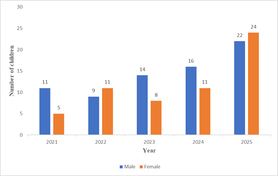
The distribution of annual number of children who had anaesthesia for MRI showed an overall rise in the corresponding values from the first to the fifth year during the period of study (Figure 1).
Neurological disorder, in comparison with non-neurological condition, constituted the highest indication for MRI. (Table II).
| Indication | N (%) |
| Neurological Disorders | |
| Seizure disorder | 30 (22.9) |
| Cerebral palsy | 20 (15.3) |
| Megalencephalic Congenital/Acquired hydrocephalus | 17 (13.0) |
| Meningitis | 10 (7.6) |
| Meningocele/Myelomeningocele | 10 (7.6) |
| Developmental Retardation | 10 (7.6) |
| Autism Spectrum Disorder | 7 (5.3) |
| Recurrent Febrile Convulsions, Dyskinesia | 7 (5.3) |
| Spasmus Nutans, Astasia-Abasia, Behavioural Disorder, Space Occupying Lesion, Sudden Weakness of Limbs | 6 (4.4) |
| Tethered Cord Syndrome, Bilateral Optic Atrophy, Space Occupying Lesion, Craniosynostosis | 4 (3.0) |
| Hypoxic Ischaemic Encephalopathy, Traumatic brain Injury with Hemiparesis, Arnold-Chiari malformation, | 3 (2.3) |
| Total | 124 (94.7) |
| Non-neurological Disorders | |
| Multiple Congenital Anomalies, Congenital Orbital Teratoma | 2 (1.5) |
| Right Hemithoraic Wall Mass, Cystic Hygroma of Neck/Chest | 2 (1.5) |
| Posterior Ureteric valve, Right Undescended Testis | 2 (1.5) |
| Retinoblastoma/Recurrent Retinoblastoma | 1 (0.8) |
| Total | 7 (5.3) |
Laryngospasm was the leading anaesthesia-related challenge and occurred in nine children post LMA removal. Of this number five were noted to have undeclared URTI. The challenges related to poor resource facility were difficulty with patient monitoring, difficulty with induction of anaesthesia, and physician Anaesthesiologist discomfort from monitoring within the cold MRI suite correspondingly as the first, second and third most frequent. There was however no mortality (Table III).
| Challenges | N (%) |
| Anaesthesia related | |
| Laryngospasm | 9 (6.9) |
| Hypoxaemia (SpO2 < 90%) | 8 (6.1) |
| Delayed recovery | 6 (4.6) |
| Inadequate depth of anaesthesia causing MRI session interruption | 3 (2.3) |
| Poor-resource related | |
| Difficulty with monitoring due to unavailability of appropriate monitoring Device | 131 (100.0) |
| Difficulty with induction due to unavailability of appropriate anaesthetic Equipment | 112 (85.5) |
| Physician discomfort from cold MRI suite | 87 (66.4) |
Discussion
In this five-year retrospective study of 131 children aged one day to five years presenting with diverse pathologies for MRI, neurological disorders constituted the greatest indication. While laryngospasm, hypoxaemia and delayed recovery correspondingly were the first, second and third leading anaesthesia-related challenges, patient monitoring difficulty, induction difficulty and physician Anaesthesiologist discomfort from cold suite emerged respectively as the first, second and third most frequent challenges related to poor resource. There was however zero anaesthesia-related mortality.
Globally, providing safe general anaesthesia for paediatric radio-diagnosis remains a desirable and recognized challenging goal arising from an interplay of MRI-related peculiarities, general anaesthesia and innate features of young children. In their study of the challenges and clinical applications of paediatric MRI Ditchfield [7] documents that general anaesthesia in young children is a source of significant challenge.
The finding in this study that seizure disorder was the highest indication for MRI agrees with the report by Mallory et al.,[8] in which same category of disorders constituted 63.7% in their retrospective analysis of 109,947 entries of pediatric sedations for MRI, in the Pediatric Sedation Research Consortium (PSRC), from 2011 to 2017, out of which 66.2% were children aged ≤ 5 years. Children with seizure disorder presenting for MRI pose peculiar challenges because a rebound or break-through seizure episode can occur periprocedurally according to Sisodiya et al.,[9] and Al-Kattan et al.,[10]. An occurrence of seizure within the MRI bore would constitute danger to the patient and a nightmare to the Anaesthesiologist due to the associated vulnerability to trauma and deleterious impact of apnoeic intervals on a pre-existing compromised cerebral neuronal integrity as reported by Khan et al.,[11]. Ensuring a non-occurrence of seizure during MRI procedure is therefore a cardinal objective.
Maintaining airway patency and functionality remains the denominator to successful periprocedural patient care. Laryngospasm in this study occurred following LMA removal. This sustained reflex partial or complete glottic closure is usually triggered in response to laryngeal stimulation by secretion or foreign body, while in the light plane of anaesthesia [12][13]. Epidemiologically, according to the report by Olsson et al.,[14] its incidence in infants is twice that in older children and three times that in adults. Evidentially, undeclared and untreated upper respiratory tract infection (URTI) increases laryngospasm risk. In this study its occurrence in children with undeclared URTI aligns with the similar finding by Otokwala et al.,[15]. The documented empirical observation of a higher incidence of laryngospasm in children is underpinned by their relative vagotonia, attributable to an innate autonomic dysequilibrium characterized by a much more developed parasympathetic nervous system (PNS) compared to the sympathetic nervous system (SNS). In their computer-aided electrophysiological study Harteveld et al.,[16] have documented that PNS activity demonstrated a cubic trend, with an exponential increase from infancy, a plateau in mid-childhood, and a decline toward adolescence, while the SNS activity followed a contrasting linear trend with a gradual decrease from infancy toward adolescence. Consequently, younger children produce more glandular secretions than older children and adults, hence while lying supine under general anaesthesia with a supraglottic device gravity-dependent trickling of oro-pharyngeal secretions into the larynx may occur triggering laryngospasm. This necessitates judicious antisialagogue administration.
Although the timing of supraglottic device removal is still controversial in paediatric anaesthesia practice, according to Engelhardt et al.,[17] the majority of practising Anaesthesiologists preferred awake to deep supraglottic extubation as was done in this study, based on the consideration that at deep plane depressed airway protective reflexes and collapsed upper airway walls might lead to risk of airflow obstruction. While awake supraglottic removal may be associated with risk of coughing and spasmodic expiration with hypoxaemia, according to the report by Von Ungern-Sternberg et al.,[18] the occurrence of laryngospasm is not different between deep and awake extubation methods. In the present study all cases of laryngospasm were promptly treated using intravenous low dose 0.1mg/kg suxamethonium, gentle suctioning and intermittent positive pressure supplemental oxygen administration. Low dose (0.1mg/kg) intravenous suxamethonium has documented prophylactic and curative efficacy against laryngospasm without associated occurrence of fasciculations or impairment of spontaneous respiration [19][20].
Hypoxaemia in this study was secondary to laryngospasm and the finding agrees with the observation by Al-alami et al.,[21]; its impact can worsen preexisting neurological derangements. Therefore, meticulous vigilance must be ensured during the conduct of paediatric anaesthesia to enable prompt detection and management of hypoxaemia, especially in young children rendered vulnerable to the development of hypoxic cerebral ischaemia by an already existing neurological pathology. Lv et al.,[22] reports an association of chronic repeated seizure with compromised dynamic cerebral autoregulation in patients with epilepsy. Supplemental Oxygen delivery, close monitoring and prompt intervention were ensured in this study.
Intra-procedural monitoring was done from within the MRI suite by the Senior Anaesthesiologist in the team, necessitated by the paucity of MRI compliant equipment for optimal cardiorespiratory monitoring as recommended by Sury et al.,[23]. According to the guidelines by the Association of Anaesthetists of Great Britain and Ireland (AAGBI) in 2015 [24], functional minimum monitoring devices for heart rate, blood pressure, peripheral oxygen saturation, end-tidal carbondioxide and anaesthetic vapour concentration must be attached prior to general anaesthetic induction, and for continued monitoring till patient recovery. However, a complete alignment with the recommended minimum monitoring standard was difficult to achieve as reported in this study, depicting the gravity of the challenges of specialist healthcare delivery in resource-poor setting and underpinning the associated relatively greater occurrence of anaesthesia-related critical events [25].
A greater proportion of the physician Anaesthesiologists reported discomfort from cold MRI suite in this study. This was a direct consequence of a particular Anaesthesiologist taking up intraoperative monitoring for multiple sessions in a day from within the MRI suite (which had to remain operational at a threshold temperature range of 20 oC to 24oC) [26], without additional staff support, replacement or thermal care. As a mandatory monitoring standard the continued presence of a trained Anaesthesiologist must be ensured during the conduct of anaesthesia for paediatric MRI [24]. In settings using MRI compliant anaesthetic gadgets, close patient monitoring could be done from an adjacent room with a transparent glass window for direct view. Alternatively, where adequate workforce of the requisite cadre of Anaesthesiologists exists in the team, alternating in-suite patient monitoring role allows occasional breaks from the cold interior hence preventing discomfort. However, such standards are difficult to achieve in LMICs due to paucity of equipment in addition to depleted workforce caused by the continuing migration of Anaesthesiologists to high-income nations. In East Africa, a cross-sectional study by Epiu et al.,[27] in 2017 reveals the density of Anaesthesiologists per 100,000 population to be remarkably low, with values ranging from 0.02 in Burundi to 0.39 in Kenya.
In the absence of venous access, smooth and rapid inhalational induction can be suitably achieved using sevoflurane or halothane for the paediatric population [28]. However, bradycardia, cardio-depression, arrhythmias and hepatitis are drawbacks of halothane [29][30], while the high cost of sevoflurane (about two and half times that of halothane) poses a limitation to its availability in LMICs. Topical application of EMLA (Eutetic Mixture of Local Anaesthetic) CreamTM also reduces the pain of venepuncture facilitating venous access in children [31], but could not be done in this study due to unavailability. Again, the impact of a resource-poor centre posing limitation to anaesthesia practice is depicted. As a consequence the administration of ketamine to tame combative children led to delayed recovery. The occurrence of prolonged sedation and delayed emergence following the use of ketamine and midazolam in same patients is documented in literature [32].
The goals of optimal general anaesthesia for MRI should include smooth, rapid onset and offset of anaesthesia of adequate depth, adequate analgesia, airway maintenance, normoxaemia, normocapnoea, amnesia, haemodynamic stability and stillness. These however could not be completely achieved in this study in contrast with what is obtainable in advanced climes. According to the observation by the WFSA [6], there is 100 to 200 times more likelihood of the occurrence of anaesthesia-related complications and consequent mortality in a child in Africa, compared to a child in high-income nations, identifying a lack of anaesthetic/monitoring equipment as one of the factors responsible. Although the pattern of indications in this retrospective study aligns with those of advanced nations, the challenges found necessitate a desperate call for the availability of technological aid to promote safety in the practice of paediatric anaesthesia for MRI in LMICs.
The data used in this study was obtained from a single centre, thus rendering the findings not fully generalizable and tainting the study with limitation. Again, manual registers/records rather than an electronic data-base were the source of data, hence there was likelihood of some inaccuracies.
In conclusion neurological pathology predominantly seizure disorder constituted the most frequent indication for MRI in under-five children over the five-year study period. While laryngospasm, hypoxaemia and delayed recovery were anaesthesia-related, induction difficulty, monitoring difficulty and Anaesthesiologist discomfort from cold MRI suite were encountered as a result of poor resource setting, however without mortality.
Declarations
Acknowledgements
The authors express appreciation to Dr. (Mrs.) Deborah Ray-Offor, Mr. Tamunoibiton Samuel and Mrs. Amara Iwuoha for assisting with the charts and records.
Source of Funding
There is no funding by any individual or organization.
Conflict of Interest
No conflict of interest exists.
Ethical Clearance
This was granted by the hospital Research Ethics Committee (Ethical Clearance: UPTH/ADM/90/S.11/VOL.XI/2247)
Authors’ Roles:
ATA: Initial study conception, study design, data interpretation, manuscript preparation, final intellectual content development and corresponding author.
CPI: Data acquisition, initial intellectual content development, integrity appraisal, final version approval and critical reviewing author.