1. Introduction
Surgery cures more patients with solid tumors than any other oncologic modality, yet it is also, paradoxically, a biological event capable of promoting the very disease it is meant to eradicate. The concept that surgical trauma itself can accelerate tumor growth is not new: experimental and clinical observations dating back several decades have shown that wound fluid, wound-associated growth factors, and the systemic stress response to surgery can stimulate the proliferation of residual or disseminated tumor cells [1-3]. What has changed in the past twenty years is the depth of mechanistic understanding of why this happens, and the emergence of concrete, testable perioperative interventions designed to interrupt it.
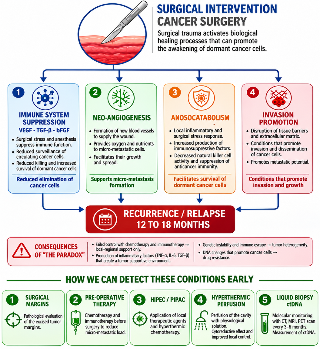
The “healing paradox” describes this counterintuitive biological reality: the very repair processes the body deploys to heal a surgical wound can simultaneously support the survival and outgrowth of any malignant cells left behind after tumor resection. Following curative-intent surgery, the operative field becomes host to a coordinated cascade of wound-healing signals—growth-factor release, new vessel formation, coagulation and fibrin deposition, and a transient period of immune suppression driven by postoperative neuroendocrine and inflammatory stress—each of which, while indispensable for tissue repair, can inadvertently nourish microscopic residual disease. This is compounded by the mechanical and biological reality of the operation itself: manipulation of the tumor, transection of lymphatics and blood vessels, and exposure of denuded serosal or peritoneal surfaces can seed viable tumor cells into the wound bed or the peritoneal cavity precisely at the moment when the local and systemic environment is most permissive for their survival [32].
Clinically, this window of vulnerability is thought to explain, at least in part, why a substantial proportion of oncologic recurrences after apparently complete (“R0”) resections cluster within the first one to two postoperative years, and why recurrence so often occurs locoregionally or on peritoneal surfaces rather than as isolated distant metastasis unrelated to the surgical field. Understanding the healing paradox is therefore not an academic exercise: it reframes the entire perioperative period—from the decision to operate, through anesthetic technique, surgical execution, and the first weeks of recovery—as a biologically active therapeutic window in which specific, evidence-based interventions may measurably improve oncologic outcomes [36].
This review updates and substantially expands an earlier summary of the concept, incorporating more recent mechanistic literature on perioperative immunosuppression, peritoneal mesothelial injury and adhesion biology, and the evolving evidence base for locoregional therapies (HIPEC/PIPAC) and ctDNA-based surveillance, with particular attention to peritoneal metastasis from gastric cancer, an area of long-standing clinical and research interest for our group [6-9].
2. Historical and Conceptual Background
The observation that wounding promotes tumor growth predates modern molecular oncology. Early animal studies demonstrated that injecting tumor cells near a healing incision, or exposing tumor cells to wound fluid, dramatically increased tumor take and growth rate compared with tumor cells injected into uninjured tissue. Hofer and colleagues showed directly that wound fluid and its constituent growth factors—particularly TGF-β and bFGF—significantly enhanced tumor growth in an animal model, and that the effect of these factors in combination approached that of whole wound fluid, implicating a multifactorial rather than single-mediator process [1]. Subsequent reviews synthesized this line of work into the broader concept that a tumor and a healing wound require biologically similar stromal microenvironments—activated fibroblasts, new vasculature, remodeled extracellular matrix, and a locally immunosuppressed milieu—such that a wound-healing stroma can effectively “host” and nourish a nascent or residual tumor focus, a notion sometimes summarized as the tumor behaving as “a wound that never heals” [34].
In parallel, within the specific context of peritoneal surface malignancy, Sugarbaker and colleagues, including our group, described the “tumor-cell entrapment phenomenon,” in which free intraperitoneal tumor cells released during surgical dissection become trapped within fibrin deposits and traumatized peritoneal surfaces at the time of operation, providing an anatomic and biological explanation for the characteristic pattern of peritoneal recurrence seen after resection of gastrointestinal and appendiceal malignancies, including pseudomyxoma peritonei [6]. This concept directly informs the current rationale for intraoperative peritoneal lavage and immediate intraoperative or early postoperative intraperitoneal chemotherapy, since the entrapment window is time-limited to the first hours to days after surgery, before fibrin organizes into mature adhesions [9].
3. Mechanisms of the Healing Paradox
3.1 Wound-Associated Growth Factors
The surgical wound—at both the visceral/intestinal and superficial levels—releases a coordinated set of mediators aimed at tissue repair, including VEGF, TGF-β, bFGF, platelet-derived growth factor (PDGF) and epidermal growth factor (EGF). These same factors, released by platelets, macrophages, and activated fibroblasts within the wound, act as trophic stimuli capable of driving the proliferation of tumor cells that remain near or distant from the resection site—a process first described mechanistically as the tumor-cell entrapment phenomenon in the peritoneal setting, but which applies more broadly to any operative field [6].
Clinical studies in breast cancer have quantified this phenomenon directly. Curigliano and colleagues showed that circulating levels of bFGF, VEGF, and TGF-β rise significantly by the fifth postoperative day, and that this rise is proportional to the extent of surgery, supporting the hypothesis that more extensive operations generate a proportionally larger systemic and local growth-factor stimulus capable of promoting residual disease [2]. Hormbrey and colleagues, studying wound fluid VEGF directly after breast cancer surgery, found that local wound VEGF concentrations markedly exceed circulating plasma levels and correlate with the angiogenic response in the healing wound, noting that the surgical site is most favorable to tumor cell implantation while actively healing and becomes progressively less permissive once healing is complete—a finding with direct implications for the timing of perioperative antiangiogenic or systemic therapy [3].
Mechanistically, TGF-β occupies a central, dual role. During normal wound healing it orchestrates inflammation resolution, collagen deposition, and stromal remodeling. In the tumor context, however, the same TGF-β-driven stromal program—myofibroblast activation, extracellular matrix deposition, and stiffening of the peritumoral stroma—can be co-opted to create a hospitable niche in which an initiated or residual malignant cell can establish and expand, a concept articulated in the “wound-healing stroma promotes tumor growth” hypothesis [33]. More recent work integrating tumor and wound biology has further shown that collagen cross-linking and increased mechanical stiffness of the healing stroma, mediated in part by lysyl oxidase (LOX) and TGF-β signaling, actively promotes tumor cell invasion and is associated with increased VEGF expression and angiogenesis in experimental models, reinforcing the mechanistic link between the physical and biochemical remodeling of the wound and tumor progression [34].
3.2 Neoangiogenesis
The formation of new vascular networks is indispensable for wound healing, supplying oxygen, nutrients, and inflammatory cells to the repairing tissue. The same angiogenic program, however, simultaneously supplies any micrometastatic focus present in or adjacent to the resection bed, facilitating its regrowth at precisely the time when systemic surveillance is otherwise weakened. Hormbrey and colleagues emphasized that because wound VEGF concentrations are so much higher locally than in plasma, circulating VEGF is a poor surrogate for the true angiogenic stimulus present at the tumor bed, a point relevant to the design and timing of antiangiogenic strategies intended to blunt this specific window rather than to treat established metastatic disease more broadly [3].
This has practical implications for the sequencing of antiangiogenic agents (e.g., bevacizumab and related compounds) around the time of surgery: because the same VEGF-driven pathway is required for normal wound healing, indiscriminate perioperative antiangiogenic therapy risks both impaired wound healing and anastomotic complications, while therapy timed to avoid the acute wound-healing window may still blunt the angiogenic support available to residual disease later in the postoperative course [3].
3.3 Perioperative Inflammation and Immunosuppression
Alongside growth-factor release and angiogenesis, the surgical stress response—comprising the local inflammatory reaction to tissue injury, the systemic neuroendocrine stress response (catecholamines, cortisol, prostaglandins), and the immunomodulatory effects of anesthesia, analgesia, blood transfusion, hypothermia, and pain—produces a transient but clinically important window of immunosuppression. This “window of vulnerability” is now recognized as one of the best-characterized and most therapeutically actionable components of the healing paradox [14,35].
Natural killer (NK) cells, which under normal conditions provide innate cytotoxic surveillance against circulating and residual tumor cells without requiring prior antigen sensitization, are profoundly and consistently suppressed in the postoperative period [14-16]. Experimental work has directly implicated postoperative NK-cell dysfunction as a causal mediator of metastasis formation after surgery, with surgical stress promoting coagulation-dependent and catecholamine/prostaglandin-dependent mechanisms of NK-cell suppression that can be pharmacologically reversed in animal models using perioperative β-adrenoceptor blockade and cyclooxygenase-2 (COX-2) inhibition. Mechanistically, postoperative NK-cell dysfunction has been linked to hypercoagulability, the release of immunosuppressive soluble factors, tissue hypoxia, and expansion of myeloid-derived suppressor cell (MDSC) populations that further blunt cytotoxic lymphocyte function and promote formation of pre-metastatic niches [15][16].
Anesthetic technique itself modulates this immunosuppressive window. Several retrospective series have reported an association between volatile (inhalational) anesthesia and reduced recurrence-free survival compared with total intravenous anesthesia (typically propofol-based), and opioid analgesia has independently been shown to suppress NK-cell, T-cell, dendritic-cell, and macrophage function via direct action on opioid receptors on immune cells, in addition to its established role in modulating the stress response. Regional anesthetic techniques, by reducing systemic opioid requirements and attenuating the neuroendocrine stress response to surgery, have been proposed—though not yet definitively proven in adequately powered randomized trials—as a means of mitigating this component of the healing paradox. In thoracic and lung cancer surgery specifically, perioperative suppression of NK-cell cytotoxicity and interferon-gamma secretion has been directly correlated with intratumoral NK-cell density and postoperative prognosis, and downregulation of chemokine signaling with associated MDSC recruitment has been linked independently to recurrence risk [37].
Taken together, this body of evidence reframes perioperative immunosuppression not as an unavoidable side effect of major surgery, but as a modifiable, mechanistically defined therapeutic target—one increasingly proposed as an “unexploited” window for perioperative immunotherapy, including cytokine-based approaches (IL-2, GM-CSF, interferon-α), checkpoint inhibition timed around surgery, and adjunctive pharmacologic blockade of the catecholamine/prostaglandin axis [36].
3.4 Fibrin Deposition, Mesothelial Injury, and Tumor-Cell Entrapment
Large traumatized surfaces—whether cutaneous, visceral, or peritoneal—are covered within the first postoperative hours to days by a fibrin-rich provisional matrix, the same structure that in normal wound healing serves as a scaffold for fibroblast migration, angiogenesis, and eventual collagen deposition. In the oncologic setting, this fibrin scaffold performs an additional, unwanted function: it allows any free tumor cells present in the operative field—released by surgical manipulation, transected lymphatics, or serosal disruption—to adhere, become physically entrapped, and begin to proliferate within a matrix that is simultaneously rich in the growth factors described above [32].The cellular biology of this process has been elucidated in detail in the closely related literature on postoperative peritoneal adhesion formation. Disruption of the mesothelial monolayer—the normally frictionless single-cell layer lining the peritoneal cavity—exposes the underlying basement membrane and is now understood to be the initiating “motor” of fibrin attachment between denuded surfaces. Within hours of peritoneal injury, stressed mesothelial cells undergo a calcium-dependent shape change, extend membrane protrusions, and begin to express pro-adhesive markers, subsequently undergoing a mesothelial-to-mesenchymal transition (MMT) that contributes directly to the myofibroblast population responsible for organizing the fibrin clot into a mature, collagen-dense structure over the following two to four weeks. Tissue hypoxia—itself a consequence of surgical trauma and impaired local perfusion—further disrupts the normal balance of mesothelial apoptosis and proliferation, favoring persistence of the fibrin matrix and impairing the fibrinolytic clearance that would otherwise resolve it. Because fibrinolytic activity (mediated by tissue plasminogen activator and its inhibitor, PAI-1) is itself suppressed at sites of peritoneal injury, fibrin deposits that would normally be degraded within days instead persist and organize, providing the temporal and physical scaffold in which entrapped tumor cells have an extended opportunity to establish themselves before the field re-mesothelializes [39][41].
This mechanistic picture directly supports the original tumor-cell entrapment concept described in the peritoneal surface malignancy literature: the risk of tumor cell entrapment and subsequent peritoneal recurrence is highest during the earliest postoperative period, when fibrin is freshly deposited and mesothelial coverage is not yet restored, and diminishes once re-mesothelialization and fibrinolysis are complete. It is precisely this time-limited window that provides the mechanistic rationale for immediate intraoperative peritoneal lavage and for locoregional intraoperative chemotherapy (HIPEC) or early postoperative intraperitoneal chemotherapy, discussed further in Section 5.4 below [6].
4. Clinical Consequences of the Paradox
The convergence of these four mechanisms—growth-factor release, neoangiogenesis, immunosuppression, and fibrin-mediated tumor-cell entrapment—has several downstream clinical consequences.
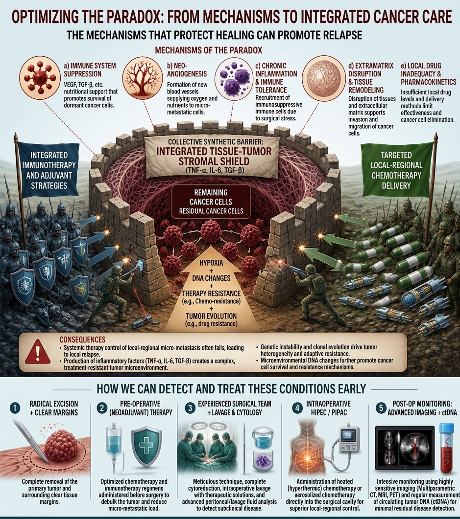
First, reduced responsiveness to chemotherapy and immunotherapy is a recognized feature of the early postoperative microenvironment, predisposing to locoregional recurrence. The presence of trophic healing mediators and enzymes, including TNF-α, IL-6, and TGF-β, generates a scar-like connective tissue matrix that functions as a physical and biological shield around the operative field, sheltering residual tumor cells from immune effector cells and limiting the penetration of chemotherapeutic agents to a therapeutically adequate concentration [6][32].
Second, this protective microenvironment appears to select for a more treatment-resistant tumor-cell phenotype. Tissue hypoxia, itself a consequence of postoperative tissue remodeling and locally suppressed perfusion, is a well-established driver of chemoresistance, both by directly limiting drug delivery and by promoting hypoxia-inducible transcriptional programs associated with a more aggressive and treatment-refractory phenotype. Cells that survive and proliferate within this hypoxic, immunologically shielded niche are therefore not simply “leftover” tumor cells behaving as they would have in the primary tumor; they represent a population shaped by a distinct microenvironment created by the healing process itself, and accumulating genomic instability under these conditions can further entrench a treatment-resistant phenotype for as long as the protective healing-associated “wall” persists [16,39].
Third, at the population level, these mechanisms are consistent with the well-documented clustering of locoregional and peritoneal recurrence within the first 12 to 24 months after apparently curative resection, a pattern that is difficult to explain by tumor biology alone and that instead implicates a perioperative, host- and wound-driven contribution to early relapse [1].
5. Reversing the Paradox: Evidence-Based Strategies
5.1 Surgical Precision and Oncologic Technique
The most direct countermeasure to the healing paradox remains rigorous adherence to oncologic surgical principles: complete tumor excision with histologically negative margins, respect for anatomical tissue planes, minimization of unnecessary tumor manipulation, and avoidance of incomplete or emergency-setting procedures wherever an elective, adequately staged operation is feasible. Because the degree of the systemic growth-factor and cytokine response is proportional to the extent and duration of surgical trauma, minimizing unnecessary dissection and operative time—without compromising oncologic completeness—is itself a mechanistically grounded strategy to blunt the magnitude of the healing-paradox stimulus [2].
5.2 Rationally Timed Perioperative Systemic and Immunotherapy
Neoadjuvant chemotherapy and immunotherapy, administered before surgery and tailored to tumor location, biological subtype, and disease extent, reduce viable tumor burden before the perioperative immunosuppressive window opens and may condition the immune system favorably before surgical stress supervenes. Building on the mechanistic understanding of the perioperative immunosuppressive window described in Section 3.3, an emerging body of translational and early clinical work has proposed the immediate perioperative period itself—rather than only the traditional adjuvant window beginning weeks after surgery—as a deliberate opportunity for immunotherapy, since tumor antigen release at the time of resection coincides with a transient window in which checkpoint blockade or NK-cell-directed cytokine therapy could, in principle, be used to counteract surgery-induced immunosuppression rather than simply arriving after it has already permitted micrometastatic establishment. Pharmacologic strategies aimed specifically at the catecholamine/prostaglandin axis (perioperative β-blockade combined with COX-2 inhibition) have shown reductions in postoperative metastasis in preclinical models and are under early clinical investigation as low-cost, low-toxicity adjuncts that could be layered onto standard perioperative care without delaying surgery [16][36].
5.3 Anesthetic and Perioperative Care Considerations
Given the evidence linking anesthetic technique, opioid exposure, and the surgical stress response to the degree of postoperative NK-cell and T-cell suppression, several modifiable elements of perioperative anesthetic management are increasingly discussed as part of a comprehensive strategy to counteract the healing paradox: preference for total intravenous (propofol-based) over volatile inhalational anesthesia where feasible, opioid-sparing multimodal analgesia, use of regional anesthetic techniques to blunt the neuroendocrine stress response, and attention to perioperative normothermia and hemodynamic stability, all of which have plausible mechanistic links to preserved NK-cell function. It should be emphasized that, while mechanistically coherent and supported by retrospective associations, definitive proof that anesthetic technique alone changes oncologic outcomes awaits confirmation from adequately powered prospective randomized trials, several of which are ongoing [14].
5.4 Selection of an Experienced Surgical Team and Thorough Intraoperative Lavage
An experienced multidisciplinary team with specific expertise in the surgical management of malignancy is essential, both to achieve the oncologic precision described in Section 5.1 and to execute the specific intraoperative measures that follow directly from the tumor-cell entrapment mechanism described in Section 3.4. On completion of the procedure, thorough irrigation of the operative field with copious normal saline is recommended, with the specific aim of mechanically clearing free tumor cells before fibrin organization can entrap them, and cytological sampling of the final lavage fluid can be used to document the adequacy of this clearance and, in some series, to identify patients at higher risk of recurrence [9].
5.5 Locoregional Intraoperative Therapies: HIPEC and PIPAC
For peritoneal surface malignancies, locoregional intraoperative or early postoperative chemotherapy directly targets the tumor-cell entrapment window described in Section 3.4, delivering cytotoxic therapy while the peritoneal cavity is still open (or via aerosolized delivery under pressure) and before fibrin organization and re-mesothelialization can shield entrapped tumor cells from systemic agents. Hyperthermic intraperitoneal chemotherapy (HIPEC) and pressurized intraperitoneal aerosol chemotherapy (PIPAC) are indicated in selected patients with peritoneal surface disease, where the natural history of untreated peritoneal metastasis is poor—historically reported at only a few months’ median survival with palliative chemotherapy alone—and where survival after cytoreductive surgery is closely linked to the completeness of cytoreduction and the peritoneal cancer index (PCI) at diagnosis [4].
The evidence base is most mature for colorectal peritoneal metastasis, where cytoreductive surgery (CRS) combined with HIPEC in patients with limited, completely resectable disease has been associated with median survival in the range of 48–63 months and five-year survival around 40–51% in selected series, compared with 5–13 months for chemotherapy alone in historical cohorts—although it should be noted that these figures largely derive from single-arm and retrospective series with strong selection effects, and ongoing randomized trials (e.g., GECOP-MMC) are specifically designed to isolate the incremental benefit of HIPEC itself once complete cytoreduction has already been achieved [43].
In gastric cancer with peritoneal metastasis specifically—the clinical context most relevant to our own group’s prior work—the evidence for CRS/HIPEC is more heterogeneous. The phase III GASTRIPEC-I trial, randomizing patients with histologically proven peritoneal metastasis from gastric cancer to perioperative chemotherapy plus CRS alone versus CRS plus HIPEC (mitomycin C and cisplatin, 42°C, 60 minutes), reported broadly similar median overall survival between arms in its published analysis, tempering earlier optimism from smaller single-arm and retrospective series and underscoring that patient selection—particularly a low PCI (≤7) and the feasibility of complete cytoreduction—remains the dominant determinant of outcome, arguably more so than the addition of HIPEC itself [21]. Consistent with this, a recent retrospective series confirmed that PCI ≥7 and major postoperative complications were independently associated with reduced survival after CRS for gastric peritoneal metastasis, reinforcing that rigorous patient selection, rather than the locoregional therapy alone, is what currently drives the benefit seen in the best-performing series [22]. Complementary approaches under investigation include PIPAC as part of multimodal, bidirectional treatment strategies for patients with low-volume (oligometastatic) peritoneal disease from gastric cancer, currently being evaluated in randomized trials such as PIPAC VEROne, which combines systemic FOLFOX chemotherapy with PIPAC using cisplatin and doxorubicin in patients with PCI ≤6 [23]. A broader systematic review of prophylactic HIPEC in gastrointestinal malignancy found a consistent peritoneal recurrence-free survival benefit specifically in gastric cancer, in contrast to a less clear signal in colorectal cancer, suggesting that the biology of peritoneal seeding—and therefore the benefit of locoregional therapy targeting the entrapment window—may differ meaningfully by primary tumor type [24].
Taken together, the current evidence supports locoregional intraperitoneal therapy as a rational, mechanistically grounded strategy to counteract the tumor-cell entrapment component of the healing paradox in carefully selected patients with limited peritoneal disease burden, while also highlighting that patient selection, completeness of cytoreduction, and trial design remain active areas requiring further prospective clarification, particularly for gastric primary tumors [24].
5.6 Structured Postoperative Surveillance and Liquid Biopsy
Because the healing paradox creates conditions favorable to the survival of minimal residual disease (MRD) that may be undetectable by conventional imaging, structured postoperative surveillance combining periodic cross-sectional imaging (CT, MRI, PET as indicated) with liquid biopsy assessment of circulating tumor DNA (ctDNA) has emerged as a complementary strategy to detect recurrence at the earliest possible, and most treatable, stage [25].
In colorectal cancer, the evidence base for ctDNA-based MRD detection is now substantial. A systematic review and meta-analysis in stage II colorectal cancer found that postoperative ctDNA positivity was associated with an approximately 3.7-fold increase in recurrence risk, and that dynamic ctDNA monitoring detected recurrence earlier than either carbohydrate/carcinoembryonic antigen (CEA) or conventional imaging [25]. The landmark PRODIGE-13 study demonstrated directly that postoperative ctDNA detection was strongly associated with recurrence risk in resected stage II colorectal cancer, a finding subsequently corroborated and substantially expanded by the observational GALAXY cohort of the CIRCULATE-Japan study, which analyzed more than 2,200 patients with stage II–IV colorectal cancer and found that serial ctDNA measurement was more sensitive than CEA, with the large majority of patients who developed radiologic recurrence having detectable ctDNA at or before the time of imaging-confirmed relapse [27][29]. A network meta-analysis across multiple ctDNA testing strategies found that both preoperative and postoperative ctDNA positivity were associated with markedly increased hazard of progression, with testing after completion of adjuvant therapy appearing most informative for identifying patients at highest risk of harboring residual disease [29]. Analogous MRD-detection utility has now been reported in pancreatic cancer and in resected colorectal liver metastases, extending the applicability of this approach beyond colorectal primary disease into other tumor types relevant to surgical oncology practice, including those managed with locoregional peritoneal therapy [30,45].
Clinically, this body of evidence supports the incorporation of ctDNA-based liquid biopsy into structured postoperative surveillance programs, both to detect recurrence earlier than imaging alone and, prospectively, to help stratify which patients may benefit from treatment escalation (e.g., intensified adjuvant therapy) versus de-escalation, although prospective interventional trials formally testing ctDNA-guided treatment decisions are still maturing and are not yet uniformly incorporated into international guidelines [25][46].
6. Integrating the Strategies: Toward a Perioperative Care Bundle
None of the strategies described in Section 5 acts in isolation, and the mechanistic overlap between them argues for their integration into a coherent perioperative care bundle rather than their adoption as isolated interventions. Meticulous surgical technique reduces the magnitude of the growth-factor and cytokine stimulus (Section 3.1); thoughtful anesthetic and analgesic choices help preserve NK-cell and T-cell function during the immunosuppressive window (Section 3.3); intraoperative lavage and, where indicated, locoregional intraperitoneal therapy directly target the fibrin-mediated tumor-cell entrapment mechanism during its narrow temporal window (Section 3.4); rationally sequenced perioperative systemic or immunotherapy addresses both the residual tumor burden and the immunosuppressive environment simultaneously; and structured ctDNA-based surveillance provides the feedback loop needed to detect early failure of the preceding measures and intervene before macroscopic recurrence is established. Framed this way, the healing paradox is not merely a mechanistic curiosity but a practical organizing principle for perioperative oncologic care.
7. Limitations and Future Directions
Several limitations of the current evidence base deserve emphasis. Much of the mechanistic literature on growth-factor release, angiogenesis, and NK-cell suppression derives from animal models or small human cohorts, and translating precise perioperative pharmacologic interventions (e.g., β-blockade/COX-2 inhibition, perioperative checkpoint blockade) into routine practice will require adequately powered randomized trials with oncologic, not just immunologic, endpoints. In the locoregional therapy literature, the discordant results of the GASTRIPEC-I trial relative to earlier retrospective gastric cancer series illustrate the broader problem that single-arm and retrospective HIPEC/PIPAC series are highly susceptible to selection bias, and that patient selection (PCI, completeness of cytoreduction) may explain more of the observed survival benefit than the locoregional therapy itself. Finally, while ctDNA-based MRD detection is now well validated as a prognostic biomarker in colorectal cancer, its role as a predictive biomarker capable of safely guiding treatment escalation or de-escalation—including whether ctDNA-guided initiation of locoregional or systemic therapy can meaningfully reduce recurrence attributable to the healing paradox—remains to be established in prospective interventional trials [13].
Future research priorities that follow directly from this review include: (i) prospective trials of perioperative immunomodulatory strategies (anesthetic technique, β-blockade/COX-2 inhibition, timed immunotherapy) powered for recurrence and survival endpoints rather than surrogate immunologic markers; (ii) better patient-selection tools—potentially incorporating ctDNA or peritoneal cytology—to identify which patients with peritoneal or locoregional disease are most likely to benefit from locoregional intraperitoneal therapy; and (iii) integration of ctDNA-guided surveillance into randomized, treatment-decision trials rather than purely observational cohorts.
8. Conclusion
The healing paradox reframes postoperative recovery not merely as a passive convalescent interval but as a biologically active window in which wound-associated growth factors, neoangiogenesis, transient perioperative immunosuppression, and fibrin-mediated tumor-cell entrapment can collectively favor the survival and outgrowth of residual disease. Recognizing this window has direct clinical implications: it argues for uncompromising surgical radicality and minimization of unnecessary trauma, rational sequencing and timing of perioperative systemic and immune-directed therapy, attention to anesthetic technique as a modifiable contributor to postoperative immune function, generous intraoperative lavage, judicious use of locoregional therapies such as HIPEC/PIPAC in appropriately selected patients—including those with peritoneal metastasis from gastric cancer—and a structured, biomarker-informed surveillance strategy incorporating ctDNA-based liquid biopsy. Further prospective, adequately powered clinical research is warranted to define the optimal combination and timing of perioperative interventions that specifically target the wound-healing–related mechanisms of recurrence described in this review.
Declarations
Ethical Approval
Not Applicable
Data availability
Not Applicable
Acknowledgements
None
Conflict of Interest
The author declares no conflicts of interest.
Funding
No external funding was received for this work.