Introduction
Back pain is common in pregnancy owing to the substantial physiological changes that accompany gestation [1]. Hormonal and biomechanical alterations particularly the effect of relaxin on ligamentous laxity alter spinal musculature and posture [1,2]. As the foetus grows, anterior protrusion of the gravid uterus creates a tendency for the mother to tip forward; to compensate, pregnant women lean backward, producing measurable changes in spinal alignment [2,3].
Pregnancy is associated with a marked increase in lumbar lordosis and thoracic kyphosis, particularly between the second and third trimesters [3]. These changes reflect an anterior shift in the centre of gravity and an increased anterior pelvic tilt, both of which correlate with a higher incidence of back pain [2,3]. This pain is frequently underestimated and undertreated, often being attributed solely to the gravid uterus [4].
Management is typically conservative analgesics, bed rest, and physiotherapy with surgery generally deferred until after delivery [5]. However, certain spinal pathologies necessitate emergency intervention: an estimated 1-2% of pregnant patients with spinal disease will require urgent surgery [6]. Before proceeding, clinicians must weigh the short- and long-term risks and benefits to both mother and foetus.
Key considerations prior to surgery include the availability of appropriate imaging, gestational age, timing of intervention, surgical setup, anaesthetic choice, maternal positioning, and foetal monitoring [7]. A multidisciplinary approach is essential to ensure that all aspects of maternal and foetal care are addressed. The American College of Obstetricians and Gynecologists (ACOG) advises that surgery should generally be deferred until after delivery, but that a valid surgical indication must never be overlooked because of pregnancy [8]. Delaying surgery in cases of acute spinal cord compression risks irreversible neurological deficit, underscoring the importance of early diagnosis and intervention.
Diagnostic imaging in pregnancy is complicated by the risks of ionising radiation, which is associated with carcinogenesis and genetic mutation [9]. Organogenesis occurs between 2 and 15 weeks of gestation, during which exposure to potentially harmful agents should be minimised [6,8]. Magnetic resonance imaging (MRI) without contrast poses no known risk to the foetus, even in the first trimester, and is the preferred modality; gadolinium-based contrast is contraindicated because it crosses the placenta and may affect foetal renal function [9]. In many low- and middle-income settings, however, urgent access to MRI is limited [4].
Spinal surgery carries a steep learning curve, and intraoperative fluoroscopy although necessary to reduce the risk of cord injury and incorrect level localisation should be minimised wherever feasible [9]. The lateral or supine position is generally preferred over prone positioning in pregnancy because it avoids abdominal and aortocaval compression, particularly in the late second and third trimesters [8]. Some authors report that the prone position can nonetheless be used safely at any gestational age with appropriate padding to offload the abdomen [6].
Intraoperative foetal monitoring is particularly challenging before 24 weeks of gestation. ACOG recommends that monitoring be individualised to the clinical scenario, with continuous monitoring performed whenever feasible [8]. Both regional and general anaesthesia are considered safe in pregnancy, but regional techniques are generally preferred as they limit foetal drug exposure [1][2]; in spinal surgery, however, regional anaesthesia is not always technically feasible. General anaesthesia has been linked to spontaneous abortion and should be used cautiously, particularly in the first trimester [4,5].
Analgesic choice in pregnancy is similarly constrained: nonsteroidal anti-inflammatory drugs should be avoided due to the risk of premature ductus arteriosus closure, and opioids should be avoided because of their teratogenic potential [1,4,5,8,9]. Paracetamol is therefore regarded as the analgesic of choice throughout pregnancy.
This study reports our institutional experience with pregnant patients requiring emergency spinal surgery, describing gestational trends, surgical positioning strategies, and foetal outcomes, with the aim of informing multidisciplinary decision-making in similar cases. The aim of the study was to evaluate the impact of emergency spinal surgery on pregnant patients by assessing gestational age at presentation, neurological status, intraoperative positioning, and foetal viability following delivery. The specific objectives were to: determine the trimester distribution and mean gestational age of pregnant patients requiring emergency spinal surgery, analyse the surgical positioning techniques used to optimise maternal and foetal safety and evaluate foetal outcomes following spinal surgery.
Methods
Study Design and Setting
This was a descriptive, retrospective review of pregnant patients with spinal pathology requiring emergency surgical management, conducted at the Spine Unit, Department of Orthopaedics, Charlotte Maxeke Johannesburg Academic Hospital (CMJAH), a tertiary referral centre. The study period spanned four years, from 1 January 2020 to 31 December 2024.
Eligibility Criteria
The inclusion criteria included female patients with confirmed pregnancy at the time of presentation with spinal pathology requiring emergency surgical treatment during pregnancy. Whereas exclusion criteria were women with known spinal pathology predating the pregnancy and those with spinal that did not require emergency surgical treatment.
Data Collection
Perinatal, perioperative, operative, and postoperative data were extracted from medical records. They included gestational age, parity, gravidity, type of anaesthesia, surgical position, intraoperative imaging, intraoperative foetal monitoring, diagnosis, surgical procedure, analgesic regimen, and foetal viability. Neurological status was classified using the American Spinal Injury Association (ASIA) Impairment Scale, and traumatic injuries were classified according to the Arbeitsgemeinschaft für Osteosynthesefragen (AO) spine classification system [4][8]. Management data were obtained from clinical notes and theatre records, and imaging was reviewed using each patient’s most recent radiographs. All patients were followed up postoperatively at a minimum interval of three months.
Ethics Approval
Ethics approval for this retrospective study was obtained from the Human Research Ethics Committee of the University of the Witwatersrand (Ethics Number: M190943).
Results
Six pregnant patients with spinal pathology were identified over the four-year study period. Four underwent surgical intervention during pregnancy, while two were managed conservatively and underwent surgery only after delivery, owing to a history of difficulty conceiving. Mean maternal age was 29.5 years and mean gestational age at presentation was 23.2 weeks. Neurological status by ASIA grade was: D in three patients, A in two patients, and E in one patient. Underlying diagnoses comprised T3 breast cancer metastasis (n = 1), spinal trauma (n = 3), and spinal tuberculosis (n = 2, involving T9/T10 and L1/L2). The absolute indications for surgery were neurological fallout, worsening neurological status, and spinal instability.
All surgical procedures were performed under general anaesthesia; three patients were positioned prone and one supine. Intraoperative imaging was used in all four surgical cases, while continuous intraoperative foetal monitoring was not performed in any of them. Paracetamol was the sole analgesic used across all six patients. All foetuses of surgically treated mothers were confirmed viable postoperatively (Table 1).
| Cases | Case 1 | Case 2 | Case 3 | Case 4 | Case 5 | Case 6 |
| Age (years) | 30 | 33 | 34 | 24 | 28 | 28 |
| Gestational age (weeks) | 13 | 28 | 24 | 32 | 7 | 35 |
| Parity | 0 | 1 | 2 | 0 | 1 | 2 |
| Gravidity | 1 | 2 | 4 | 1 | 7 | 2 |
| Anaesthesia | GA | GA | GA | GA | N/A | N/A |
| Position | Prone | Prone | Prone | Supine | N/A | N/A |
| Intraoperative foetal monitoring | No | No | No | No | N/A | N/A |
| Intraoperative imaging | Yes | Yes | Yes | Yes | N/A | N/A |
| Neurological status (ASIA) | D | A | A | E | D | D |
| Diagnosis | AO Type C, T12/L1 (L1: A4) | T3 breast cancer metastasis | T9–T10 tuberculosis | AO Type C, C3–C4 | AO Type A4, T7 | Tuberculosis, L1–L2 |
| Treatment | ACDF | N/A (conservative) | N/A (conservative) | |||
| Analgesia | Paracetamol | Paracetamol | Paracetamol | Paracetamol | Paracetamol | Paracetamol |
| Foetal viability | Viable | Viable | Viable | Viable | Viable | Viable |
GA = general anaesthesia; N/A = not applicable (conservatively managed during pregnancy); PDF = posterior decompression and fusion; ACDF = anterior cervical discectomy and fusion; ASIA = American Spinal Injury Association Impairment Scale.
Selected Case Presentations
Case 1
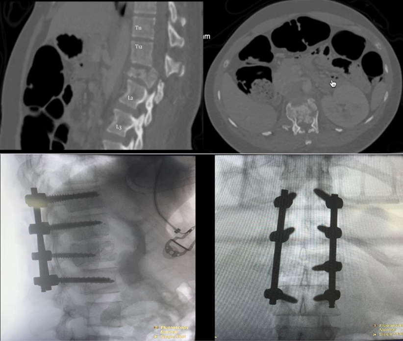
A 30-year-old nulliparous woman at 13 weeks’ gestation presented after a motor vehicle accident with an AO Type C fracture at T12/L1 (L1: A4), an ipsilateral clavicle fracture, and bilateral haemopneumothoraces. She was managed according to Advanced Trauma Life Support (ATLS) principles. Examination revealed saddle anaesthesia and 4/5 power at the L2 level. CT imaging demonstrated a right mid-shaft clavicle fracture (managed conservatively), bilateral haemopneumothoraces (treated with bilateral intercostal drains), and an L1 burst fracture with a retropulsed bony fragment. She underwent urgent posterior decompression and stabilisation under general anaesthesia in the prone position, with intraoperative fluoroscopy used to confirm spinal level and instrumentation placement. The procedure lasted 120 minutes. There were no postoperative complications, and the foetus remained viable (Figure 1).
Case 2
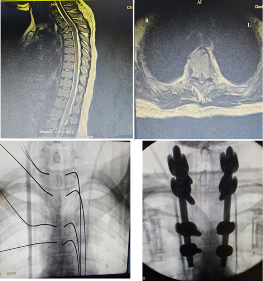
A 33-year-old uniparous woman at 28 weeks’ gestation, newly diagnosed with breast cancer, presented with a seven-day history of lower-limb weakness and loss of bowel and bladder control. Examination revealed 0/5 power and absent sensation from L2 to S1. MRI demonstrated thoracic spinal metastases with cord compression at T3. Given the risk of autonomic dysreflexia and her inability to perceive uterine and foetal movement, she was advised to proceed with surgical intervention during pregnancy. She underwent T3 laminectomy with T1-T5 minimally invasive posterior stabilisation under general anaesthesia in the prone position, with intraoperative radiography used to confirm spinal level and instrumentation. The procedure lasted 90 minutes. Postoperatively, she had no surgical complications and cardiotocography (CTG) was normal. She subsequently delivered a healthy baby but died six months later from progression of her underlying malignancy (Figure 2).
Discussion
Prompt diagnosis and management of spinal pathology in pregnancy is essential given the risk of permanent neurological injury. Paralysis may precipitate autonomic dysreflexia, impair the mother’s ability to perceive uterine and foetal movement, and complicate the initiation of breastfeeding [10]. Approximately 1-2% of pregnant women undergo non-obstetric surgery, most commonly for appendicitis or cholecystitis [11,12]. Spinal surgery during pregnancy remains rare because of the compounded risks associated with positioning, anaesthesia, fluoroscopy, surgical approach, and most critically foetal wellbeing [13,14]. In this series, four patients underwent successful spinal surgery during pregnancy with good maternal and foetal outcomes, supporting the feasibility of surgical intervention when clinically indicated.
Imaging
MRI without contrast remains the imaging modality of choice in pregnancy. However, in resource-constrained settings such as ours, it is often unavailable for urgent cases [15]. The risks associated with x-ray and CT imaging must therefore be discussed with patients as part of informed consent.
In this series, CT was used to characterise the L1 burst fracture in Case 1, including its retropulsed fragment and relationship to the gravid uterus (Figure 1), while MRI was used to delineate the extent of metastatic cord compression at T3 in Case 2 (Figure 2); this reflects individualised imaging selection guided by clinical urgency, local resource availability, and gestational risk rather than a fixed protocol.
Spinal surgery carries a steep learning curve, and intraoperative fluoroscopy is necessary to reduce complication rates [16]. Hence, should be performed by the most senior available surgeon to minimise fluoroscopy time, given its teratogenic potential at different gestational stages. Organogenesis occurs between weeks 2 and 15, during which maternal radiation exposure should be minimised and all risks discussed with the patient and multidisciplinary team [17]. Depending on gestational age, radiation exposure can result in congenital abnormality, growth restriction, or intellectual disability [17,18]. Despite intraoperative imaging in all four surgical cases in this series, all foetuses were viable with no congenital abnormalities identified. However, the long-term effects of this radiation exposure remain unknown and warrant continued follow-up.
Positioning
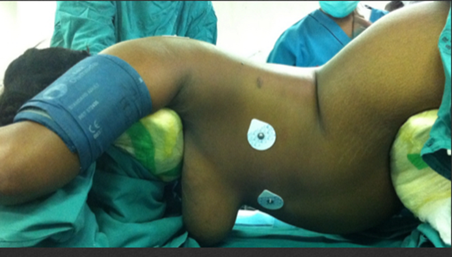
The lateral position is generally preferred over prone positioning in pregnancy because it avoids abdominal compression and, in turn, aortocaval compression is a particular concern in the late second and third trimesters [1,19,20]. In this series, three posterior decompression and fusion procedures were performed prone using a Jackson table, which allows the abdomen to hang free [21]. The use of specialised padding in the prone position to prevent aortocaval compression has been described in previous studies [6] and consistent with the padded prone set-up used in our unit (Figure 3).
Intraoperative Foetal Monitoring
This is technically difficult, particularly at gestational ages below 24 weeks. ACOG recommends that monitoring decisions be individualised to each patient’s clinical presentation but performed whenever feasible [4,22]. In this series, pre- and postoperative sonography was used to confirm foetal viability; all foetuses remained viable, with ongoing obstetric follow-up continued through to delivery.
Anaesthesia
Neither regional nor general anaesthesia is contraindicated in pregnancy, though regional techniques are generally preferred because they limit foetal drug exposure [7,23]. Regional anaesthesia is not always feasible in spinal surgery, and studies similarly reported general anaesthesia as the preferred technique in this context [6]. General anaesthesia has nonetheless been associated with spontaneous abortion and should be used cautiously, particularly in the first trimester [24] which is a risk profile that did not manifest in this cohort. All patients in this series received general anaesthesia after multidisciplinary discussion of the associated risks.
Analgesia
Analgesic choice in pregnancy must follow a specific risk-mitigation protocol. Nonsteroidal anti-inflammatory drugs should be avoided owing to the risk of premature ductus arteriosus closure, and opioids should be avoided because of their teratogenic potential [5,7,25]. Paracetamol is accordingly the analgesic of choice in pregnancy [25]. Our institutional protocol used intravenous paracetamol intraoperatively and oral paracetamol postoperatively.
Limitations
This study has several limitations. Its retrospective, single-centre design and reliance on existing clinical records carry a risk of selection and information bias, with some variables notably intraoperative foetal monitoring inconsistently documented. The sample size was small, limiting analysis to descriptive statistics. Hence, no comparative or inferential testing was performed and findings should be regarded as hypothesis-generating rather than definitive evidence of safety. Follow-up was short and variable (minimum three months), insufficient to capture delayed maternal complications or long-term developmental outcomes in children exposed in utero to radiation and general anaesthesia. Finally, as findings arise from a tertiary centre with dedicated multidisciplinary support, generalisability to lower-resource settings with limited access to senior surgical input, MRI, or multidisciplinary care may be restricted. Larger, prospective, multicentre studies with extended follow-up are needed.
Conclusion
Spine surgery can be performed safely during pregnancy in the prone position when managed through a coordinated multidisciplinary approach. Elective surgery should nonetheless be deferred until after delivery wherever possible, to create the safest possible environment for both mother and foetus. However, pregnancy alone should never be a reason to withhold urgent surgical intervention when clinically indicated.
Declarations
No declaration to be made.
Acknowledgement
The authors would like to acknowledge all the clinical staff of the Spine unit, for their assistance.
Funding
No funding was received for this study.
Conflict of Interest
The authors declare that they have no competing interests.
Data Availability Interest
Data sharing is not applicable to this article as no new data were created or analysed in this study.
Authors’ contributions
F.U.N: Conceptualisation, Methodology, Investigation, Data Curation, Formal Analysis, Writing Original Draft, Writing Review & Editing, Administration. Z.M.K: Investigation, Original Draft, Writing Review and Editing. N.E: Writing Original Draft, Writing Review and Editing.
Ethics approval and consent to participate
All study protocols were approved by the Human Research Ethics Committee of the University of the Witwatersrand (Study number: M190943).