Introduction
Dermatofibrosarcoma protuberans (DFSP) is an uncommon cutaneous tumor that originates from dermal fibroblasts. The condition was initially characterized in 1924 by Darier and Ferrand as a recurrent dermatofibroma and subsequently named as dermatofibrosarcoma protuberans in 1925 by Hoffmann [1,2]. Breast DFSP has typically been described as a superficial, oval or lobulated, relatively well-circumscribed mass without calcification [3]. It manifests as a painless, gradually enlarging subcutaneous nodule [4]. DFSP is locally aggressive with a tendency for local recurrence but rarely metastasizes [5]. It predominantly occurs in young to middle-aged adults with range of 17–48 years [6,7,8]. The disease and the symptoms may mimic those of breast cancer, and this poses a challenge on the diagnosis [9]. Mammograms and biopsy are required to assess the disease condition. Histopathology and immunohistochemistry (IHC) are essential investigations to confirm the disease [6]. When the diagnosis is confirmed, treatment is mainly surgical resection [4,10]. Main objective of the study is to share our experience with literature review of the Fungating Dermatofibrosarcoma protuberans of the breast. This case report has been structured in line with the CARE guidelines.
Case Report
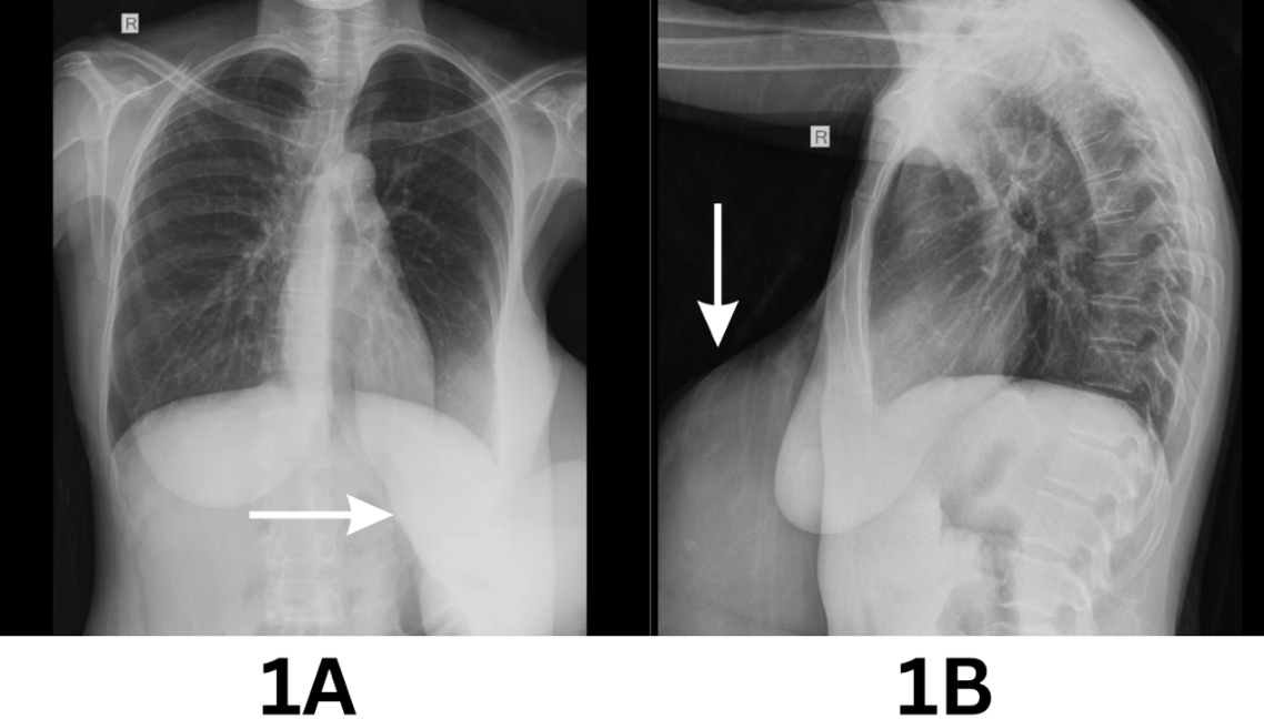
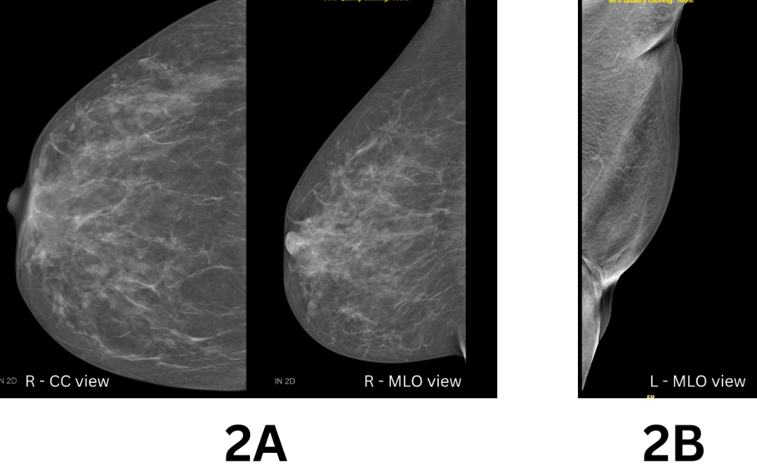
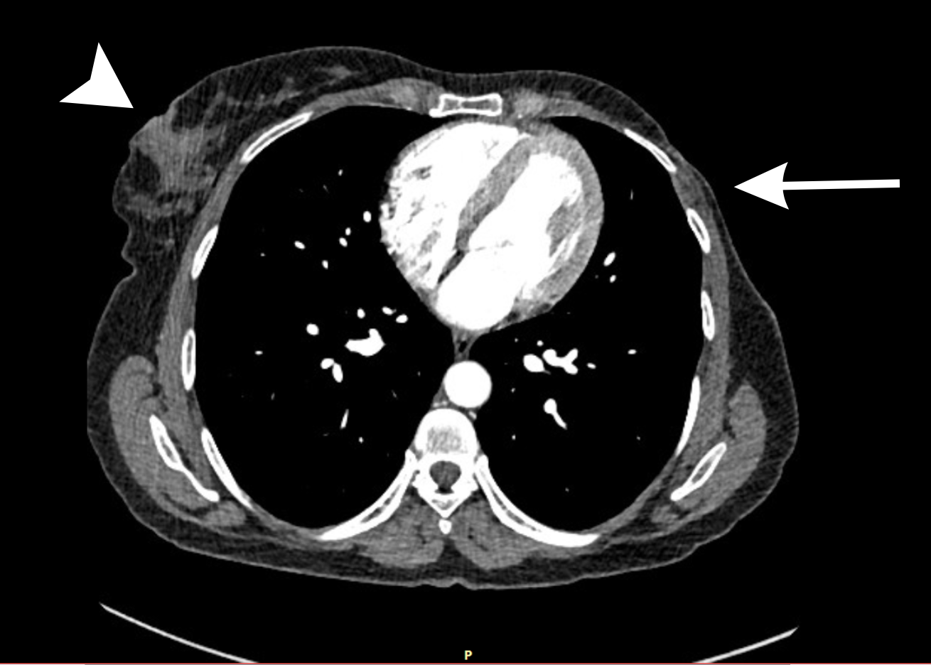
A 45-year-old female patient presented with painless left breast mass which was growing slowly over 5 years. The last 2 years it turned into ulcerative lesion, and she observed that there was bleeding at times in past few months. The patient did not attend any hospital for breast lumps until the ulcerative lesion started bleeding. She received blood transfusion at the local hospital and later referred to Pietersburg academic hospital for further intervention. On physical examination she was anemic and there was a big fungating ulcerating mass (10 cm x 15 cm) in left breast involved all four quadrants of the breast. It was foul smelling and bleeding from ulcerative lesions was observed. There was no obvious enlargement of regional axillary lymph node on clinical examination. On plain Chest X-ray there was no metastasis in the lung and the left breast shadow was observed to be much bigger than the right (Fig 1). Mastectomy was performed. The gross histopathology revealed a breast tissue of 3100g with ulcerating polypoid tumor present on the surface of the skin measuring 300mm x185mm x 100mm. Histological examination demonstrated a spindle cell proliferation which displayed a herringbone growth pattern and involvement of the epidermis. 11 mitoses identified in ten high power fields with no representation of malignant glandular elements. There were areas of haemorrhage and necrosis. The tumour was 1mm from the deep resection margin. Immunohistochemical profile reflected CD34: diffuse membranous and cytoplasmic positivity within the tumour cells. BCL2: strong and diffuse cytoplasmic positive within the tumour cells, S100: isolated tumour cell positivity. The Diagnosis of DFSP was contemplated on histology. The post-operative course was uneventful and wound healed. Patient was discharged and advised to come for follow up in the surgical clinic and referred to oncology clinic as well. However, the patient defaulted to attend surgical clinic and oncology department for almost 2 years. Currently she visited the general surgery clinic for follow up. At present there was no obvious complaint. On clinical examinations, there are no signs of mass or recurrence observed on mastectomy sites. This time, Mammogram and Computer tomography (CT) scan were performed. The mammogram reported normal breast finding on the right breast and normal scar tissue in the left post mastectomy side. No sign of recurrence or no obvious chest wall mass was observed (Figure 2). CT (Figure 3) reflected no pulmonary metastasis and normal breast tissue (right) and post mastectomy scar (left). Patient was given follow up date for oncology and surgical clinic.
Discussion
Dermatofibrosarcoma protuberans (DFSP) can develop in various parts of the body. However, DFSP is rarely found in the breast, and only a few cases have been reported in the literature [6]. The symptoms of DFSP may mimic those of breast cancer [9]. and modality of treatment of DFSP different from carcinoma of breast. Though DFSP is uncommon in breast, the typical mean age of presentation is approximately 30 years. [6]. In our case, the patient was 45-year-old and presented very late to the local hospital with fungating breast mass which mimic as breast cancer. Previous study from Limpopo found that most of the patients with breast mass presented late in health care facility and the time diagnosis breast cancer already advances stage (70%). The main reasons for the delay were lack of breast cancer knowledge, ignorance and painless breast mass [11]. Similarly, we see this patient also presented late until bleeding started from the fungating ulcerative lesion of the breast. Mammograms and biopsy are required to assess the condition. As this patient was anemic and presented with bleeding from ulcerative lesion, mastectomy was offered before mammogram was performed. Histopathological and immunohistochemistry (IHC) investigations of the surgical specimen are required to confirm the pathological diagnosis [6]. The tumor is usually composed of spindle cells arranged in a storiform pattern, showing minimal nuclear pleomorphism and low mitotic activity on histopathological specimen. In immunohistochemistry about 90% of cases, DFSP demonstrates CD34 positivity, while it is negative for other markers such as S-100, actin, desmin and limited studies have indicated an association between up-regulation of derived growth factor subunit B(PDGFB) gene and DFSP [12]. In our case 11 mitoses identified in ten high power fields with no representation of malignant glandular elements and there was positive CD34 and S100 were demonstrated on immunohistochemical profile. The current recommended first-line treatment for resectable DFSP is surgical excision, with the goal of achieving complete microscopic clearance and tumor free margin of 2 - 3 cm [4.10]. Mastectomy may be considered in some circumstances particularly when the tumor is big and involves all quadrant of breast. Rouhani et al. reported that patients with DFSP who underwent complete excision had an excellent prognosis with a 5-year-survival rate of 99% [13]. Our case demonstrated a locally aggressive ulcerative lesion involving all four quadrants of the breast with bleeding. Mastectomy was offered with the aim to achieve histologically tumor free margins to prevent local recurrence. The overall efficacy of adjuvant radiotherapy has not been well studied because of the rarity of DFSP [5]. Health education and breast cancer awareness campaign is highly important for women particularly when they come to attend primary health care for any other condition. Though no sign of recurrence was observed within two years in this case. Nevertheless, further follow up is necessary to detect any recurrence.
Conclusion
Dermatofibrosarcoma protuberans of the breast is a rare condition and symptoms may mimic those of breast cancer. Dermatofibrosarcoma protuberans involves all four quadrants of the breast, necessitating a mastectomy. The recommended first-line treatment for resectable Dermatofibrosarcoma protuberans is surgical excision, with tumor free margin.
Declarations
Author Contribution
All authors involved conceptualization; methodology; project administration; visualization; writing of original draft and editing. All authors read and approved of the final manuscript.
Ethics statement
Written informed consent was obtained from the patient’s family for publication of this case report and the accompanying clinical, radiological, operative, and pathological images. Permission was taken from the Clinical Executive Director of Mankweng Academic hospital.
Conflicts of interest
The Authors have no conflict of interests
Funding
No funding was utilized in the preparation of this manuscript
Data availability and Material
No new data was generated or analyzed in this manuscript; Data sharing is not applicable.
Acknowledgement
None