Introduction
Spontaneous intramural hematoma of the small intestine (SIHSI) is a rare condition, most commonly associated with anticoagulant therapy [1,2]. The first case of spontaneous bowel intramural hematoma (IMH) was reported by McLauchlan in 1838 [2,3]. The jejunum is the most frequently affected segment [4], while abdominal pain and vomiting are the most common presenting symptoms [5]. Computed tomography Scan (CT) is the investigation of choice [2], and conservative management is the mainstay of treatment in hemodynamically stable patients without complications [6]. Complete resolution of the hematoma may take 5-7 days to 2 months [2,4,7,8]. Surgical intervention is reserved for patients with complications such as perforation, peritonitis, ischemia, intra-abdominal hemorrhage, or persistent intestinal obstruction [1]. Here, we report a rare case of spontaneous intramural hematoma of the ileum with no identifiable underlying cause.
Case Report
A 33-year-old African male presented to the emergency department with a 3-day history of abdominal pain, multiple episodes of vomiting, and absolute constipation. The abdominal pain had an insidious onset without any preceding history of trauma. It was generalized in distribution and associated with recurrent episodes of vomiting. Initially intermittent, the pain progressively became continuous and increased in severity. There were no identifiable aggravating or relieving factors. There was no history of recent medication use or bleeding disorders. The patient denied any history of skin rashes, joint pain, or weakness of the limbs. He had no previous episodes of per rectal bleeding or unintentional weight loss. There was also no history of recent interventions, including endoscopic procedures.
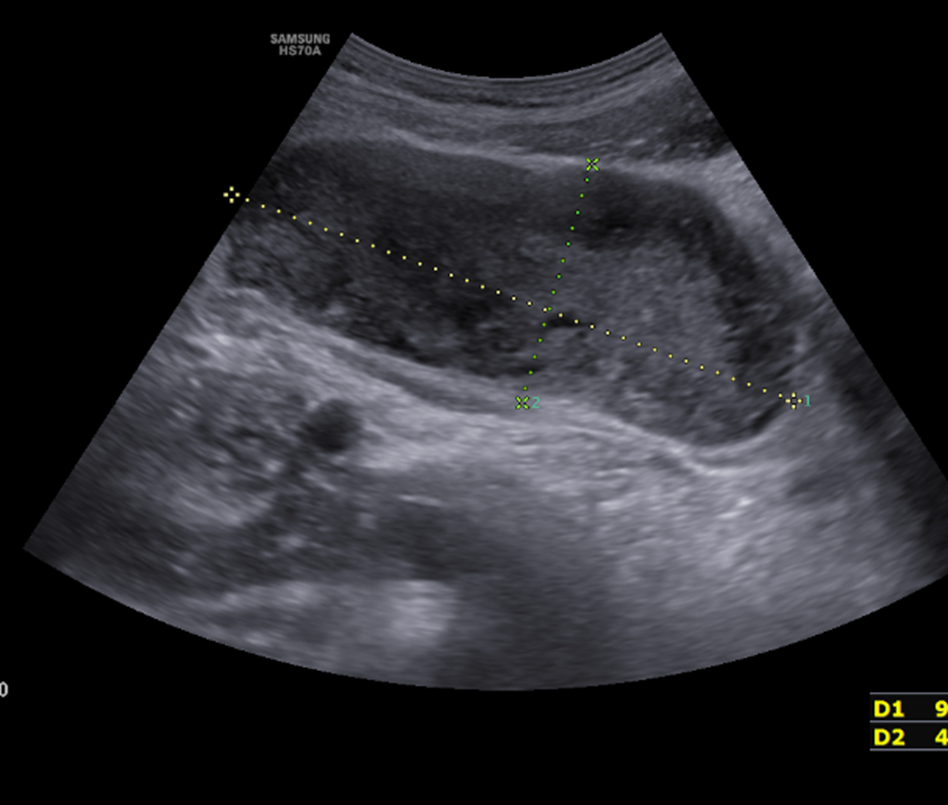
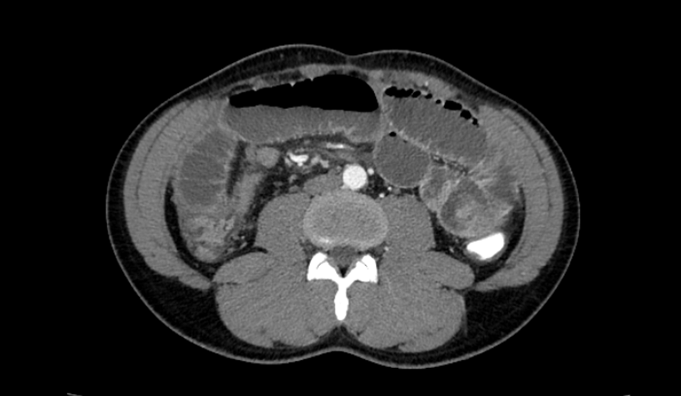
On abdominal examination, there was moderate abdominal distension with no visible bowel loops or peristaltic movements. Tenderness was present in the lower abdomen with hyperactive bowel sounds. Laboratory investigations, including complete blood count, erythrocyte sedimentation rate, renal function tests, liver function tests, and C-reactive protein, were within normal limits, except for mildly reduced hemoglobin and hematocrit levels. Abdominal ultrasonography revealed a large, well-defined, thick-walled, organized hypoechoic collection measuring 98 × 41 mm, containing thick, mobile internal echoes. Multiple dilated small-bowel loops were visualized throughout the abdomen, with a maximum diameter of 31–35 mm (Figure 1). CT of the abdomen demonstrated grossly dilated jejunal and ileal loops, measuring up to 4.0 cm in diameter, with air-fluid levels and mildly enhancing concentric wall thickening. A large, well-defined, sharply demarcated, non-enhancing, hyperdense, oval-shaped lesion measuring approximately 95 × 50 mm was identified along the course of the distal ileal loops in the pelvis. The lesion caused severe luminal narrowing, with resultant marked upstream dilatation of the remaining small-bowel loops. These findings were suggestive of an ileal wall hematoma (Figure 2).
Despite conservative treatment, the patient's symptoms did not resolve. Serial abdominal examinations demonstrated progressive abdominal distension with an increasing abdominal girth. In view of the worsening clinical condition and features of intestinal obstruction, surgical intervention was planned. Intraoperatively, an intramural hematoma involving approximately 13–15 cm of the ileum was identified, located 25 cm proximal to the ileocecal junction (Unfortunately, the intraoperative photograph could not be included because the device used to capture the image was subsequently damaged, resulting in the loss of the photograph). The remaining bowel appeared normal, and no other intra-abdominal abnormalities were identified. Segmental resection of the affected ileum with primary anastomosis was performed. The patient had an uneventful postoperative recovery and was discharged on postoperative day 5. On postoperative day 8, he presented with symptoms of subacute intestinal obstruction, which were managed conservatively with a good clinical response. He subsequently attended two follow-up visits and remained asymptomatic at both visits.
Discussion
Epidemiology
Non-traumatic intramural hematomas of the small intestine are referred to as SIHSI [2]. SIHSI is a rare condition [4], with an incidence of 0.003% per year [9]. It is encountered in approximately 1 in 2,500 patients receiving anticoagulant medication [4,8,10,11]. The incidence is 50 times higher with warfarin than with heparin [10]. Reported incidence varies across different journals, but the average age ranges from 57.6 to 74.1 years [4,9,12], with a male predominance [1].
Although SIHSI remains a rare condition, the diagnosis and reporting of IMH have increased with the widespread availability of advanced imaging modalities and the growing use of anticoagulant therapy [12].
History
The earliest recorded description of an IMH was published by McLouchlan in 1838 [2–5,12]. He reported a case of a duodenal hematoma, probably secondary to a pseudoaneurysm, causing intestinal obstruction [5,12)]. However, due to the uncertainty regarding its etiology, the condition was initially believed to be traumatic. In 1904, Sutherland reported a case of a non-traumatic IMH in a child. The hematoma was probably associated with Henoch–Schönlein purpura, resulting in intussusception [3, 5].
Site
Intestinal hematoma may occur from the oesophagus to the rectum [3,4]. The most common cause of SIHSI is over-anticoagulation with warfarin [13], and the small intestine is affected in up to 85% of cases [12,13]. SIHSI is most commonly seen in the jejunum [1,4,5,9], followed by the ileum and duodenum [1,5,9]. The colon is a rare site for hematoma formation, probably due to the taeniae coli, which prevent the dispersal of hemorrhage through the bowel wall [5].
SIHSI in the duodenum is rare, probably due to its short length and surrounding organs, which help in compressing and absorbing the hematoma, contrary to the jejunum or ileum [9]. On the contrary, the most common site of traumatic IMH is the duodenum [5]. The probable reasons may include its rich vascular supply [7,14], its fixation in the retroperitoneal space [12,15], its close relationship to the lumbar spine [12], and the lack of a mesentery [12,15], and the duodenojejunal junction at the ligament of Treitz, again due to its fixity [15].
The most common location of hemorrhage is the submucosal layer of the bowel. Intramural, intraluminal, intramesenteric, and retroperitoneal involvement can occur, especially when the duodenum is involved [16]. Lai CC et al. reported the first case of SIHSI in the efferent loop of a Billroth II anastomosis [17].
Etiology
The most common cause of IMH is trauma (90%) [8], following blunt abdominal trauma [8,18], child abuse [18], endoscopy and biopsy [18], and abdominal massage [19].
The most common cause of SIHSI is anticoagulation and antiplatelet therapy (78.6%), followed by hemophilia (4.9%). Among anticoagulant agents, SIHSI is frequently reported with warfarin (73.8%) [9]. The other causes of SIHSI are vasculitis [1,2,4],idiopathic thrombocytopenic purpura [2-5,9], hemobilia [4], von Willebrand disease [9], lupus [9], liver failure [9], peptic ulcer disease [9], alcoholism [20], bone marrow transplantation [5], Henoch–Schönlein purpura [5], polyarteritis nodosa [5], pancreaticoduodenal aneurysm [21], malignancy [2] including leukemia [3,4,5,8], lymphoma [3,4,5], and myeloma [4,5], pancreatic cancer [4,5], chemotherapy [3,4,5], pancreatitis [4,5,20], and Glanzmann’s thrombasthenia [8] or Idiopathic [22].
The use of oral anticoagulants has increased markedly worldwide. Navar et al. reported an increase in overall anticoagulant use from 56.3% in 2011 to 64.7% in 2020 among patients with atrial fibrillation [23]. Bleeding is a major complication of anticoagulant therapy, with reported rates ranging from 5% to 48%, while gastrointestinal bleeding accounts for approximately 3%-4% of bleeding events [24]. Although the use of oral anticoagulants has increased substantially in recent years, warfarin remains an important anticoagulant, with bleeding being its most common and clinically significant complication [9,25].
Mechanism
The hematoma is thought to originate from the rupture of the terminal vessels. These vessels arise from the mesentery and penetrate the muscular layer of the intestine. Bleeding within this space causes separation of the muscularis mucosae from the underlying muscular layer. As the hematoma enlarges, it may result in further vascular disruption and extension, leading to hemorrhage and edema. These changes can cause mucosal thickening and, in severe cases, may result in luminal obstruction [2,4,26].
Samir R et al. reported that, in traumatic cases, hematoma formation may result from rupture of the terminal blood vessels. In patients receiving anticoagulation therapy, they hypothesized that a reduction in intra-abdominal pressure, possibly following decompression-although not specifically described in the article—could lead to abrupt decompression of the splanchnic circulation and rapid refilling of the splanchnic vascular system. This may subsequently result in rupture of the terminal vessels and subsequent bleeding [6].
Abbas AM et al. noted that several authors have suggested that trauma may be the underlying cause of all the intestinal hematoma, even in the absence of a documented history of trauma, particularly in children [5]. However, several findings suggest a possible non-traumatic etiology:
-
Anatomical distribution: Spontaneous hematomas more commonly involve the jejunum, whereas traumatic hemtomas are commonly involve the duodenum.
-
Patient characteristics: Spontaneous hematomas tend to occur in elderly patients, particularly those with underlying risk factors for bleeding.
-
Anticoagulation status: In the reported cases, spontaneous hematoma was associated with warfarin toxicity, most likely due to supratherapeutic anticoagulation or overdose.
-
Extent of bowel involvement: Unlike focal hematomas associated with trauma, spontaneous hematomas may involve longer segments of the bowel, possibly because impaired coagulation prevents effective clot formation and allows bleeding to extend along the bowel wall.
-
Supratherapeutic anticoagulation: Spontaneous hematomas appear to be more commonly associated with excessive anticoagulation rather than with appropriately titrated therapeutic doses. However, the precise mechanism by which supratherapeutic anticoagulation predisposes to intestinal hematoma remains unclear [5].
Post-endoscopic duodenal hematoma is a rare complication [14]. Shearing forces can cause separation of the mucosal and submucosal layers, resulting in rupture of the submucosal vessels and subsequent bleeding [27].
Clinical Features
The most common initial symptoms are abdominal pain, with nausea and vomiting [1,4,5,8,28]. Broadly the presentation of SIHSI can vary from mild nonspecific symptoms [2,8] to acute abdomen [2] and intestinal obstruction [2,8] and peritonitis [8] depending upon the site and extent of the hematoma [8].
The reason for the delayed presentation in patients with SIH remains unclear [5]. One proposed hypothesis for the variability in clinical presentation and timing of presentation is that the hematoma within the bowel wall creates a high osmotic gradient, resulting in an increase in hematoma size due to the influx of fluid from the surrounding tissues [2,5].
Sorbello MP et al reported abdominal pain as the most common presenting features (97.5%) along with nausea in half of the cases. Vomiting was present in 40% of t cases. Both the latter symptoms are due to obstruction at the level of duodenum or proximal jejunum [12]. Zhu Y et al reported that the average time from the onset of the symptoms until medical attention is approximately 2.5 days [28] whereas Schiller B et al reported that the symptoms developed 3 days after endoscopy and biopsy [18]. Limmer AM et al. reported that approximately 40% of patients with intramural hematoma (IMH) develop intraluminal bleeding, which may manifest as hematemesis, frank rectal bleeding, or melena following rupture of the hematoma. They also reported that leukocytosis and a supratherapeutic INR are common, while the prevalence of anemia ranges from 7.6% to 84.6% [8].
Early complications include gastrointestinal bleeding (GIB) which may result from rupture of the hematoma [4], while intestinal obstruction and intussusception [6], peritonitis due to intra-abdominal bleeding, and bowel gangrene [4,6] may also occur. Intestinal obstruction due to fibrosis and stenosis may present as a late complication [6].
The risk factors for GIB in patients receiving warfarin include a history of previous GIB, older age, comorbidities such as atrial fibrillation, renal insufficiency, and chronic liver disease, as well as concomitant use of antiplatelet agents and medications that compete for CYP3A4-mediated metabolism [8].
The clinical manifestations of colonic hematoma are often nonspecific [6] and less pronounced, possibly owing to the relatively larger diameter of the colonic lumen [3]. Patients most commonly present with nausea and vomiting, which may be attributed to intestinal obstruction [6].
Investigations
Plain abdominal radiography has limited diagnostic specificity, while barium studies have become largely obsolete with the advent and widespread use of ultrasonography (USG) and CT [2]. The classic coil-spring sign on barium study is pathognomonic of IMH [29].
USG has limited diagnostic value because of its nonspecific findings [8,27]. However, it can be used as a scout investigation [27,30] owing to its rapid availability, short examination time, non-invasive nature [14, 30], and absence of ionizing radiation [14]. These advantages make USG the preferred initial imaging modality in children presenting with abdominal symptoms [14]. It is also the investigation of choice for follow-up assessment of hematoma resolution [15,27], with the hematoma appearing as a thickened and echogenic submucosal layer [27].
CT is the imaging modality of choice for diagnosing IMH [1,2,12,27]. Sorbello et al. reported that combining USG with CT Scan achieved 100% diagnostic accuracy [12]. Although MRI provides superior soft-tissue resolution [14,18], CT is preferred over MRI because of its wider availability and cost-effectiveness [20]. Findings suggestive of IMH are circumferential wall thickening, Luminal narrowing, hyperdensity, and intestinal obstruction [1,4,7,27]. Carkman S et al. stated that the extent of the hemorrhage may produce characteristic imaging features, including the coil-spring and pseudo-kidney signs in CT [2]. In SIHSI, the affected bowel segment is typically longer, with an average length of approximately 23 cm. Nevertheless, bowel wall thickening is a nonspecific finding and can also be seen in various other conditions, including inflammatory bowel disease and malignancy [31]. Several articles recommend performing a non-contrast CT before contrast administration, as IMH is inherently hyperdense. Subsequent administration of contrast may further increase the attenuation of the hemorrhagic component, potentially obscuring the lesion and making the diagnosis more challenging [2,12,13]. On a plain CT, IMH typically appears as homogeneous, symmetrical intramural thickening with hyperdense material, measuring approximately 30-80 Hounsfield units [32]. CT is generally avoided in children but may be performed when clinically indicated, as radiation exposure has not been reported to cause malignancy when using a low-dose protocol, as observed in a few reports [15].
MRI provides superior soft-tissue resolution and eliminates the risk of radiation exposure [14,18]. Coronal and axial planes provide a more accurate assessment of the size and extent of the IMH [15]. In the hyperacute stage, the hematoma contains oxyhemoglobin, which subsequently transforms into deoxyhemoglobin. Consequently, the hematoma appears predominantly isointense to hypointense on T1-weighted images and hypointense on T2-weighted images. Later, with the formation of methemoglobin, the hematoma becomes hyperintense on T1-weighted images and remains hypointense on T2-weighted images [14,15]. In the very late stage, the signal intensity decreases on both T1- and T2-weighted images [15].
Treatment
The most important aspect of managing IMH is early detection, which can help prevent intestinal obstruction and, consequently, avoid the need for surgery to manage obstruction-related complications [6].
Management should be individualized based on the underlying etiology and patient’s general condition [6] however, the mainstay of treatment is cessation of anticoagulant therapy [4,12,25], correction of coagulopathy (PT) [25-27,30] with vitamin K injection [2,4,12,25,26] and fresh frozen plasma [2,4,12,25, 26], bowel rest with nil per oral intake [2,4,18,25-27, 30], IVF [27], Correction of electrolyte disturbances [2,12], TPN [12,18,30], Nasogastric decompression [2,4,26,27,30], analgesics [18], and correction of anemia [2,25,30], if any.
Discontinuation of anticoagulation may result in resolution of symptoms in approximately 30% of cases [3]. Ho MP et al. suggested that, in elderly patients on anticoagulation, we should pay attention to the appropriate drugs and dosage [25]. The patient should be closely monitored for a rapid decrease in hemoglobin, evidence of hematoma expansion, and adverse peritoneal signs such as abdominal tenderness, guarding, or rigidity [6]. Conservative treatment of SIHSI is associated with favorable outcomes [13,18] and low mortality [18].
Schiller B et al. noted that an interventional approach should be reserved for cases of SIHSI associated with complications, with available options including endoscopic incision and drainage, percutaneous drainage, laparoscopic drainage, and endoscopic balloon catheter dilatation [18].
Conservative management is typically favored as the first-line treatment for SIHSI, especially in uncomplicated cases [2,30] but it comes with the price of longer hospital stay [21]. Carkman S et al stated that in addition to the supportive measures, they highlighted the role of factor VIII replacement therapy in cases where factor VIII deficiency contributes to hematoma formation [2].
Surgery
Historically, exploratory laparotomy (EL) was the primary diagnostic approach for intestinal hematoma [2]. In addition to establishing the diagnosis, EL allows therapeutic intervention through hematoma evacuation, bypass procedures, or bowel resection with anastomosis [3,5]. However, with advances in diagnostic imaging, earlier recognition of the condition, and the availability of effective conservative management, surgery has become less frequently indicated [3,5,6]. The potential morbidity associated with operative intervention has further contributed to the preference for non-operative management in appropriately selected patients [3,5].
Vecchio R et al. supported the use of EL in emergency settings, particularly when the diagnosis remains uncertain and cannot be established promptly by non-invasive investigations. The authors mentioned that relief of the compression effect following conservative treatment may lead to recurrent bleeding and, consequently, increase the risk of infective complications [3].
The indications for surgery in SIHSI include bowel ischemia [1,2], perforation [1,2], peritonitis [1,2,6,17], Intra-abdominal hemorrhage [2], ongoing bleeding within the hematoma [6], and failure of conservative treatment, such as persistent intestinal obstruction [1,2,6].
Loganathan AK et al. suggested that, in patients with intestinal obstruction due to a large hematoma, simple evacuation of the hematoma may be insufficient, hence primary resection with anastomosis or an additional bypass procedure should therefore be considered in such patients [27].
Endoscopy
Samra M et al. successfully relieved duodenal obstruction secondary to IMH by simply passing the endoscope across the obstructed part of the duodenum and proposed considering endoscopic balloon dilatation as one of the options in the therapeutic armamentarium for relieving luminal obstruction in selected cases [29].
Samir R et al. reported that endoscopy combined with USG can have both diagnostic and therapeutic roles by identifying the site, thickness, and characteristics of the hematoma, while also facilitating hematoma drainage, stent placement, and clipping of bleeding vessels [6].
Valerii G et al. reported a case in which a lumen-apposing metal stent was used in a patient with SIHSI involving the second part of the duodenum after failure of conservative treatment. One end of the stent was placed within the submucosa, while the other end was positioned in the duodenal lumen. Stent placement facilitated drainage of the intramural contents, resulting in resolution of the hematoma-related bulging within 3 weeks [33].
Selective arterial embolization in cases of ongoing bleeding is also one of the options available with a success rate of 85% [6]. Oliveira JHB highlighted that the endoscopic approach is minimally invasive and avoids radiation exposure; however, its limitation lies in controlling bleeding from areas that are inaccessible within the bowel wall. The authors recommended arterial embolization in such cases, particularly in hemodynamically stable patients, whereas laparotomy should be reserved for hemodynamically unstable patients [21].
Eichele DD et al. argued that minimally invasive, image-guided drainage is not recommended because of technical difficulties related to accessibility and the potential risk of bowel perforation [30].
Recovery
In cases of uncomplicated SIHSI, symptomatic improvement can be observed within 5-7 days with conservative treatment, whereas complete resolution of the hematoma usually takes approximately 2 months [2,4,7,8]. During follow-up, clinical assessment and USG assessment to see the progress of resolution in terms of the size of the IMH, its characteristics, and luminal compression [18]. If the IMH persists beyond 2 months, further investigation should be performed to exclude other causes of bowel wall thickening [2,4].
Anticoagulation should be resumed after resolution of the acute phase and within the therapeutic limit [7,8], or with regression of the hematoma [4,7].
Conclusion
Spontaneous intramural small-bowel hematoma is an uncommon condition that may be difficult to diagnose at initial hospital presentation because of its nonspecific clinical manifestations and the wide range of possible differential diagnoses. A high index of suspicion, particularly in patients with relevant risk factors, should prompt timely imaging investigations. Early recognition is essential to differentiate intramural hematoma from other causes of acute abdomen and intestinal obstruction and, where appropriate, to avoid unnecessary surgical intervention. Clinical awareness, together with a high index of suspicion and prompt diagnostic evaluation, can facilitate timely intervention in the form of conservative management and thereby improve patient outcomes.
Declarations
Ethical Approval
Not Applicable
Author Contributions
Devajit Chowlek Shyam: Conceptualization and formulation of the research idea; Literature search and collection of relevant articles; Data collection, extraction, and analysis; Initial drafting of the manuscript; Preparation and revision of text and figures; Proofreading.
Ranjit Chowlek Shyam: Contribution to the study concept; Assistance with literature search and review; Review and verification of collected data; review and editing of the manuscript; Proofreading and final revision.
Conflict of Interest
Nil
Funding
Nil
Acknowledgment
This work is dedicated to the pursuit of better patient care, with the hope that it contributes to improved health outcomes and quality of life.