Introduction
Endometrial carcinoma is the most common gynecologic malignancy in developed countries, and its global incidence continues to increase owing to aging populations, obesity, and metabolic disorders [1][2][3]. While most endometrial carcinomas are low-grade endometrioid neoplasms with favorable outcomes, a relatively small subset comprises high-grade histological variants responsible for a disproportionate burden of recurrence, metastasis, and disease-related mortality [1][2]. Accurate histopathological classification of these tumors is therefore essential for prognostic assessment, therapeutic planning, and appropriate patient management.
Uterine carcinosarcoma (UCS), dedifferentiated endometrial carcinoma (DDEC), and undifferentiated endometrial carcinoma (UDEC) are rare but highly aggressive endometrial malignancies that frequently present at an advanced stage and are associated with poor survival [1][3]. Although these tumors often present with similar clinical manifestations, most commonly postmenopausal bleeding, they differ considerably in histomorphology, immunophenotype, molecular alterations, and biological behavior. Accurate distinction among these entities is therefore essential because diagnosis directly influences prognostic assessment and therapeutic decision-making.
The understanding of UCS has undergone substantial evolution over the past two decades. Previously regarded as malignant mixed Müllerian tumors, UCS are now recognized by the World Health Organization (WHO) as metaplastic endometrial carcinomas arising through epithelial-mesenchymal transition from a common epithelial precursor [1]. Molecular studies demonstrating shared TP53 mutations and other driver alterations within both epithelial and sarcomatous components strongly support this concept and have fundamentally altered current management strategies, which now parallel those of high-grade endometrial carcinoma rather than primary uterine sarcoma [4][5].
Dedifferentiated and undifferentiated endometrial carcinomas represent a distinct spectrum characterized by partial or complete loss of epithelial differentiation. DDEC comprises a low-grade endometrioid carcinoma abruptly transitioning to an undifferentiated component, whereas UDEC lacks any recognizable differentiated epithelial component [1][6]. Because the undifferentiated component may closely resemble sarcoma, lymphoma, neuroendocrine carcinoma, or FIGO grade 3 endometrioid carcinoma, these tumors remain among the most challenging diagnoses in gynecologic pathology, particularly in limited biopsy specimens. Careful morphologic assessment supported by a targeted immunohistochemical panel is therefore indispensable for accurate diagnosis [6][7].
Recent advances in molecular pathology have further refined the classification of high-grade endometrial malignancies. The Cancer Genome Atlas (TCGA) established four molecular subgroups with distinct prognostic and therapeutic implications [8]. In addition, loss of SWI/SNF chromatin-remodeling complex proteins, particularly SMARCB1 (INI1) and SMARCA4 (BRG1), has emerged as a characteristic finding in a subset of DDEC and UDEC and is associated with aggressive clinical behaviour [9]. Consequently, evaluation of p53, mismatch repair proteins, and selected SWI/SNF markers has become an important adjunct to conventional histopathological diagnosis.
Comparative clinicopathological studies evaluating UCS, DDEC, and UDEC within a single institutional cohort remain limited, particularly from the Indian subcontinent [10][11]. Such analyses are valuable because these tumors frequently enter the same differential diagnosis in routine surgical pathology practice. The present study retrospectively analyzes the clinicopathological characteristics, histomorphological spectrum, immunohistochemical findings, treatment, and clinical outcomes of 22 patients with UCS, DDEC, and UDEC diagnosed at a tertiary cancer center and compares the findings with contemporary published literature.
Materials and Methods
Study Design
This retrospective observational study was conducted in the Department of Pathology at a tertiary cancer center and included patients diagnosed with uterine carcinosarcoma (UCS), dedifferentiated endometrial carcinoma (DDEC), or undifferentiated endometrial carcinoma (UDEC) between January 2022 and June 2026. The study aimed to evaluate the clinicopathological characteristics, histomorphological spectrum, immunohistochemical findings, treatment, and clinical outcomes of these uncommon high-grade endometrial malignancies.
Patient Selection
Twenty-two consecutive cases fulfilling the diagnostic criteria for UCS, DDEC, or UDEC were identified from the departmental archives. Histopathological slides, pathology reports, immunohistochemistry (IHC) results, and clinical records were reviewed.
Patients with confirmed histopathological diagnoses and adequate clinicopathological information were included. Cases with inadequate tissue for review or incomplete clinical records that precluded meaningful evaluation were excluded.
Histopathological Evaluation
All available hematoxylin and eosin-stained slides were reviewed. Tumours were classified according to the 2020 World Health Organization (WHO) Classification of Female Genital Tumours [1]. Endometrioid carcinomas were graded according to the FIGO grading system [1] and tumour staging was performed according to the 2023 International Federation of Gynecology and Obstetrics (FIGO) staging system for endometrial carcinoma [12].
For UCS, both epithelial and mesenchymal components were identified and further subclassified. Sarcomatous elements were categorized as homologous or heterologous.
DDEC was diagnosed by demonstrating an abrupt transition between a differentiated endometrioid carcinoma and an undifferentiated carcinoma component. UDEC was diagnosed when the tumour consisted entirely of undifferentiated malignant cells without an identifiable differentiated epithelial component [1][7].
Pathological parameters assessed included tumour size, histological subtype, depth of myometrial invasion, cervical stromal involvement, serosal involvement, adnexal involvement, parametrial involvement, lymphovascular space invasion (LVSI), lymph node metastasis, and FIGO stage.
Immunohistochemistry
Immunohistochemistry was performed selectively according to the morphological differential diagnosis.
The antibody panel included epithelial markers (AE1/AE3, EMA, PAX8), hormone receptors (ER and PR), mismatch repair proteins (MLH1, PMS2, MSH2, and MSH6), p53, Ki-67, SWI/SNF complex proteins (INI1/SMARCB1 and BRG1/SMARCA4), and lineage-specific mesenchymal markers, including desmin, smooth muscle actin (SMA), myogenin, S100, and vimentin, whenever indicated.
The final diagnosis was established by integrating morphology with the immunophenotypic findings.
Clinical Data and Follow-up
Clinical information, including age, presenting symptoms, operative findings, treatment received, and follow-up status wherever available, was retrieved from the institutional electronic medical records.
Clinical outcome at the last available follow-up was categorized as alive without disease, alive with recurrent disease, died of disease, or lost to follow-up.
Statistical Analysis
Data were analysed using descriptive statistics. Continuous variables are presented as mean ± standard deviation or median (range), whereas categorical variables are expressed as frequencies and percentages.
Because of the relatively small cohort size and incomplete follow-up in several patients, survival analysis was not performed.
Results
Patient Demographics
Twenty-two patients with high-grade endometrial malignancies were included in the study, comprising 14 uterine carcinosarcomas (63.6%), five dedifferentiated endometrial carcinomas (22.7%), and three undifferentiated endometrial carcinomas (13.6%).
The median age at diagnosis was 61 years (range, 44-76 years), with a mean age of 60.5 ± 9.1 years. The majority of patients were postmenopausal. Abnormal uterine bleeding was the most common presenting symptom, occurring in 18 patients (81.8%), while the remaining patients presented predominantly with abdominal pain or pelvic discomfort.
The detailed clinicopathological characteristics of individual patients are summarized in Table 1, while the overall clinicopathological profile is presented in Table 2.
| Case No. | Age (years) | Presenting complaint | Histological diagnosis | FIGO stage | Treatment | Adjuvant Treatment | Outcome |
| 1 | 52 | White discharge, pain | CS | IIC | TAH BSO + BPLND | CT | Alive without disease |
| 2 | 61 | Bleeding per vaginum | CS | IIC | TAH BSO + BPLND | CT | Lost to follow up |
| 3 | 56 | Endometrial polyp | CS | IIC | TAH BSO + BPLND + RPLND + Omentectomy | CT + RT | Lost to follow up |
| 4 | 61 | Endometrial polyp | CS | IIIA | TAH BSO + BPLND + RPLND + Omentectomy | CT + RT | Lost to follow up |
| 5 | 76 | Bleeding per vaginum | CS | IIC | TAH BSO + BPLND | CT | Lost to follow up |
| 6 | 61 | Endometrial polyp | CS | IIC | TAH BSO + BPLND | CT | Alive with recurrent disease (vault recurrence) |
| 7 | 57 | Bleeding per vaginum | CS | IIIC2 | TAH BSO + BPLND+ RPLND + Omentectomy | CT + RT | Alive without disease |
| 8 | 55 | Bleeding per vaginum, white discharge | CS | IIC | TAH BSO + BPLND | CT + RT | Alive without disease |
| 9 | 52 | Prolapse with bleeding | CS | IIC | TAH BSO + BPLND | CT | Alive without disease |
| 10 | 61 | Bleeding per vaginum | CS | NA | TAH BSO + BPLND | CT + RT | Alive without disease |
| 11 | 76 | Bleeding per vaginum | CS | IIC | TAH BSO + BPLND + RPLND + Omentectomy | CT + RT | Alive without disease |
| 12 | 61 | Pain abdomen | CS | IIC | TAH BSO + BPLND | CT | Alive without disease |
| 13 | 64 | Bleeding per vaginum | CS | NA | TAH BSO + BPLND | CT + RT | Alive without disease |
| 14 | 65 | Bleeding per vaginum | CS | IIIA | TAH BSO + BPLND+ RPLND + Omentectomy | CT + RT | Died of disease (seven months after diagnosis) |
| 15 | 64 | Pain abdomen | UDEC | IIC | TAH BSO + BPLND | CT | Lost to follow up |
| 16 | 44 | Heavy menstrual bleeding | UDEC | IIIC1 | TAH BSO + BPLND+ RPLND + Omentectomy | CT + RT | Alive with recurrent disease (vault recurrence) |
| 17 | 50 | Bleeding per vaginum | UDEC | IIIA | TAH BSO + BPLND+ RPLND + Omentectomy | CT + RT | Alive without disease |
| 18 | 50 | Heavy menstrual bleeding | DDEC | IIIC2 | TAH BSO + BPLND+ RPLND + Omentectomy | CT + RT | Died of disease (nine months after diagnosis) |
| 19 | 68 | Bleeding per vaginum | DDEC | IIC | TAH BSO + BPLND+ RPLND + Omentectomy | CT + RT | Alive without disease |
| 20 | 67 | Bleeding per vaginum, foul discharge | DDEC | IIIA | TAH BSO + BPLND+ RPLND + Omentectomy | CT + RT | Lost to follow up |
| 21 | 67 | Bleeding per vaginum, foul discharge | DDEC | IIIA | TAH BSO + BPLND+ RPLND + Omentectomy | CT + RT | Lost to follow up |
| 22 | 70 | Bleeding per vaginum | DDEC | IVC | TAH BSO + BPLND + RPLND + Omentectomy | CT + RT | Died of disease (Ten months after diagnosis) |
Abbreviations: CS, Carcinosarcoma; UDEC, Undifferentiated Endometrial Carcinoma; DDEC, Dedifferentiated Endometrial Carcinoma; TAH BSO, Total abdominal Hysterectomy with bilateral Salpingo-opherectomy; BPLND, Bilateral Pelvic Lymph node dissection; RPLND; Retroperitoneal Lymph node dissection; CT, Chemotherapy; RT, Radiotherapy
| Variable | n (%) |
| Age (years) | |
| Mean ± SD | 60.5 ± 9.1 |
| Median (range) | 61 (44–76) |
| Clinical presentation | |
| Abnormal uterine bleeding/bleeding per vaginum | 18 (82.0) |
| Pain abdomen | 4 (18.0) |
| Histological diagnosis | |
| CS | 14 (64.0) |
| DDEC | 5 (22.0) |
| UDEC | 3 (14.0) |
| Depth of myometrial invasion | |
| <50% | 4 (18.0) |
| ≥50% | 18 (82.0) |
| Lymphovascular space invasion | |
| Present | 11 (50.0) |
| Absent | 11 (50.0) |
| Cervical stromal involvement | |
| Present | 8 (37.0) |
| Absent | 13 (59.0) |
| Not available | 1 (4.0) |
| Serosal involvement | |
| Present | 7 (32.0) |
| Absent | 15 (68.0) |
| Adnexal involvement | |
| Present | 4 (18.0) |
| Absent | 16 (73.0) |
| Not available | 2(9.0) |
| Parametrial involvement | |
| Present | 3(14.0) |
| Absent | 17(77.0) |
| Not available | 2(9.0) |
| Lymph nodal Involvement | |
| Present | 5(22.7%) |
| Absent | 17(77.2%) |
| FIGO stage | |
| Stage I | 0(0.0) |
| Stage II | 11(50.0) |
| Stage III | 8(36.3) |
| Stage IV | 1(5.0) |
| Not available | 2(9.0) |
| Outcome | |
| Alive without disease | 10 (46.0) |
| Alive with recurrent disease | 2(9.0) |
| Died of disease | 3 (14.0) |
| Lost to follow-up | 7 (31.0) |
Pathological Findings
Most tumours demonstrated adverse pathological features at presentation. Deep myometrial invasion (≥50%) was identified in 18 patients (81.8%), LVSI in 11 patients (50.0%), cervical stromal involvement in eight (36.4%), serosal involvement in seven (31.8%), adnexal involvement in four (18.2%), parametrial involvement in three (13.6%), and lymph node metastasis in five patients (22.7%).
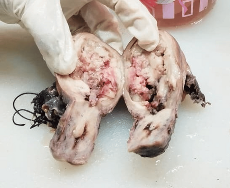
Eleven patients (50.0%) presented with FIGO stage II disease, eight (36.4%) with stage III disease, and one (4.5%) with stage IV disease. FIGO stage was unavailable in two patients (9.1%). Figure 1: Gross specimen of uterine carcinosarcoma - A large polypoid.
Histological Spectrum of Uterine Carcinosarcoma
Among the fourteen UCS cases, the epithelial component consisted predominantly of high-grade endometrioid carcinoma (6/14, 42.9%) and serous carcinoma (6/14, 42.9%), while grade 2 endometrioid carcinoma was identified in two cases (14.2%).
The mesenchymal component demonstrated considerable diversity. Homologous sarcoma was identified in eight cases (57.1%), whereas heterologous differentiation was present in six cases (42.9%). Chondrosarcoma was the most common heterologous component, occurring in five of 14 cases (35.7%) (Figure 2), followed by rhabdomyosarcoma in one of 14 cases (7.1%) (Figure 3).
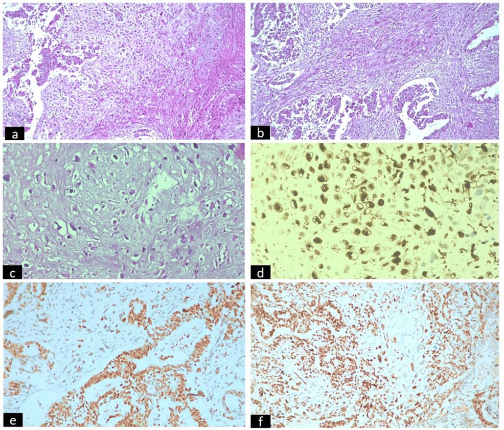
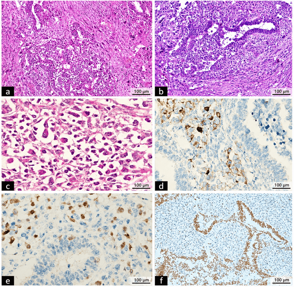
The complete histological spectrum is summarized in Table 3 and 4
| Carcinoma component | n (%) |
| Endometrioid carcinoma, FIGO grade 3 | 6 (42.8) |
| Serous carcinoma | 6 (42.8) |
| Endometrioid carcinoma, FIGO grade 2 | 2 (14.2) |
| Mesenchymal (sarcomatous) component | |
| Chondrosarcoma (Heterologous) | 5 (36.0) |
| Rhabdomyosarcoma (Heterologous) | 1 (7.0) |
| Sarcoma, NOS (Homologous) | 8 (57.1) |
Abbreviations: NOS, not otherwise specified
| Differentiated epithelial component | |
| Histological subtype | n (%) |
| Endometrioid carcinoma, FIGO grade 2 | 2 (40.0) |
| Endometrioid carcinoma, FIGO grade 1 | 2 (40.0) |
| Serous carcinoma | 1 (20.0) |
| Undifferentiated component | |
| Undifferentiated carcinoma | 5 (100) |
Histological Findings in DDEC and UDEC
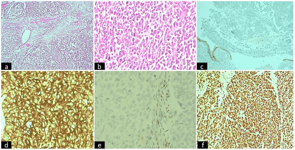
All five DDEC demonstrated the characteristic abrupt transition between a differentiated epithelial component and an undifferentiated carcinoma component. The differentiated component comprised FIGO grade 1 endometrioid carcinoma in two cases, FIGO grade 2 endometrioid carcinoma in two cases, and serous carcinoma in one case (Figure 4).
All three UDEC consisted entirely of diffuse sheets of discohesive undifferentiated tumour cells without an identifiable differentiated epithelial component. Histologically, these tumours showed diffuse solid growth, marked nuclear pleomorphism, brisk mitotic activity, and variable geographic necrosis (Figure 5).
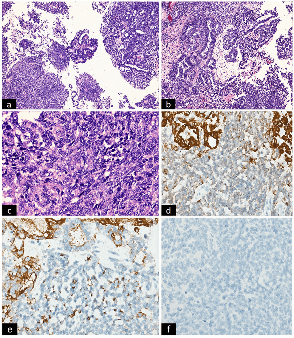
Immunohistochemical Findings
Immunohistochemistry proved valuable in establishing the diagnosis in morphologically challenging cases (Table 5).
In UCS, epithelial differentiation was confirmed by cytokeratin and epithelial membrane antigen expression, while lineage-specific mesenchymal markers facilitated characterization of the sarcomatous component.
In DDEC and UDEC, AE1/AE3, EMA, and PAX8 confirmed epithelial differentiation in the majority of cases. Mismatch repair protein expression was retained in most tumours evaluated. One DDEC demonstrated complete loss of INI1 (SMARCB1) expression, consistent with SWI/SNF complex deficiency, whereas BRG1 expression was retained in the evaluated UDEC cases. Aberrant p53 expression and high Ki-67 proliferative indices were observed in several high-grade tumours.
| Case No. | Diagnosis | Carcinoma component positive markers | Sarcomatous component positive markers |
| 1 | CS | ER, PR, P53 Wild type, MMR proficient | SMA, Vimentin |
| 2 | CS | P53 mutant, ER focal, MMR proficient | SMA, Desmin, Vimentin |
| 3 | CS | P53 mutant, ER focal, MMR proficient | SMA, Desmin, Vimentin |
| 4 | CS | ER, PR, P53 Wild type, MMR Deficient | S100 |
| 5 | CS | ER, PR, P53 Wild type, MMR proficient | SMA, Desmin, Vimentin |
| 6 | CS | ER, PR, P53 mutant, MMR deficient | Desmin, Myogenin |
| 7 | CS | P53 mutant, MMR proficient | SMA, Vimentin |
| 8 | CS | ER, PR, P53 mutant, MMR proficient | S100 |
| 9 | CS | Mutant p53, MMR proficient | SMA, Desmin, Vimentin |
| 10 | CS | Mutant p53, MMR proficient | SMA, Desmin, Vimentin |
| 11 | CS | ER, PR mutant p53, MMR proficient | S100 |
| 12 | CS | Mutant p53, MMR proficient | S100 |
| 13 | CS | ER, PR, P53 Wild type, MMR Proficient | SMA, Desmin, Vimentin |
| 14 | CS | ER, PR, P53 mutant, MMR proficient | S100 |
| Positive markers | Negative markers | ||
| 15 | UDEC | CK, EMA, PAX8 focal, ER focal, P53 wild type, MMR deficient | Vimentin, SMA, |
| 16 | UDEC | CK, EMA, BRG1, MMR Proficient, P53 wild type, MMR Proficient | ER, PR, PAX8 |
| 17 | UDEC | CK, EMA, BRG1, MMR Proficient, Ki-67 80%, Synaptophysin, P53 wild type | Vimentin, ER, PR, PAX8 |
| Differentiated component positive markers | Undifferentiated component positive markers | ||
| 18 | DDEC | ER, PR, P53 wild type, MMR proficient | CK, EMA, focal synaptophysin |
| 19 | DDEC | ER, PR, P53 wild type, MMR deficient | CK, EMA |
| 20 | DDEC | P53 mutant, MMR proficient | CK, EMA, Ki67 80% |
| 21 | DDEC | ER, PR, P53 wild type, MMR proficient | CK, EMA, P53 mutant |
| 22 | DDEC | ER, PR, P53 wild type, MMR proficient | CK, EMA, INI loss |
Abbreviations: CS, Carcinosarcoma; UDEC, Undifferentiated Endometrial Carcinoma; DDEC, Dedifferentiated Endometrial Carcinoma; MMR, Mismatch Repair
Treatment and Clinical Outcome
All patients underwent primary surgical treatment followed by adjuvant chemotherapy and/or radiotherapy according to tumour stage and multidisciplinary tumour board recommendations.
At the last available follow-up, 12 patients (54.5%) were alive, including two patients with vault recurrence, three patients (13.6%) had died of disease, and seven patients (31.8%) were lost to follow-up. Disease recurrence and mortality were observed predominantly among patients with advanced-stage disease and adverse pathological features.
Discussion
Uterine carcinosarcoma (UCS), dedifferentiated endometrial carcinoma (DDEC), and undifferentiated endometrial carcinoma (UDEC) are uncommon but highly aggressive endometrial malignancies that frequently present with advanced disease and are associated with poor clinical outcomes. Although these tumors differ in their histomorphological features and molecular alterations, they often pose overlapping diagnostic challenges, particularly in limited biopsy specimens. Accurate classification is therefore essential because prognosis, therapeutic decisions, and eligibility for molecularly targeted therapies depend on the correct pathological diagnosis [1][3].
In the present study, 14 of 22 cases (63.6%) were UCS, five (22.7%) were DDEC, and three (13.6%) were UDEC. The median age at diagnosis was 61 years, and abnormal uterine bleeding was the most common presenting symptom. These findings are consistent with previously published institutional and multicenter series, which have reported that these tumors predominantly affect postmenopausal women presenting with abnormal uterine bleeding [2][10].
Our cohort demonstrated a high frequency of adverse pathological features, including deep myometrial invasion in 18 of 22 patients (81.8%), lymphovascular space invasion in 11 of 22 patients (50.0%), cervical stromal involvement in eight of 22 patients (36.4%), and lymph node metastasis in five of 22 patients (22.7%). These pathological parameters are recognized predictors of recurrence and reduced survival and have consistently been associated with aggressive clinical behavior in high-grade endometrial malignancies [4,13-16]. Although survival analysis was not performed because of the relatively small sample size and incomplete follow-up, disease recurrence and mortality in our cohort occurred predominantly in patients with advanced-stage tumors and multiple adverse pathological features.
Uterine Carcinosarcoma
Current WHO classification recognizes UCS as a metaplastic carcinoma rather than a true malignant mixed Müllerian tumor, based on accumulating molecular evidence supporting a monoclonal epithelial origin with subsequent epithelial-mesenchymal transition [1, 5, 8]. This concept has important clinical implications because current management strategies align more closely with those for high-grade endometrial carcinoma than for primary uterine sarcoma.
In our series, high-grade endometrioid carcinoma and serous carcinoma were the predominant epithelial components, while homologous sarcoma was more common than heterologous differentiation. Chondrosarcoma represented the most frequent heterologous component. Similar histomorphological patterns have been reported in previous institutional studies, although the relative proportions of epithelial and mesenchymal elements vary among published cohorts [10,17-19].
Dedifferentiated and Undifferentiated Endometrial Carcinomas
DDEC and UDEC remain among the most diagnostically challenging endometrial malignancies because of their variable morphology and overlap with several high-grade neoplasms. According to the WHO classification, DDEC is characterized by an abrupt transition between a differentiated endometrioid carcinoma and an undifferentiated carcinoma component, whereas UDEC lacks an identifiable differentiated epithelial component [1][6]. All DDEC cases in our cohort demonstrated the characteristic abrupt transition, while all UDEC cases showed diffuse sheets of discohesive undifferentiated tumor cells without recognizable glandular differentiation, supporting the current diagnostic criteria.
These tumors may be misinterpreted as FIGO grade 3 endometrioid carcinoma, undifferentiated uterine sarcoma, neuroendocrine carcinoma, lymphoma, or metastatic malignancy, particularly in curettage or small biopsy specimens [6,7,20]. Recognition of the characteristic morphology and appropriate use of immunohistochemistry are therefore critical to avoid diagnostic errors.
Diagnostic Utility of Immunohistochemistry
Immunohistochemistry played an important role in confirming the diagnosis in morphologically challenging cases. Broad-spectrum cytokeratin, EMA, and PAX8 were useful in establishing epithelial differentiation, while lineage-specific mesenchymal markers facilitated characterization of sarcomatous components in UCS. Evaluation of mismatch repair proteins and p53 provided additional information regarding tumor classification and may have therapeutic implications, particularly in the era of molecularly guided management [9,21].
One DDEC in our study demonstrated complete loss of INI1 (SMARCB1) expression. Loss of SWI/SNF complex proteins, including INI1 and BRG1, has been increasingly recognized in dedifferentiated and undifferentiated endometrial carcinomas and is associated with aggressive biological behavior [9,22-24]. Although comprehensive molecular testing was not available in the present study, incorporation of selected SWI/SNF markers into the immunohistochemical panel proved valuable in resolving diagnostically difficult cases.
Comparison with Published Literature
The overall clinicopathological profile observed in our cohort is comparable with recently published international and Indian series (Table 6) [10,11,14,16,22,23,25,26-29].
Similar distributions of age, presenting symptoms, high rates of deep myometrial invasion, LVSI, and advanced-stage disease have been reported by Smyth et al. in patients with uterine carcinosarcoma and by Reshmi et al. in dedifferentiated and undifferentiated endometrial carcinomas [10][11]. While our cohort is smaller, the findings reinforce the aggressive biological behavior of these tumors and support the applicability of contemporary WHO diagnostic criteria in routine clinical practice.
| Study | Tumour type | No. of cases | Median/ Mean age (years) | Epithelial component (%) | Sarcomatoid component (%) | Advanced stage (III/IV) (%) | Outcome |
| Smyth et al, 2025 [10] | UCS | 77 | 70 | SC-36 (53.7%), HG EEC-17 (25.4%), Mixed-9 (13.4%), UC-3(4.5%), LG EEC-1(1.5%), CC-1(1.5%) | Homologous -56(75.5%), Heterologous-18(24.3%) | 32(41.6%) | Recurrence-33 (42.86%), DOD-45 (58.4%) |
| Reshmi J.S., 2025 [11] | DDEC/UDEC | 11 | 57 | 7-DDEC,4-UEC | 5 (45.4%) | Alive-3, DOD- 1, Progressive disease-2, LFU-1 | |
| Terblanche et al, 2022 [15] | UCS | 61 | 66.8 | NA | Homologous-11 (18.0%), Heterologous 40 (65.6%) | 38(62.3%) | Recurrence-17, 5-year OS- 19.7% |
| RA de Jong et al, 2011 [17] | UCS | 40 | 6 | EEC-25 (68%), SC-7 (19%), CC-2, UC-3 (8%) | Homologous-27 (75%), Heterologous -9 (25%) | 20 (50%) | Alive-9 (22%), DOD-23 (58%), Death due to other disease- 8 (20%) |
| Li Z et al, 2016 [23] | DDEC/UDEC | 9 | 61 | 9-DDEC | 8 (92%) | 10% DFS | |
| Stewart CJ et al, 2015 [24] | DDEC/UDEC | 22 | 67.6 | 5-UEC,17-DDEC | 11 (50%) | Not reported | |
| Efared et al, 2024 [25] | UCS | 9 | 58.88 | SC-6, CC-1, EEC-2 | Sarcoma-5, RMS-3, CS-1 | 5(55.55%) | Alive-2, DOD-3, LFU-4 |
| Zhu J et al, 2016 [26] | UCS | 60 | 59.5 | NA | Heterologous-12 (20.0%), Homologous-44 patient (73.3%) | 23(38.3%) | DOD-20,5-year OS rates 54.2% |
| Ganju et al, 2018 [27] | DDEC/UDEC | 24 | 66 | 5-UDEC, 19-DDEC | 10 (42%) | DOD-1%, Progressive disease-11% | |
| Ureyan et al, 2015 [28] | DDEC/UDEC | 18 | 61 | 18-UDEC | 14(77.7%) | 16.6%-DOD | |
| Onder et al, 2017 [29] | DDEC/UDEC | 11 | 59 | 2-UDEC,9-DDEC | 6 (54.5%) | 80%-DOD | |
| Present study, 2026 | UCS/DDEC /UDEC | 22 | 61 | UCS-14, DDEC-5, UDEC-3 | CS-5, RMS-1, Homologous-8 | 9 (40.9%) | Alive-10, Recurrent disease-2, DOD-3, LFU-7 |
Abbreviations: UCS, Uterine CS; DDEC, Dedifferentiated Endometrial Carcinoma; UDEC, Undifferentiated Endometrial Carcinoma; SC, SC; HG EEC, High Grade Endometrioid Endometrial Carcinoma; LG EEC, Low Grade Endometrioid Endometrial Carcinoma; UC- UDEC; CC- Clear cell Carcinoma; DOD- dead of disease; LFU- Lost on follow up; DFS- Disease free survival; OS- Overall Survival
Strengths and Limitations
The principal strength of this study is the comprehensive evaluation of three uncommon but biologically related high-grade endometrial malignancies within a single institutional cohort using contemporary WHO diagnostic criteria. Correlation of clinicopathological findings with immunohistochemical features provides practical information relevant to routine diagnostic practice.
The study is limited by its retrospective design, relatively small sample size, and incomplete follow-up, which precluded meaningful survival analysis. In addition, comprehensive molecular profiling, including POLE sequencing and next-generation sequencing, was not available. Future multicenter studies incorporating molecular classification and long-term clinical follow-up are required to further define prognostic factors and optimize individualized treatment strategies.
Conclusion
Uterine carcinosarcoma, dedifferentiated endometrial carcinoma, and undifferentiated endometrial carcinoma constitute a spectrum of highly aggressive endometrial malignancies with overlapping clinical presentations but distinct pathological characteristics. Accurate diagnosis requires integration of meticulous histomorphological assessment with appropriately selected immunohistochemical markers, particularly in tumors with undifferentiated morphology or unusual histological features.
Our findings demonstrate that deep myometrial invasion, lymphovascular space invasion, cervical stromal involvement, and advanced-stage disease are frequent in these neoplasms and underscore the importance of comprehensive pathological evaluation. This study adds clinicopathological data from an Indian tertiary cancer center and supports the application of contemporary WHO diagnostic criteria in routine practice. Larger multicenter studies incorporating standardized molecular profiling and long-term follow-up are required to further clarify prognostic factors and optimize patient management.
List of Abbreviations
CC: Carcinosarcoma
DDEC: Dedifferentiated endometrial carcinoma
UEC: Undifferentiated endometrial carcinoma
EC: Endometrial carcinoma
IHC: Immunohistochemistry
MMR: Mismatch repair
MSI: Microsatellite instability
p53: Tumor protein p53
POLE: DNA polymerase epsilon
FIGO: International Federation of Gynecology and Obstetrics
TNM: Tumor, node, metastasis
LVSI: Lymphovascular space invasion
NACT: Neoadjuvant chemotherapy;
OS: Overall survival
DFS: Disease-free survival
HR: Hazard ratio
CI: Confidence interval
SD: Standard deviation;
WHO: World Health Organization.
Declarations
Acknowledgements
The authors would like to thank the Department of Pathology and the Department of Gynecologic Oncology for their support in the conduct of this study. The authors also acknowledge the technical staff and medical records personnel for their assistance in retrieving and processing the relevant clinical and pathological data.
Conflict of interest
None
Funding/ financial support
None
Ethical Clearance
Taken (BBCI IEC no 13001017)
Informed Consent
Waived by the Institutional Ethics Committee in view of retrospective nature of the study.
Trial details
Not Applicable
Data Availability
The datasets generated and/or analyzed during the current study are not publicly available due to patient privacy and confidentiality considerations but are available from the corresponding author on reasonable request, subject to institutional policies and applicable ethical requirements.
Authors’ Contributions
Sukanya Gogoi: Conceptualization, Methodology, Data curation, Investigation, Formal analysis, Writing – original draft, Writing – review and editing.
Soumi Bhattacharyya: Conceptualization, Methodology, Investigation, Data curation, Writing – review and editing.
Shiraj Ahmed: Investigation, Data curation, Histopathological evaluation, Writing – review and editing.
Lopa Mudra Kakoti: Investigation, Histopathological evaluation, Data curation, Writing – review and editing.
Anupam Sarma: Supervision, Validation, Investigation, Writing – review and editing.
Debabrata Barmon: Clinical evaluation, Data interpretation, Supervision, Writing – review and editing.
All authors approved the final version of the manuscript and agreed to be accountable for all aspects of the work.