Introduction
Premature ovarian insufficiency (POI) is characterized by the loss of normal ovarian function before the age of 40 years, affecting approximately 1% of women of reproductive age [1]. It is clinically defined by oligo/amenorrhea for at least four months, accompanied by elevated follicle-stimulating hormone (FSH) levels and reduced estradiol concentrations [2]. POI is associated not only with infertility but also with long-term health consequences, including osteoporosis, cardiovascular disease, and psychological distress, thereby posing a significant clinical and public health challenge [3].
The etiology of POI is multifactorial and often remains idiopathic, although genetic abnormalities, autoimmune disorders, iatrogenic causes, and environmental factors have been implicated [4]. Current management strategies primarily focus on hormone replacement therapy (HRT) to alleviate symptoms and prevent long-term complications; however, these approaches primarily provide symptomatic relief and do not reliably restore ovarian function or fertility [2].
Assisted reproductive techniques, particularly oocyte donation, remain the most effective option for achieving pregnancy in these patients, although their acceptability and accessibility may be limited for some individuals [5]. In recent years, regenerative medicine approaches for ovarian rejuvenation have gained increasing attention. Among these, platelet-rich plasma (PRP) has been investigated for its potential to improve ovarian function through the release of growth factors involved in angiogenesis, tissue repair, and follicular development [6].
Platelet-derived preparations contain bioactive growth factors, including platelet-derived growth factor (PDGF) and transforming growth factor-beta (TGF-β), which are involved in tissue repair and cellular proliferation [7]. Intraovarian administration of platelet-derived growth factors has been proposed as a strategy to directly target ovarian tissue, with the potential to activate residual follicles and improve ovarian reserve markers. Preliminary studies using platelet-rich plasma (PRP) have reported encouraging findings in terms of hormonal improvement and follicular development; however, the available evidence remains limited, and robust randomized controlled trials are still lacking, particularly in women with POI [8].
The therapeutic rationale for platelet-derived growth factor preparations lies in their ability to deliver bioactive molecules involved in angiogenesis, cellular proliferation, and tissue remodeling. Preparations rich in growth factors contain mediators such as platelet-derived growth factor (PDGF), vascular endothelial growth factor (VEGF), and transforming growth factor-beta (TGF-β), which may contribute to tissue regeneration [9].
Direct intra-ovarian administration of platelet-derived growth factors may enhance the ovarian microenvironment by improving vascularization and modulating local paracrine signaling pathways that could contribute to follicular activation and tissue repair. Compared with systemic regenerative approaches, intra-ovarian delivery provides targeted exposure of ovarian tissue to bioactive growth factors, which may have therapeutic potential in ovarian rejuvenation. In this context, the present study was designed to evaluate the effect of laparoscopic intra-ovarian infusion of autologous growth factor concentrate on ovarian reserve parameters, hormonal profile, and clinical outcomes in women with premature ovarian insufficiency through a randomized controlled trial.
Materials and Methods
Study Design
This prospective, randomized controlled study was conducted in the Department of Obstetrics and Gynecology at Upper India Sugar Exchange Maternity Hospital, GSVM Medical College, Kanpur, India. The study was carried out over a period of 18 months after obtaining approval from the Institutional Ethics Committee. The study was conducted in accordance with the ethical principles of the Declaration of Helsinki and written informed consent was obtained from all participants prior to enrollment.
Study Population
A total of 112 women aged 25 - 40 years diagnosed with premature ovarian insufficiency (POI) were enrolled in the study. Eligible participants were infertile women presenting with clinical features of POI, including serum anti-Müllerian hormone (AMH) levels <1 ng/mL, antral follicle count (AFC) <5 on transvaginal ultrasonography, and elevated serum follicle-stimulating hormone (FSH) levels (>25 IU/L) on two occasions at least four weeks apart.
Women with genetic causes of ovarian failure, platelet dysfunction, active infections, severe male or tubal factor infertility, moderate to severe endometriosis, Müllerian anomalies, active pelvic infection, significant systemic illness, or recent use of anticoagulants or nonsteroidal anti-inflammatory drugs were excluded from the study.
All participants underwent a detailed clinical evaluation, including history taking, physical examination, and baseline investigations. Hormonal assessment (AMH, FSH, LH) and transvaginal ultrasonography for AFC were performed at baseline prior to intervention.
Randomization and Allocation
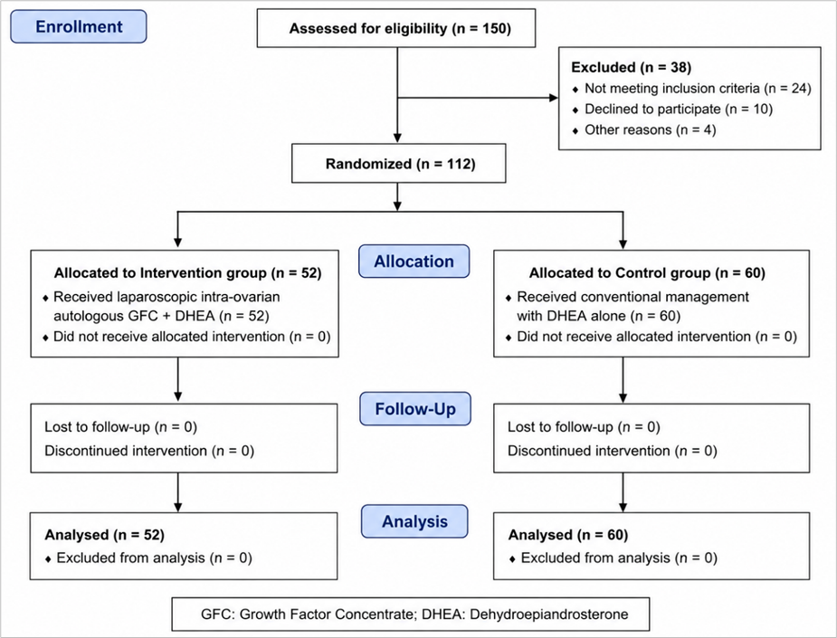
After enrollment and baseline assessment, participants were allocated into two groups: the intervention (case) group and the control group. A total of 112 participants were assigned, with 52 women in the intervention group and 60 women in the control group (Fig 1).
Randomization was performed using a simple random allocation method. Participants were assigned sequentially into either group based on availability and eligibility at the time of recruitment. Due to the nature of the intervention, blinding was not feasible, and the study was conducted as an open-label trial.
Intervention Procedure
Participants in the intervention group underwent laparoscopic intra-ovarian infusion of autologous growth factor concentrate (GFC) under aseptic conditions.
Preparation of Growth Factor Concentrate
Autologous GFC was prepared from peripheral venous blood collected from each participant. Blood samples were centrifuged to separate components, and platelet activation was induced to release growth factors. The supernatant rich in growth factors were then aspirated under sterile conditions for intra-ovarian use.
Laparoscopic Intra-Ovarian Infusion
The procedure was performed under general anesthesia using standard laparoscopic techniques. GFC was injected into the ovarian cortex of both ovaries under direct visualization using a fine needle.
Control Group
Participants in the control group received dehydroepiandrosterone (DHEA) 75 mg sustained release once daily for three months.
Outcome Measures
Primary Outcomes
The primary outcomes of the study were changes in ovarian reserve markers, including serum anti-Müllerian hormone (AMH) levels and antral follicle count (AFC), assessed before and after the intervention.
Secondary Outcomes
Secondary outcomes included changes in serum follicle-stimulating hormone (FSH) and luteinizing hormone (LH) levels, menstrual pattern (resumption or improvement of menses), and pregnancy outcomes during the follow-up period.
Follow-up
Participants in both groups were followed up for a period of three months after the intervention. Hormonal parameters (AMH, FSH, LH) and antral follicle count (AFC) were reassessed at the end of the follow-up period. Menstrual history was recorded during follow-up to assess any improvement or resumption of menses. Pregnancy outcomes, if any, were also documented during this period.
Statistical Analysis
Data were entered into Microsoft Excel and analyzed using Statistical Package for the Social Sciences (SPSS) software (IBM Corp., Armonk, NY, USA). Continuous variables were expressed as mean ± standard deviation (SD), while categorical variables were presented as frequencies and percentages.
For comparison of continuous variables between the two groups, the independent sample t-test was used. Paired t-test was applied for within-group comparisons before and after the intervention. Categorical variables were analyzed using the chi-square test or Fisher’s exact test, as appropriate. A p-value of less than 0.05 was considered statistically significant.
Results
Participant Characteristics
A total of 112 women diagnosed with premature ovarian insufficiency were enrolled in the study. Among them, 52 participants were allocated to the intervention group and 60 to the control group. All participants completed the study and were included in the final analysis.
The baseline demographic and clinical characteristics of the participants in both groups were comparable, with no statistically significant differences observed (Table 1). The mean age of participants in the intervention and control groups was similar. Baseline ovarian reserve parameters, including serum AMH levels and antral follicle count (AFC), as well as hormonal parameters such as FSH and LH, were also comparable between the two groups.
| Parameter | Intervention (n = 52) | Control (n =60) | p-value |
| Age (Years) | 32.6 ± 3.3 | 32.1 ± 3.3 | 0.36 |
| BMI (Kg/m²) | 24.1 ± 2.5 | 23.8 ± 1.1 | 0.39 |
| Duration of infertility (years) | 3.0 ± 1.1 | 2.9 ± 1.1 | 0.74 |
| Type of infertility (Primary) | 30 (57.7%) | 30 (50.0%) | 0.36 |
Ovarian Reserve Outcomes
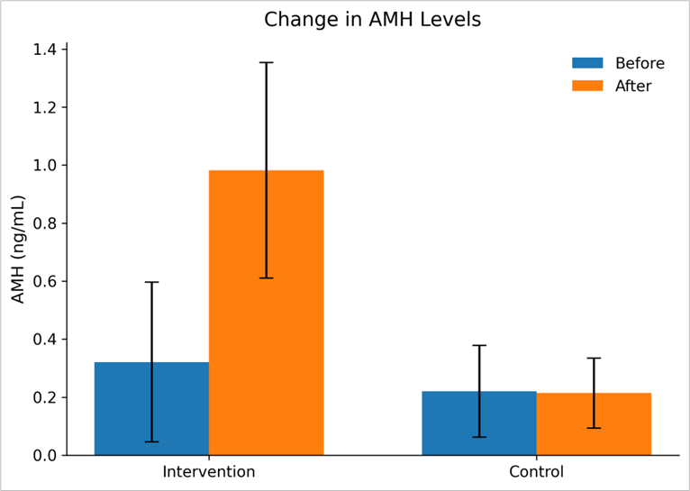
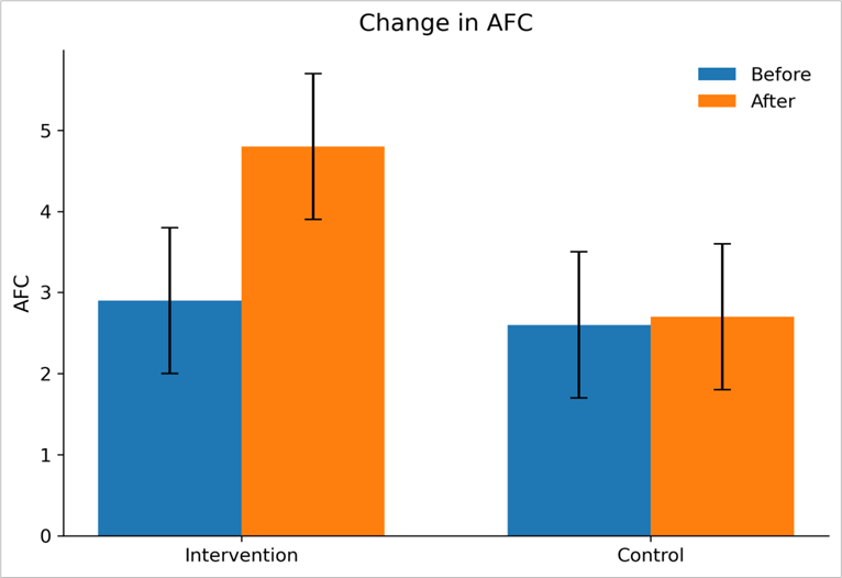
Significant improvement in ovarian reserve parameters was observed in the intervention group following intra-ovarian GFC infusion. Serum AMH levels showed a statistically significant change in the intervention group compared to the control group. Similarly, antral follicle count (AFC) demonstrated a significant increase in the intervention group, whereas no meaningful change was observed in the control group. The intervention group demonstrated significant improvement in ovarian reserve parameters compared to the control group (Table 2, Fig. 2 and 3).
| Parameter | Group | Before (mean ± SD) | After (mean ± SD) | p-value |
| AMH | Intervention control | 0.321 ± 0.275 0.220 ± 0.158 | 0.983 ± 0.372 0.214 ± 0.121 | 0.01* 0.779 |
| AFC | Intervention control | 2.9 ± 0.9 2.6 ± 0.9 | 4.8 ± 0.9 2.7 ± 0.9 | 0.01* 0.180 |
AMH: Anti-Müllerian Hormone; AFC: Antral Follicle Count
Hormonal Outcomes
A significant improvement in hormonal parameters was observed in the intervention group following intra-ovarian GFC infusion. Serum follicle-stimulating hormone (FSH) levels demonstrated a statistically significant reduction in the intervention group after treatment, whereas no significant change was observed in the control group.
Similarly, luteinizing hormone (LH) levels showed a significant decrease in the intervention group, while the control group did not exhibit any meaningful change. On intergroup comparison, the reduction in both FSH and LH levels was statistically significant in the intervention group compared to the control group (Table 3).
| Parameter | Group | Before (mean ± SD) | After (mean ± SD) | p-value |
| FSH (IU/L) | Intervention Control | 33.57 ± 4.03 32.41 ± 3.86 | 20.27 ± 3.57 31.37 ± 3.69 | 0.01* 0.654 |
| LH (IU/L) | Intervention Control | 14.20 ± 2.73 14.67 ± 1.18 | 11.39 ± 1.92 14.17 ± 1.90 | 0.01* 0.28 |
FSH: Follicle Stimulating Hormone; LH: Luteinizing Hormone
Menstrual and Pregnancy Outcomes
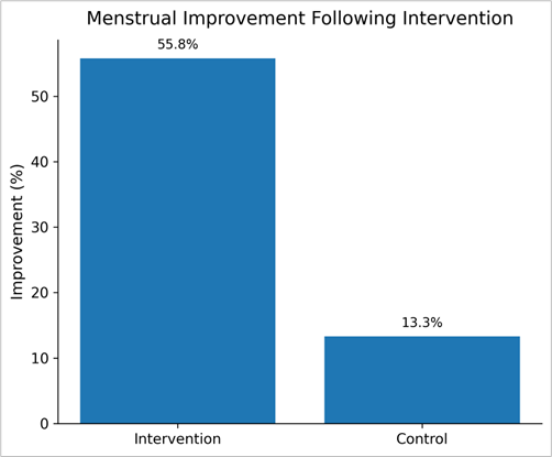
Improvement in menstrual pattern was observed in the intervention group following intra-ovarian GFC infusion. A higher proportion of participants in the intervention group reported resumption or improvement of menses compared to the control group. In contrast, minimal or no improvement was observed in the control group. The difference between the two groups was statistically significant (Table 4, Figure 4).
Pregnancy was observed in a small proportion of participants in the intervention group during the follow-up period, whereas few or no pregnancies were reported in the control group. However, the difference between the two groups was not statistically significant.
| Outcome | Intervention (n =52) | Control (n = 60) | p-value |
| Menstrual improvement | 29 | 8 | 0.01* |
| No change Pregnancy (conceived) | 23 4 | 52 1 | 0.12 |
Discussion
The present randomized controlled study evaluated the effect of laparoscopic intra-ovarian infusion of autologous growth factor concentrate (GFC) in women with premature ovarian insufficiency (POI). The findings of the study demonstrated significant improvement in ovarian reserve markers, including serum anti-Müllerian hormone (AMH) levels and antral follicle count (AFC), along with significant reduction in serum follicle-stimulating hormone (FSH) and luteinizing hormone (LH) levels in the intervention group compared to the control group. In addition, improvement in menstrual pattern was observed more frequently among women receiving GFC infusion. Although pregnancy was achieved in a small proportion of participants in the intervention group, the difference was not statistically significant.
POI remains one of the most challenging reproductive disorders because of its multifactorial etiology and limited therapeutic options. Conventional management primarily involves hormone replacement therapy and assisted reproductive techniques, which provide symptomatic and reproductive support but do not reliably restore intrinsic ovarian function [3,10]. In recent years, regenerative medicine approaches, including platelet-rich plasma (PRP) and other growth factor-based therapies, have gained increasing attention because of their potential role in ovarian rejuvenation [6].
The observed improvement in AMH and AFC in the present study suggests a potential positive effect of intra-ovarian GFC on ovarian reserve. AMH is considered one of the most reliable biomarkers of ovarian reserve, while AFC reflects the pool of recruitable follicles [10]. The increase in these parameters following GFC infusion may indicate enhanced follicular activity and improvement in the ovarian microenvironment. These findings are consistent with the study by Cakiroglu et al., who reported improvement in ovarian reserve markers and ovarian response parameters following intra-ovarian PRP administration in women with POI [8]. Similarly, Aflatoonian et al. observed improvement in ovarian reserve parameters and follicular response after intra-ovarian PRP injection in women with poor ovarian reserve and POI [11].
The mechanism underlying ovarian rejuvenation following GFC therapy is not completely understood; however, it is thought to involve the action of multiple growth factors released from activated platelets. Growth factors such as platelet-derived growth factor (PDGF), vascular endothelial growth factor (VEGF), insulin-like growth factor (IGF), and transforming growth factor-beta (TGF-β) are involved in angiogenesis, cellular proliferation, tissue remodeling, and regenerative processes [9]. Improved ovarian vascularity and local paracrine signaling have been hypothesized to contribute to follicular activation and enhancement of ovarian function. Compared with conventional PRP preparations, GFC is designed to deliver a concentrated preparation of platelet-derived growth factors, which may provide more consistent biological activity.
A significant reduction in serum FSH and LH levels was also observed in the intervention group in the present study. Elevated gonadotropin levels are characteristic of ovarian insufficiency because of diminished ovarian negative feedback mechanisms [4]. The reduction in these hormonal parameters following treatment may indicate partial restoration of ovarian endocrine activity. Similar hormonal improvements after intra-ovarian PRP therapy have been reported in previous studies evaluating ovarian rejuvenation approaches [8,11,12].
Menstrual improvement observed in the intervention group further supports the possibility of partial restoration of ovarian activity following GFC infusion. Resumption or improvement of menstrual cycles has also been reported in earlier studies investigating PRP-based ovarian rejuvenation therapies [6,13]. Restoration of menstrual function may represent an important clinical benefit in women with POI, even in the absence of immediate conception.
Although pregnancy was achieved in a few participants in the intervention group, the difference between groups was not statistically significant. This finding should be interpreted cautiously. Fertility restoration in women with POI is influenced by multiple factors, including residual follicular reserve, oocyte quality, age, and duration of ovarian insufficiency [3,5]. The relatively short follow-up period and limited sample size in the present study may also have influenced pregnancy outcomes. Similar observations have been reported in previous studies, where improvement in ovarian reserve parameters did not always translate into significantly higher pregnancy rates [14,15]. Therefore, while GFC therapy appears promising in improving ovarian function, its definitive impact on fertility outcomes requires further investigation through larger multicentric trials with longer follow-up.
One of the major strengths of the present study is its randomized controlled design, which provides stronger clinical evidence compared with many previously published observational studies and case series in this field. Furthermore, laparoscopic intra-ovarian administration allowed precise delivery of growth factors into ovarian tissue under direct visualization. However, the study also has certain limitations. The follow-up duration was relatively short; therefore, the long-term sustainability of ovarian improvement could not be assessed, and pregnancy outcomes remained limited. In addition, the exact molecular mechanisms underlying ovarian rejuvenation following GFC therapy remain unclear and warrant further investigation.
Overall, the findings of the present study suggest that intra-ovarian infusion of autologous GFC may represent a promising regenerative therapeutic approach for improving ovarian reserve and hormonal parameters in women with POI. Nevertheless, larger randomized studies with standardized treatment protocols and longer follow-up are required before this intervention can be routinely recommended in clinical practice.
Declarations
Ethical Clearance
Ethical approval was obtained from the Ethics Committee (For Biomedical Health & Research), GSVM Medical College, Kanpur (Ref No.: EC/BMHR/2024/177). Written informed consent was obtained from all participants prior to enrolment.
Funding/ financial support
This research received no specific grant from any funding agency in the public, commercial, or not-for-profit sectors.
Conflict of interest declaration
The authors declare that there are no conflicts of interest related to this study.
Authors’ Contributions
All authors contributed substantially to the conception of the study, data acquisition, analysis, drafting, and critical revision of the manuscript. All authors have read and approved the final version of the manuscript.
Acknowledgements
The authors would like to thank all participants who took part in this study and the staff of the Department of Obstetrics and Gynaecology and laboratory personnel of the participating institution for their support.
Trial Details
Not Applicable.
Contributors
Dr. Priyanka Choudhary, Department of Obstetrics and Gynaecology, Ganesh Shankar Vidyarthi Memorial Medical College, Kanpur, Uttar Pradesh, India
Dr. Uruj Jahan, Department of Obstetrics and Gynaecology, Ganesh Shankar Vidyarthi Memorial Medical College, Kanpur, Uttar Pradesh, India
Dr. Neena Gupta, Department of Obstetrics and Gynaecology, Ganesh Shankar Vidyarthi Memorial Medical College, Kanpur, Uttar Pradesh, India
Dr. Rashmi Yadav, Department of Obstetrics and Gynaecology, Ganesh Shankar Vidyarthi Memorial Medical College, Kanpur, Uttar Pradesh, India
Dr. Seema Dwivedi, Department of Obstetrics and Gynaecology, Ganesh Shankar Vidyarthi Memorial Medical College, Kanpur, Uttar Pradesh, India
Dr Renu Gupta, Department of Obstetrics and Gynaecology, Ganesh Shankar Vidyarthi Memorial Medical College, Kanpur, Uttar Pradesh, India