Introduction
Cirrhosis is associated with profound cardiovascular alterations, collectively referred to as hyperdynamic circulation. This state is characterized by increased heart rate and cardiac output, together with reduced systemic vascular resistance and arterial blood pressure [1][4]. The primary mechanism underlying hyperdynamic circulation is peripheral and splanchnic vasodilatation, mediated by increased production or activity of vasodilatory substances, including nitric oxide, carbon monoxide, and endogenous cannabinoids, along with reduced vascular responsiveness to vasoconstrictors [4][6]. The resulting reduction in effective arterial blood volume activates the renin-angiotensin-aldosterone system, sympathetic nervous system, and antidiuretic hormone pathways, leading to renal vasoconstriction, sodium and water retention, and expansion of plasma volume [4][6].
Although cardiovascular abnormalities in cirrhosis were initially attributed largely to alcohol-related cardiotoxicity, subsequent evidence demonstrated that cardiac dysfunction can occur independently of alcohol consumption and is associated with chronic liver disease itself. Early observations by Kowalski and Abelmann described increased resting cardiac output and stroke volume, with relatively preserved blood pressure and reduced systemic vascular resistance in patients with alcoholism and cirrhosis [1]. Subsequent reports of cardiac failure following liver transplantation, transjugular intrahepatic portosystemic shunt placement, and surgical portocaval shunting further highlighted the clinical significance of previously unrecognized myocardial dysfunction in patients with cirrhosis [1].
Cardiac involvement in chronic liver disease is characterized by a spectrum of structural, functional, and electrophysiological abnormalities. These include increased resting cardiac output, impaired myocardial contractile reserve, particularly during stress, left ventricular hypertrophy, diastolic dysfunction, and electrocardiographic abnormalities such as prolongation of the corrected QT interval [7][8]. The absence of overt cardiac symptoms may mask clinically important dysfunction because the reduced systemic vascular resistance characteristic of cirrhosis decreases cardiac afterload and may compensate for impaired myocardial performance at rest [7]. These observations led to the recognition of cirrhotic cardiomyopathy, defined as cardiac dysfunction in patients with cirrhosis in the absence of other established cardiac disease and characterized predominantly by impaired contractile responsiveness to stress, diastolic dysfunction, and electrophysiological abnormalities [9][12].
At the 2005 World Congress of Gastroenterology in Montreal, proposed criteria for cirrhotic cardiomyopathy included systolic dysfunction, demonstrated by a blunted increase in cardiac output following exercise, volume challenge, or pharmacological stimulation, or a resting left ventricular ejection fraction below 55%. Diastolic dysfunction was characterized by an age-adjusted E/A ratio below 1.0, prolonged deceleration time, or prolonged isovolumetric relaxation time. Supportive features included electrophysiological abnormalities, impaired chronotropic response, electromechanical dyssynchrony, prolonged QTc interval, left atrial enlargement, increased myocardial mass, and elevated cardiac biomarkers such as brain natriuretic peptide, proBNP, or troponin I [4].
Cirrhotic cardiomyopathy may remain clinically silent because conventional measures of cardiac function can appear relatively preserved at rest. Consequently, its true prevalence and clinical significance may be underestimated. Cardiac dysfunction has been reported in a substantial proportion of patients undergoing liver transplantation, and clinically significant cardiac complications may contribute to morbidity and mortality during the post-transplant period [13][14]. Identification of subclinical cardiac abnormalities is therefore important, particularly in patients with advanced liver disease who may be exposed to physiological stress during disease progression or therapeutic interventions.
Echocardiography provides a non-invasive means of assessing cardiac structure and function and can identify abnormalities in systolic and diastolic performance in patients with chronic liver disease. Conventional echocardiographic parameters, together with newer deformation imaging techniques, may facilitate the detection of otherwise clinically inapparent myocardial dysfunction. Assessment of these abnormalities may also provide insight into the relationship between cardiac involvement and the severity of underlying liver disease.
In view of the potential for clinically silent cardiac involvement in chronic liver disease, the present study was undertaken to determine the prevalence and characterize the spectrum of cardiac changes detectable by echocardiography in affected patients. The study further aimed to evaluate the association of these echocardiographic abnormalities with the severity of chronic liver disease and to assess the morbidity and mortality attributable to cardiac involvement. Specifically, the study sought to determine the prevalence of cardiac changes in patients with chronic liver disease using echocardiography, assess the nature and extent of these cardiac changes, and evaluate morbidity and mortality associated with cardiac involvement.
Materials and Methods
The present study was designed as a prospective observational study and was conducted in the Department of Medicine of a tertiary care multidisciplinary hospital from January 2023 to June 2024. Prior to commencement of the study, the thesis protocol was reviewed and approved by the institutional ethics committee of the tertiary care centre and Government Medical College. Ethical approval was obtained prior to recruitment of participants, and confidentiality of all study participants was maintained throughout the study.
The sample size was calculated using the formula Z² × p × q/l², where Z represents the standard normal deviate, p represents the expected prevalence, q represents 1 − p, and l represents the relative error. Based on previous literature reporting a prevalence of cardiac involvement of approximately 20% among patients with chronic liver disease in India [15], with a relative error of 10% and a Z value of 1.96, the calculated sample size was approximately 61 participants. A total of 60 patients with chronic liver disease were included in the study. Consecutive sampling was used for recruitment, and all eligible patients presenting during the study period from January 2023 to June 2024 were considered for inclusion until the required sample size was achieved.
Patients with liver cirrhosis diagnosed on the basis of clinical features and ultrasonographic findings, with or without evidence of portal hypertension on upper gastrointestinal endoscopy, were eligible for inclusion. The underlying etiologies included alcoholic liver disease, non-alcoholic steatohepatitis, hepatitis B, hepatitis C, drug-related liver disease, Wilson's disease, and other causes of chronic liver disease. Patients with known primary cardiac disease, including ischemic heart disease, congenital heart disease, rheumatic heart disease, pericardial disease, or hypertension, were excluded. Patients with diseases known to affect cardiac function, including hypothyroidism and thyrotoxicosis, as well as patients with known diabetes mellitus, were also excluded.
Demographic characteristics, clinical history, and relevant laboratory investigations were obtained from hospital medical records and clinical assessment. The principal outcomes of the study were echocardiographic parameters reflecting cardiac systolic and diastolic function, including ejection fraction, systolic and diastolic annular velocities, and other conventional Doppler and tissue Doppler measurements.
Conventional two-dimensional and Doppler echocardiography was performed using a 2.5-MHz multiphase array transducer in standard parasternal and apical views, in accordance with recommendations of the American Society of Echocardiography [16]. Left ventricular ejection fraction was assessed using the modified biplane Simpson's method from apical two-chamber and four-chamber views. Measurements were obtained from three consecutive cardiac cycles, and the mean of the three measurements was used for analysis. Doppler assessment of diastolic mitral inflow was performed from the apical four-chamber view, with measurements averaged over two consecutive cardiac cycles [16].
Transmitral early and late diastolic velocities and transtricuspid early and late diastolic velocities were recorded, and the corresponding E/A ratios were calculated. Pulsed-wave tissue Doppler imaging was used to assess the direction, timing, and synchrony of myocardial motion and to quantify systolic and diastolic myocardial function [16]. Mitral annular peak systolic velocity (Sm), early diastolic annular velocity (E′m), and late diastolic annular velocity (A′m) were measured at the anterior, inferior, lateral, and septal sites of the mitral annulus. The mean mitral annular velocity was calculated from these four measurements. Similarly, tricuspid annular peak systolic velocity (St), early diastolic annular velocity (E′t), and late diastolic annular velocity (A′t) were measured at the lateral and septal sites of the tricuspid annulus, and the mean values were calculated. Mitral inflow measurements during the Valsalva maneuver were not performed.
Additional echocardiographic parameters included left atrial volume, isovolumetric relaxation time, and deceleration time. Peak early diastolic annular velocity (e′) was measured at the septal and lateral mitral annular sites, and the average of the two measurements was calculated. The E/e′ ratio was subsequently determined. Left ventricular diastolic dysfunction was assessed and graded according to the American Society of Echocardiography recommendations [16], using relevant parameters including mitral inflow velocities, annular e′ velocity, E/e′ ratio, left atrial volume, deceleration time, and other echocardiographic indices.
Liver cirrhosis was defined on the basis of evidence of hepatocellular failure together with characteristic ultrasonographic findings and/or evidence of portal hypertension on upper gastrointestinal endoscopy. Ultrasonographic findings suggestive of cirrhosis included coarse hepatic echotexture, a nodular liver surface, and features of portal hypertension such as ascites, splenomegaly, portal vein dilatation, defined as a portal vein diameter greater than 13 mm, and portosystemic collateral vessels. Clinical manifestations of hepatocellular failure included jaundice, hepatic encephalopathy, cutaneous manifestations such as spider angiomas and palmar erythema, and endocrine manifestations such as breast atrophy in females and gynecomastia or testicular atrophy in males. Clinical evidence of portal hypertension included gastroesophageal or rectal varices, splenomegaly, and ascites.
Data were entered and analyzed using IBM Statistical Package for the Social Sciences (SPSS), version 21.0. Data entry checks were performed periodically to ensure the accuracy and validity of the recorded information. Descriptive analysis was performed for all study variables. Categorical variables were expressed as frequencies and proportions, whereas continuous variables were summarized using means and standard deviations. Bivariate analysis was performed to assess associations between relevant clinical and echocardiographic variables. The chi-square test was used for categorical variables and the independent-samples t-test was used for comparison of continuous variables, as appropriate. A p-value of less than 0.05 was considered statistically significant.
Results
The present prospective observational study included 60 patients with chronic liver disease who underwent clinical evaluation and echocardiographic assessment. The age of the study participants ranged from 40 to 79 years. The largest proportion of patients belonged to the 50–59-year age group, comprising 24 patients (40.00%), followed by the 60–69-year age group with 15 patients (25.00%), the 70–79-year age group with 12 patients (20.00%), and the 40–49-year age group with 9 patients (15.00%). There was a marked male predominance among the study participants. Of the 60 patients, 51 (85.00%) were males and 9 (15.00%) were females. With respect to occupation, 23 patients (38.34%) were professional workers, 19 (31.66%) were skilled workers, and 18 (30.00%) were unskilled workers.
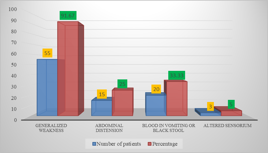
Generalized weakness was the most frequently reported symptom at presentation, occurring in 55 patients (91.67%). Blood in vomitus or black stools was reported in 20 patients (33.33%), while abdominal distension was present in 15 patients (25.00%). Altered sensorium was reported in 3 patients (5.00%). Symptoms were overlapping, and individual patients could present with more than one symptom (Figure 1).
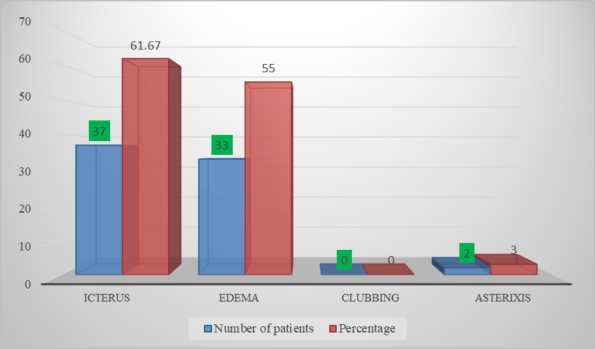
Among the clinical signs, ascites was the most frequently observed, being present in 40 patients (66.67%), followed by icterus in 37 patients (61.67%) and edema in 33 patients (55.00%). Asterixis was observed in 2 patients (3.00%), while none of the patients had clubbing. Clinical signs were overlapping among the study participants (Figure 2).
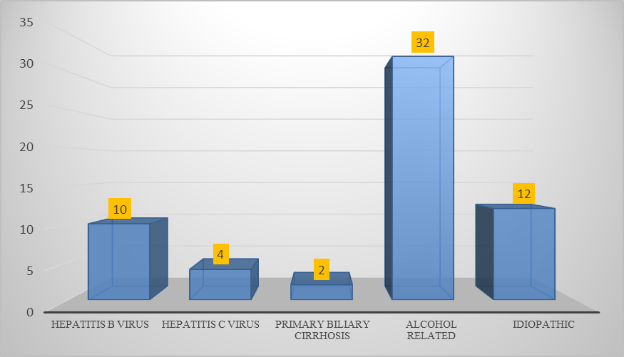
Alcohol-related liver disease was the most common etiology, accounting for 32 patients (53.33%), followed by idiopathic chronic liver disease in 12 patients (20.00%), hepatitis B virus infection in 10 patients (16.67%), hepatitis C virus infection in 4 patients (6.67%), and primary biliary cirrhosis in 2 patients (3.33%) (Figure 3).
According to the Child-Turcotte-Pugh classification, 33 patients (55.00%) belonged to Class B, 16 patients (26.67%) belonged to Class A, and 11 patients (18.33%) belonged to Class C. Thus, the majority of patients had Child-Turcotte-Pugh Class B disease.
Ascites was present in 40 patients (66.67%) and absent in 20 patients (33.33%). Varices of any grade were present in 20 patients (33.33%), whereas 40 patients (66.67%) had no varices. The distribution of ascites and varices was further assessed in relation to Child-Turcotte-Pugh class.
Among the electrocardiographic parameters assessed, a QTc interval greater than 440 ms was observed in 35 patients (58.33%), whereas 25 patients (41.67%) had a QTc interval below 440 ms. Thus, prolongation of the QTc interval was observed in more than half of the study population.
Diastolic dysfunction was detected in 33 patients (55.00%). Grade I diastolic dysfunction was present in 18 patients (30.00%), Grade II in 13 patients (21.67%), and Grade III in 2 patients (3.33%). No evidence of diastolic dysfunction was observed in 27 patients (45.00%). End-diastolic volume was above 90 in 28 patients (46.67%) and below 90 in 32 patients (53.33%). End-systolic volume was above 38 in 14 patients (23.33%) and below 38 in 46 patients (76.67%). Ejection fraction was above 60% in 36 patients (60.00%), whereas 24 patients (40.00%) had an ejection fraction below 60%.
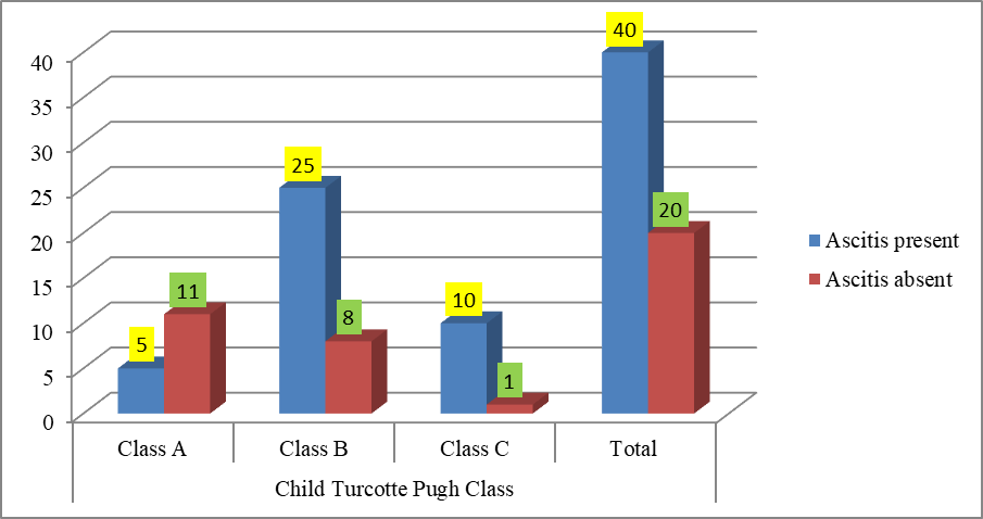
The relationship between Child-Turcotte-Pugh class and ascites was statistically significant. Ascites was present in 5 of 16 patients (31.25%) with Class A disease, 25 of 33 patients (75.76%) with Class B disease, and 10 of 11 patients (90.91%) with Class C disease. The association between Child-Turcotte-Pugh class and ascites was significant (χ² = 13.1676, p = 0.001383) (Figure 4).
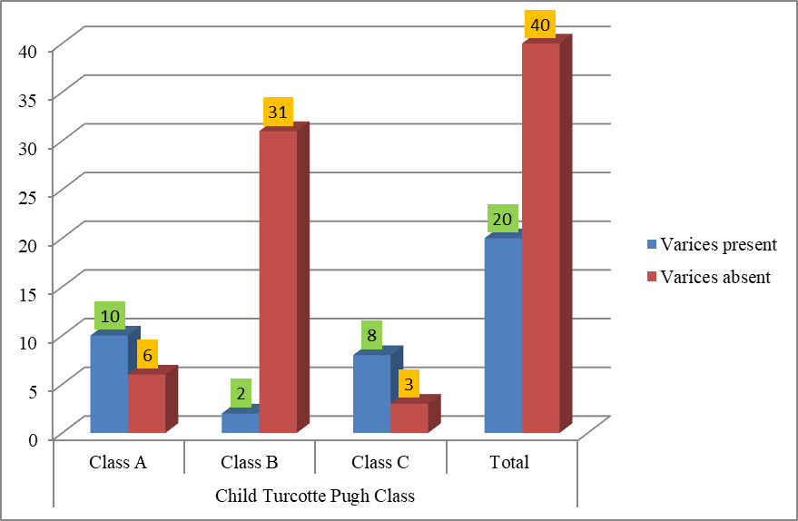
A statistically significant association was also observed between Child-Turcotte-Pugh class and the presence of varices (χ² = 24.8523, p < 0.00001). Varices were present in 10 of 16 patients with Class A disease, 2 of 33 patients with Class B disease, and 8 of 11 patients with Class C disease. The highest proportion of patients with varices was observed in Child-Turcotte-Pugh Class C, although the largest absolute number of patients with varices belonged to Class A (Figure 5).
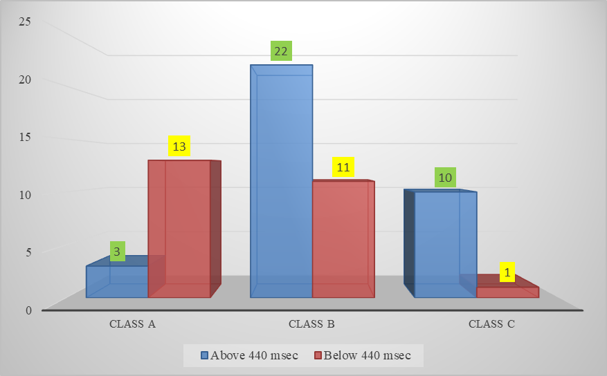
The distribution of QTc prolongation across Child-Turcotte-Pugh classes demonstrated a statistically significant association (χ² = 16.0597, p = 0.000326). A QTc interval greater than 440 ms was observed in 3 patients with Class A disease, 22 patients with Class B disease, and 10 patients with Class C disease. In terms of proportion within each Child-Turcotte-Pugh class, QTc prolongation was observed in 18.75% of Class A, 66.67% of Class B, and 90.91% of Class C patients (Figure 6).
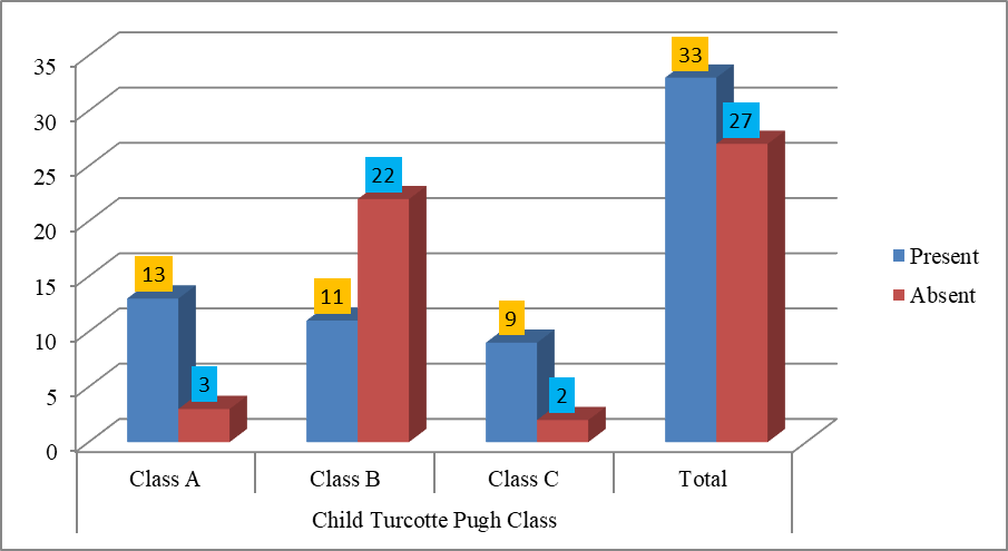
A statistically significant association was observed between Child-Turcotte-Pugh class and diastolic dysfunction (χ² = 13.9103, p = 0.000954). Diastolic dysfunction was present in 13 of 16 patients with Class A disease, 11 of 33 patients with Class B disease, and 9 of 11 patients with Class C disease. Thus, the highest proportion of patients with diastolic dysfunction was observed in Child-Turcotte-Pugh Class C, although the largest absolute number of affected patients was observed in Class A (Figure 7).
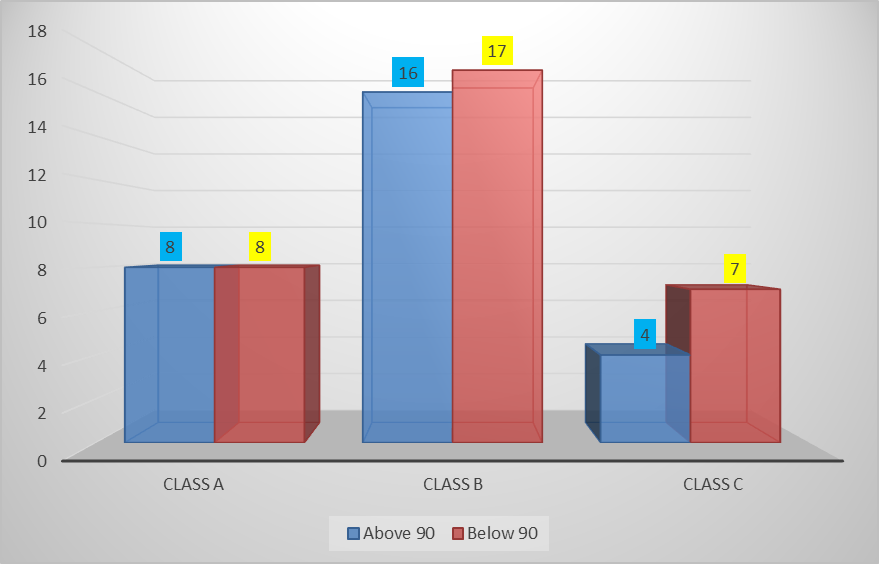
There was no statistically significant association between Child-Turcotte-Pugh class and end-diastolic volume (χ² = 0.5844, p = 0.746613). An end-diastolic volume above 90 was observed in 8 patients with Class A disease, 16 patients with Class B disease, and 4 patients with Class C disease, while values below 90 were observed in 8, 17, and 7 patients, respectively (Figure 8).
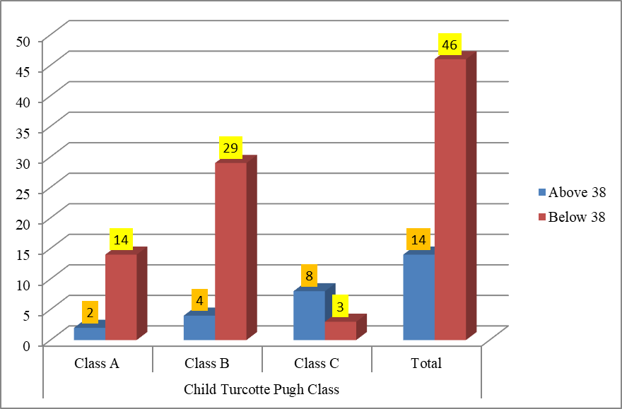
A statistically significant association was observed between Child-Turcotte-Pugh class and end-systolic volume (χ² = 18.371, p = 0.000103). An end-systolic volume above 38 was observed in 2 patients with Class A disease, 4 patients with Class B disease, and 8 patients with Class C disease. Conversely, end-systolic volume below 38 was observed in 14 patients with Class A disease, 29 patients with Class B disease, and 3 patients with Class C disease (Figure 9).
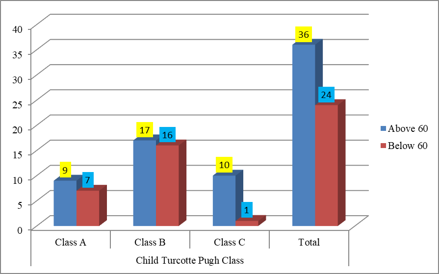
The association between Child-Turcotte-Pugh class and ejection fraction was not statistically significant (χ² = 5.4624, p = 0.06514). An ejection fraction above 60% was observed in 9 patients with Class A disease, 17 patients with Class B disease, and 10 patients with Class C disease, whereas an ejection fraction below 60% was observed in 7, 16, and 1 patient, respectively. Although patients with Child-Turcotte-Pugh Class B constituted the largest group with an ejection fraction below 60%, the overall association between Child-Turcotte-Pugh class and ejection fraction did not reach statistical significance (Figure 10).
Discussion
The present study evaluated echocardiographic abnormalities among 60 patients with chronic liver disease and assessed their relationship with the severity of liver disease according to the Child-Turcotte-Pugh classification. The findings demonstrated that cardiac abnormalities, particularly QTc prolongation and diastolic dysfunction, were common among patients with chronic liver disease despite the exclusion of patients with known primary cardiac disease. These findings support the presence of clinically relevant cardiovascular involvement in chronic liver disease and highlight the role of echocardiography in identifying otherwise unrecognized cardiac abnormalities.
In the present study, the largest proportion of patients belonged to the 50–59-year age group, accounting for 40.00% of the study population. This finding was broadly comparable with the study by Satish G et al., in which 47.29% of patients with alcoholic liver cirrhosis belonged to the 46–55-year age group [17]. Chandey M et al. reported that the maximum number of patients were between 41 and 60 years of age, accounting for 56.7% of their study population [18]. Kini R et al. reported 10 patients below 40 years, 11 between 40 and 50 years, and 9 above 50 years [19]. Keshav R et al. reported a mean age of 42.30 ± 8.42 years [20], while Venu AH et al. included patients between 40 and 70 years of age [21]. The predominance of middle-aged patients in the present study may reflect the cumulative effects of chronic liver disease and its underlying etiological factors over time.
A marked male predominance was observed in the present study, with males accounting for 85.00% of the study population. This finding was consistent with Chandey M et al., who reported that 91.1% of patients with cirrhosis were male [18], and Keshav R et al., who reported an identical male proportion of 85% [20]. Venu AH et al. also reported a substantial male predominance, with males constituting 93.3% of their study population [21]. In contrast, Kini R et al. reported a female predominance, with 22 females and 8 males in their study [19]. The marked male predominance in the present study may partly be related to the high proportion of alcohol-related liver disease in the study population.
With respect to occupation, professional workers constituted the largest occupational group in the present study, accounting for 38.34% of participants, followed by skilled and unskilled workers. This differed from the findings of Venu AH et al., in which farmers constituted the largest occupational group, accounting for 40% of the study population [21]. Differences in occupational distribution may reflect the demographic and socioeconomic characteristics of the population served by the respective study centres and are unlikely to directly account for the echocardiographic abnormalities observed in the present study.
Generalized weakness was the most frequent presenting symptom in the present study, occurring in 91.67% of patients. This finding was comparable with Venu AH et al., who reported generalized weakness in 86.6% of patients [21]. Mallik S et al. similarly reported fatigue as the most common symptom, occurring in 85.71% of patients [16]. In contrast, Chandey M et al. reported abdominal distension as the most frequent presenting symptom, followed by hematemesis and jaundice [18]. Differences in presenting symptoms may be attributable to variations in disease severity, etiology, referral patterns, and the clinical characteristics of the study populations.
Ascites was the most frequently observed clinical sign in the present study, occurring in 66.67% of patients, followed by icterus in 61.67% and edema in 55.00%. Venu AH et al. reported icterus as the most common clinical sign, followed by edema and mild ascites [21]. Mallik S et al. reported ascites in approximately 45.71% of patients [16], while Chandey M et al. reported ascites in 80% of their study population [18]. The relatively high prevalence of ascites in the present study is consistent with the substantial proportion of patients with clinically significant chronic liver disease.
Alcohol-related liver disease was the predominant etiology in the present study, accounting for 53.33% of cases, followed by idiopathic chronic liver disease in 20.00%, hepatitis B in 16.67%, hepatitis C in 6.67%, and primary biliary cirrhosis in 3.33%. This etiological pattern was broadly comparable with Venu AH et al., in which alcohol was the underlying etiology in 66.7% of patients [21]. Chandey M et al. also reported alcohol as the predominant etiology, accounting for 75.6% of cases [18]. In contrast, Mallik S et al. reported hepatitis B as the most frequent etiology, while Kini R et al. reported cases related to hepatitis B, hepatitis C, primary biliary cirrhosis, non-alcoholic fatty liver disease, and idiopathic disease [16][19]. The differences between studies may reflect regional variations in the prevalence of alcohol-related and viral liver disease as well as differences in the populations studied.
More than half of the patients in the present study belonged to Child-Turcotte-Pugh Class B (55.00%), followed by Class A (26.67%) and Class C (18.33%). This finding was comparable with Venu AH et al., in which Child-Turcotte-Pugh Class B represented the largest severity group at 43.3% [21]. Keshav R et al. similarly reported Class B as the most common category, accounting for 48.66% of patients, followed by Class C and Class A [20]. In contrast, Kini R et al. reported a predominance of Class A disease, with 66.67% of patients belonging to this category [19]. The predominance of Class B disease in the present study indicates that a considerable proportion of patients had clinically significant hepatic dysfunction at the time of evaluation.
Ascites was present in 40 of the 60 patients (66.67%) in the present study. This was higher than the proportion reported by Venu AH et al. and Mallik S et al., who reported ascites in 53.3% and 45.71% of patients, respectively, but lower than the 80% reported by Chandey M et al. [16][18][21]. A statistically significant association was observed between Child-Turcotte-Pugh class and ascites in the present study (χ² = 13.1676, p = 0.001383). Ascites was present in 31.25% of Class A patients, 75.76% of Class B patients, and 90.91% of Class C patients. The progressive increase in the proportion of patients with ascites across the Child-Turcotte-Pugh classes is consistent with increasing hepatic dysfunction and complications related to portal hypertension.
Varices were present in 20 patients (33.33%) in the present study. Venu AH et al. reported bleeding varices causing hematemesis in 26.7% of patients, while Mallik S et al. reported a similar frequency of 20% [16][21]. In the present study, the association between Child-Turcotte-Pugh class and the presence of varices was statistically significant (χ² = 24.8523, p < 0.00001). However, the distribution did not demonstrate a simple progressive increase with worsening Child-Turcotte-Pugh class, as the largest absolute number of patients with varices was observed in Class A. This finding should be interpreted cautiously because of the relatively small number of patients within individual severity groups and the possibility of variation related to the underlying causes and duration of portal hypertension.
QTc prolongation was one of the prominent cardiac abnormalities observed in the present study. A QTc interval greater than 440 ms was observed in 35 patients (58.33%). This finding is consistent with the electrophysiological abnormalities described in chronic liver disease and cirrhotic cardiomyopathy. Chandey M et al. reported that the mean QTc value was higher among patients with ascites [18]. Kini R et al. also reported QTc prolongation among patients with non-alcoholic cirrhosis, with 50% of females and 62.5% of males having QTc intervals above 440 ms [19]. In the present study, QTc prolongation showed a statistically significant association with Child-Turcotte-Pugh class (χ² = 16.0597, p = 0.000326). QTc prolongation was observed in 18.75% of Class A patients, 66.67% of Class B patients, and 90.91% of Class C patients. This increasing proportion suggests that electrophysiological abnormalities may become more prominent with increasing severity of liver disease.
Diastolic dysfunction was detected in 33 patients (55.00%) in the present study. Grade I dysfunction was present in 18 patients (30.00%), Grade II in 13 patients (21.67%), and Grade III in 2 patients (3.33%). The high frequency of diastolic dysfunction highlights the importance of echocardiographic evaluation in chronic liver disease, particularly because cardiac dysfunction may occur despite relatively preserved systolic function. Chandey M et al. also demonstrated diastolic dysfunction across different Child-Pugh classes and reported a statistically significant association between the severity of liver disease and diastolic dysfunction [18]. In the present study, the association between Child-Turcotte-Pugh class and diastolic dysfunction was statistically significant (χ² = 13.9103, p = 0.000954).
The distribution of diastolic dysfunction across Child-Turcotte-Pugh classes requires careful interpretation. Although the largest absolute number of patients with diastolic dysfunction was observed in Class A, the proportion within each class was 81.25% for Class A, 33.33% for Class B, and 81.82% for Class C. Therefore, the statistically significant association should not be interpreted as a simple linear progression of diastolic dysfunction with increasing Child-Turcotte-Pugh class. The relatively small number of Class C patients may also have influenced the observed distribution. Nevertheless, the overall association indicates that hepatic disease severity was significantly related to the presence of echocardiographic diastolic abnormalities in this cohort. These findings are broadly comparable with those of Chandey M et al., who also reported a significant association between Child-Pugh class and diastolic dysfunction [18].
End-diastolic volume was below 90 in 32 patients (53.33%) and above 90 in 28 patients (46.67%). There was no statistically significant association between end-diastolic volume and Child-Turcotte-Pugh class (χ² = 0.5844, p = 0.746613). In the present study, an end-diastolic volume above 90 was observed in 8 Class A patients, 16 Class B patients, and 4 Class C patients. These findings can be compared with those of Kini R et al., who reported 14 patients with end-diastolic volume above 90 and 16 with values below 90 [19]. Their study also showed that both categories of end-diastolic volume occurred among patients with diastolic dysfunction.
End-systolic volume was below 38 in 46 patients (76.67%) and above 38 in 14 patients (23.33%). Unlike end-diastolic volume, end-systolic volume demonstrated a statistically significant association with Child-Turcotte-Pugh class (χ² = 18.371, p = 0.000103). An end-systolic volume above 38 was observed in 2 Class A patients, 4 Class B patients, and 8 Class C patients. Thus, an increased end-systolic volume was proportionally more frequent among patients with Class C disease. This finding may suggest altered ventricular systolic performance with more advanced hepatic dysfunction. Ratnakar Kini P et al. reported 3 cases with end-systolic volume above 38 and 27 cases with values below 38, demonstrating a different distribution from that observed in the present study [22].
Left ventricular ejection fraction was above 60% in 36 patients (60.00%) and below 60% in 24 patients (40.00%). The predominance of preserved or relatively high ejection fraction is consistent with the observation that cardiac dysfunction in chronic liver disease may occur predominantly through impaired relaxation and altered contractile reserve rather than overt resting systolic failure. Ratnakar Kini P et al. reported that most patients in their study had an ejection fraction above 60% [22]. Venu AH et al. similarly reported a slightly increased mean left ventricular ejection fraction in their study group, although the difference was not statistically significant [21].
The association between Child-Turcotte-Pugh class and ejection fraction was not statistically significant in the present study (χ² = 5.4624, p = 0.06514). An ejection fraction below 60% was observed in 7 Class A patients, 16 Class B patients, and 1 Class C patient. Although Class B contained the largest absolute number of patients with an ejection fraction below 60%, the overall association did not reach statistical significance. This finding suggests that resting ejection fraction alone may not adequately reflect the extent of cardiac involvement in chronic liver disease. A preserved ejection fraction does not necessarily exclude cirrhotic cardiomyopathy, particularly when diastolic dysfunction and electrophysiological abnormalities are present. Ratnakar Kini P et al. similarly reported that most patients with cirrhotic cardiomyopathy had an ejection fraction above 60%, supporting the limitation of ejection fraction as an isolated marker of cardiac dysfunction [22].
The findings of the present study demonstrate that cardiac abnormalities in chronic liver disease are not restricted to overt systolic dysfunction. QTc prolongation and diastolic dysfunction were frequent findings, while end-systolic volume also demonstrated a significant association with Child-Turcotte-Pugh class. In contrast, end-diastolic volume and ejection fraction did not show statistically significant associations with disease severity. This pattern suggests that cardiac involvement may be detected more readily through diastolic and electrophysiological parameters than through conventional assessment of resting systolic function alone.
The present study has certain limitations. The sample size was limited to 60 patients and the study was conducted at a single tertiary care centre, which may limit the generalizability of the findings. The relatively small number of patients in Child-Turcotte-Pugh Class C may also have influenced subgroup comparisons. Furthermore, although morbidity and mortality due to cardiac involvement were included among the study objectives, the available results do not provide sufficient follow-up data to establish the relationship between the identified echocardiographic abnormalities and cardiac-related morbidity or mortality. Therefore, conclusions regarding this objective should be made only if corresponding follow-up outcome data are available.
Overall, the present study demonstrates that cardiac abnormalities are frequent among patients with chronic liver disease, with diastolic dysfunction and QTc prolongation being particularly prominent. The significant associations observed between Child-Turcotte-Pugh class and several cardiac parameters suggest that hepatic disease severity is associated with cardiovascular involvement. At the same time, the absence of a significant association between Child-Turcotte-Pugh class and ejection fraction emphasizes that preserved resting systolic function does not exclude clinically relevant cardiac abnormalities. Echocardiographic evaluation may therefore have an important role in identifying subclinical cardiac involvement in patients with chronic liver disease. Further studies involving larger populations, longer follow-up, and serial assessment of cardiac function are required to clarify the relationship between cardiac abnormalities, progression of liver disease, and clinical outcomes.
Conclusion
The present study evaluated cardiac abnormalities in patients with chronic liver disease using echocardiography and assessed their relationship with the severity of liver disease. Cardiac involvement was common in the study population, with diastolic dysfunction and QTc prolongation being prominent findings. Several cardiac parameters showed a significant association with Child-Turcotte-Pugh class, suggesting that cardiac abnormalities may increase with worsening hepatic dysfunction. However, resting left ventricular ejection fraction was not significantly associated with Child-Turcotte-Pugh class, indicating that preserved systolic function does not necessarily exclude cardiac involvement in chronic liver disease.
The findings highlight the importance of recognizing subclinical myocardial dysfunction in patients with chronic liver disease. Echocardiographic evaluation may facilitate early identification of cardiac abnormalities, particularly diastolic dysfunction, before the development of overt cardiac failure. Careful cardiac assessment is therefore particularly relevant before surgical procedures, liver-directed interventions, or other situations involving significant physiological stress, where previously unrecognized cardiac dysfunction may become clinically apparent.
Patients with chronic liver disease and evidence of cardiac dysfunction should be appropriately assessed and monitored, with attention to factors that may aggravate cardiac dysfunction, including fluid overload and excessive sodium intake. Further studies involving larger sample sizes, longer follow-up, and serial assessment of cardiac function are required to better establish the relationship between cardiac abnormalities and the progression and severity of chronic liver disease.
Declarations
Ethical Approval and Consent to Participate
All procedures performed in this case series were conducted in accordance with institutional ethical standards and the principles of the Declaration of Helsinki. Ethical approval was obtained from the appropriate institutional review board where required. Written informed consent was obtained from all patients or their legal guardians prior to the procedures.
Consent for Publication
Written informed consent for publication of clinical details and images was obtained from the patients or their legal guardians. All identifying information has been anonymized to protect patient confidentiality.
Availability of Supporting Data
The data supporting the findings of this study are available from the corresponding author upon reasonable request, subject to institutional and ethical regulations.
Competing Interests
The authors declare that they have no competing interests related to this work.
Funding
This study received no specific grant from any funding agency in the public, commercial, or not-for-profit sectors.
Authors’ Contributions
All authors contributed substantially to the conception, data acquisition, analysis, drafting, and critical revision of the manuscript. All authors have read and approved the final version of the manuscript.