Introduction
Thoracic malignancies form a diverse group of neoplasms arising from the lungs, pleura, mediastinum, oesophagus, and chest wall. Lung carcinoma remains the most common and lethal of these, accounting for a disproportionate share of global cancer mortality [5]. However, a subset of thoracic malignancies is exceptionally rare, occurring in atypical anatomic compartments with imaging features that may mimic more common entities, leading to diagnostic delay and suboptimal management [1,2].
Early and accurate diagnosis is essential, since many of these tumours are biologically aggressive and carry a poor prognosis when identified late. Imaging - particularly multidetector computed tomography (MDCT) with intravenous contrast - plays a central role in detection, characterisation, local staging, and treatment planning of thoracic masses. Several key imaging parameters must be evaluated systematically when reporting a thoracic or mediastinal mass:
-
Location, size, and morphological configuration of the lesion, including the mediastinal compartment of origin [1,2].
-
Presence of intralesional fat, cystic change, calcification, or haemorrhage
-
Relationship to, and invasion of, adjacent mediastinal structures, great vessels, airways, and the chest wall
This paper presents a retrospective case series of eight patients with histopathologically confirmed rare thoracic malignancies, with emphasis on their characteristic imaging features, and differential diagnoses
Aims and Objectives
-
To describe the spectrum of imaging findings in rare thoracic malignancies encountered at a tertiary care centre.
-
To correlate imaging features with histopathological diagnoses.
-
To outline a systematic CT reporting framework applicable to mediastinal and thoracic masses, consistent with current classification systems.
-
To improve radiological awareness of rare thoracic neoplasms and aid their differentiation from common pathologies.
Materials and Methods
This retrospective case series was conducted at the Department of Radiodiagnosis, apollo Institute of Medical Sciences and Research (AIMSR), Hyderabad. Eight patients with rare thoracic malignancies were identified from the departmental imaging. Inclusion criteria required availability of cross-sectional imaging (chest radiograph and CECT chest) together with a confirmed histopathological diagnosis.
All CT examinations were performed on a multidetector CT scanner. Histopathological confirmation was obtained by CT-guided biopsy, surgical resection, or thoracoscopic sampling in each case.
Patient demographics, presenting symptoms, imaging characteristics, and final histopathological diagnoses were tabulated for analysis.
Results
Eight cases of rare thoracic malignancy were identified over the study period. Patient demographics, imaging modalities used, key imaging findings, and final histopathological diagnoses are summarised in Table 1. The cohort ranged from 12 to 60 years of age, with a predominance of young adults - a pattern consistent with the demographic distribution reported for several of these entities in the literature [4,6,12] (Table 1).
| Case | Age/Sex | Location | Key Imaging Finding | Histopathological Diagnosis |
| 1 | 38/M | Anterior mediastinum | Large soft-tissue mass with calcification, necrosis, heterogeneous enhancement | Primary mediastinal choriocarcinoma |
| 2 | 40/M | Anterior mediastinum | Soft-tissue mass with fatty attenuation and calcification | Thymoliposarcoma |
| 3 | 35/F | Right pleura / upper lobe | Pleural-based homogeneously enhancing mass with non-enhancing foci; pleural effusion | Hemangiopericytoma (solitary fibrous tumour) |
| 4 | 23/F | Right upper hemithorax (pleuro-parenchymal) | Large well-defined cystic lesion, wall enhancement, tracheal displacement | Synovial sarcoma |
| 5 | 31/M | Right hemithorax / mediastinum | Large heterogeneous mass encasing aorta and carina; tracheal displacement | Synovial sarcoma |
| 6 | 60/M | Left upper lobe | Heterogeneous solid-cystic mass; bronchial cut-off; necrosis; peripheral wall enhancement | Sarcomatoid carcinoma |
| 7 | 12/M | Posterior mediastinum | Large posterior mediastinal mass; neural foraminal widening; loss of fat planes with cord | malignant peripheral nerve sheath tumour MPNST |
| 8 | 35/M | Chest wall (7th/8th rib) | Soft-tissue mass from chest wall, heterogeneous enhancement | Spindle cell sarcoma |
Discussion
The following section discusses, for each rare thoracic malignancy encountered, the clinical context, imaging findings, differential diagnosis, and correlation with published literature.
Case 1
Primary Mediastinal Choriocarcinoma
A 38-year-old male presented with symptoms prompting chest evaluation.
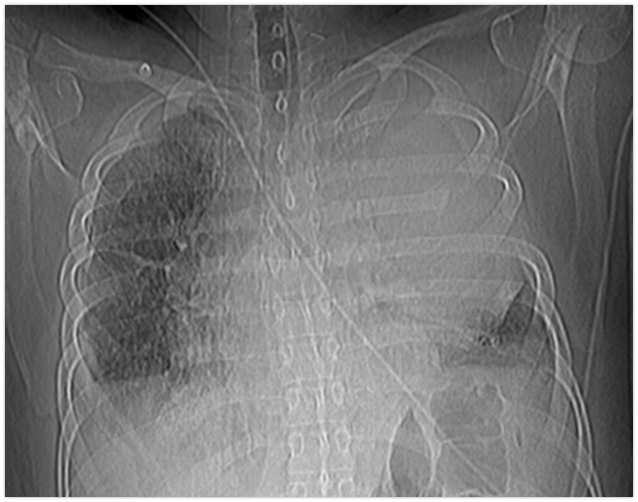
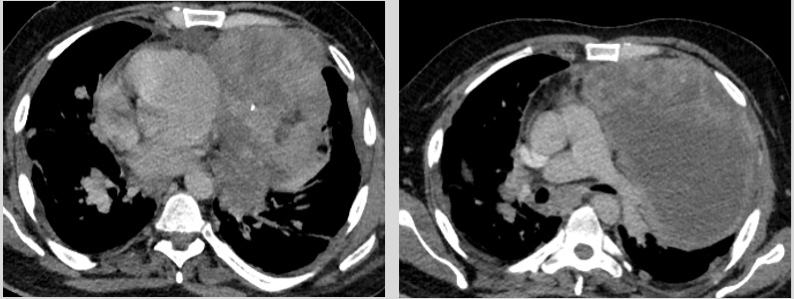
Primary mediastinal choriocarcinoma is exceedingly rare, representing 1–4% of mediastinal tumours and under 5% of mediastinal germ cell malignancies, and is among the most aggressive NSGCTs [6,7]. It occurs more frequently in young men, and elevated serum beta-hCG is a characteristic biochemical marker, although non-gestational choriocarcinoma can occasionally present with normal hCG levels and require histological confirmation [6,8].
The differential diagnosis of anterior mediastinal germ cell tumours includes:
-
Seminoma/germinoma: large, well-marginated, lobulated mass with homogeneous attenuation that may extend across the midline
-
Non-seminomatous GCTs (choriocarcinoma, yolk sac tumour): large, heterogeneous mass with irregular margins; haemorrhage and necrosis common; frequent lung invasion [2,6]
-
Teratoma: sharply marginated, round or lobulated mass; calcification in 20–40% of cases; usually unilateral; malignant teratomas show a predominantly solid component
Case 2
Thymoliposarcoma
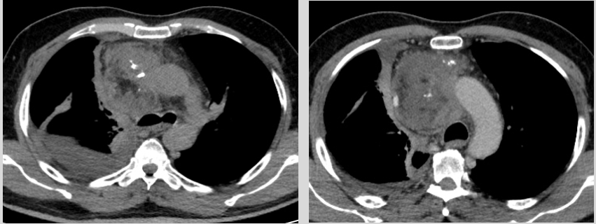
Thymoliposarcoma is an exceptionally rare malignant neoplasm of the anterior mediastinum, first described by Havlicek and Rosai, with fewer than a dozen cases reported in the literature to date [9]. It closely mimics thymolipoma on imaging, and the mean age at diagnosis is in the sixth decade with a slight male predominance [9]. The differential diagnosis of fat-containing mediastinal lesions includes:
-
Lipoma: homogeneous fat attenuation without a soft-tissue component
-
Teratoma: complex cystic lesion with fluid, fat, calcification, or dental elements
-
Thymolipoma: encapsulated lesion containing fat and strands of thymic tissue; conforms to adjacent structures and is non-invasive
-
Liposarcoma/thymoliposarcoma: may resemble thymolipoma but typically insinuates between and splays mediastinal structures rather than conforming to them [10,11].
-
Lipoblastoma: predominantly seen in children younger than 3 years
Distinction between thymolipoma and thymoliposarcoma is ultimately histopathological; imaging features that raise concern for malignancy include irregular margins, invasion of adjacent structures, and a prominent soft-tissue component interspersed with fat [9,11].
Case 3
Hemangiopericytoma (Solitary Fibrous Tumour of the Pleura)
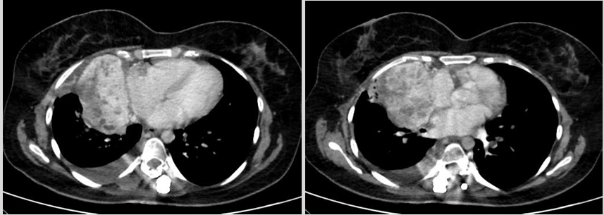
Solitary fibrous tumours of the thorax are pleural-based neoplasms accounting for fewer than 5% of all primary pleural tumours [3]. They arise predominantly from the visceral pleura (approximately 80% of cases), are usually indolent and benign in behaviour, and most commonly present in the sixth to seventh decades of life without a clear gender predilection; malignant forms are associated with large size, high mitotic rate, and necrosis [3].
Reported radiographic and CT features include a pleural-based mass that is relatively circumscribed; soft-tissue attenuation; relatively homogeneous post-contrast enhancement reflecting the tumour's rich vascularity; and non-enhancing foci corresponding to necrosis, myxoid degeneration, or haemorrhage [3]. The differential diagnosis includes peripheral lung carcinoma and localised mesothelioma.
Case 4
Synovial Sarcoma - Pleuro-Parenchymal (Young Female)
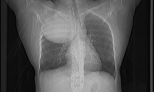
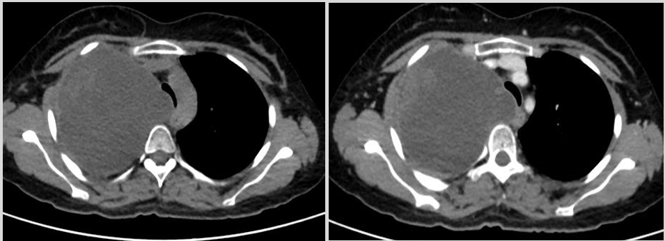
Primary pulmonary and pleural synovial sarcoma is rare, accounting for a small minority of intrathoracic sarcomas, and is characteristically biphasic or monophasic histologically, with confirmation requiring demonstration of the SS18 (SYT-SSX) gene fusion on molecular analysis [4,12]. Intrathoracic synovial sarcoma typically presents as a large, well-circumscribed, heterogeneously enhancing pleural or pulmonary mass with patchy low-density (cystic/necrotic) areas and a notable absence of calcification or significant lymphadenopathy [13,14]. A predominantly cystic appearance with wall enhancement in a young patient, as in this case, is a recognised imaging pattern [14,15]. The mass effect with tracheal displacement underscores the importance of pre-operative airway assessment and surgical planning.
Case 5
Synovial Sarcoma - Large Mediastinal Involvement (Young Male)
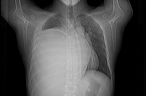
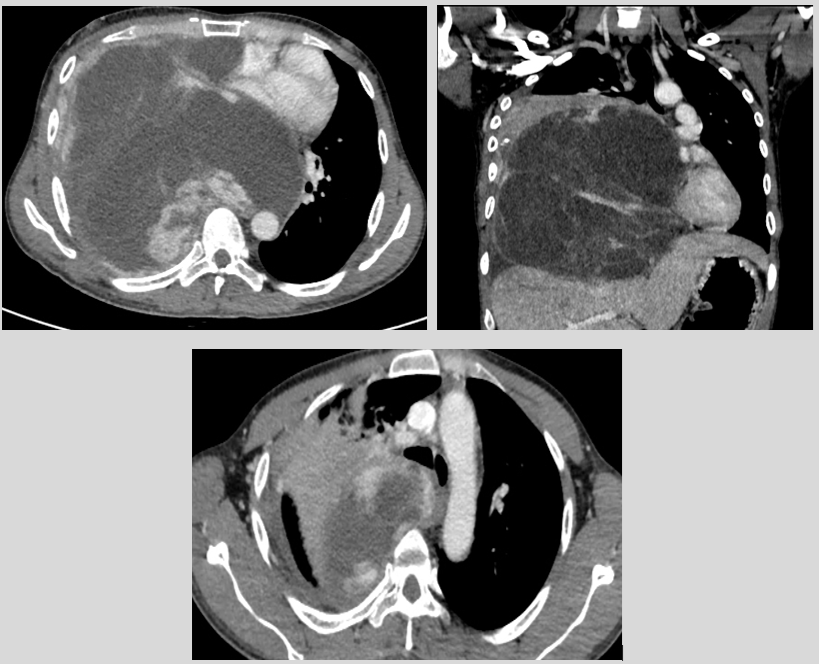
Compared with Case 4, this case illustrates the locally invasive variant of thoracic synovial sarcoma in a young adult male. Encasement of major vascular structures and central airways without overt destruction is a recognised feature of intrathoracic sarcomas and is an important imaging clue distinguishing sarcomatous encasement from more aggressive destructive invasion [13,21]. The degree of vascular encasement has direct implications for resectability and surgical planning.
Case 6
Sarcomatoid Carcinoma of the Lung
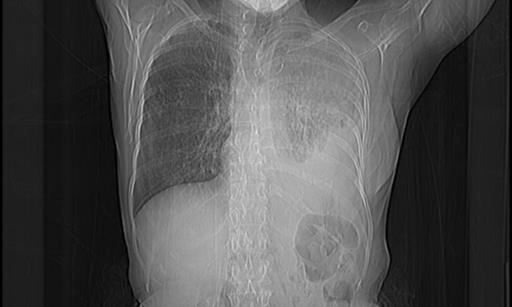
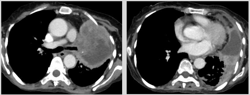
Histopathology confirmed sarcomatoid carcinoma, a rare and aggressive subtype accounting for approximately 0.1-1.3% of all lung cancers [16,17]. Pulmonary sarcomatoid carcinomas are poorly differentiated non-small cell lung carcinomas (NSCLCs) exhibiting sarcomatous differentiation and, per the WHO classification, include pleomorphic carcinoma, spindle cell carcinoma, giant cell carcinoma, carcinosarcoma, and pulmonary blastoma [5,16]. On CT, these tumours tend to present as large, peripheral or central, lobulated masses with poorly defined margins, frequent cavitation or necrosis, and may show greater low-attenuation area and pleural invasion compared with conventional NSCLC of similar size [17,18]. The bronchial cut-off sign, extensive necrosis, and irregular peripheral enhancement seen in this case are characteristic features. Median survival is poor (approximately 9–12 months), underscoring the importance of early recognition [17,18].
Case 7
Malignant Peripheral Nerve Sheath Tumour (MPNST)
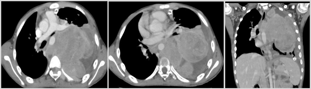
MPNST is a rare, high-grade soft-tissue sarcoma arising from peripheral nerve sheaths or Schwann cells, accounting for roughly 5–10% of all soft-tissue sarcomas; approximately 50% of cases are associated with neurofibromatosis type 1 (NF1) [19,20]. Posterior mediastinal location with neural foraminal widening and intraspinal extension is a characteristic pattern, reflecting origin along the intercostal nerves or sympathetic chain [2,19,20]. The cervicothoracic sign reflects a lesion whose bulk lies predominantly above the clavicles on a frontal radiograph, supporting a posterior mediastinal/paravertebral origin. The combination of young age, a posterior mediastinal mass, neural foraminal widening, and intraspinal involvement - as in this case - should prompt strong consideration of MPNST, particularly when an underlying history of neurofibromatosis is present [19,20].
Case 8
Spindle Cell Sarcoma of the Chest Wall
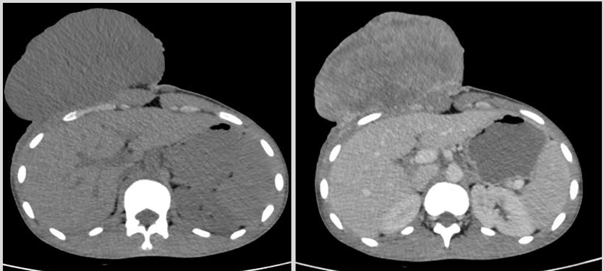
Primary chest wall sarcomas are uncommon, and spindle cell sarcoma represents a broad histological category encompassing several tumour subtypes [22,23]. A sarcoma should be suspected when an intrathoracic or chest-wall mass exceeds approximately 7 cm, shows well-defined or lobulated margins, intratumoural vessels, and associated pleural effusion without significant lymphadenopathy [21]. CT features of malignant chest wall masses include cortical bone destruction, heterogeneous enhancement, and extrapleural fat-pad displacement [22,23]; cross-sectional imaging (CT and MRI) is essential for local staging, while MRI offers superior soft-tissue contrast for assessing neurovascular and multicompartment involvement [22]. Differentiation from chondrosarcoma, Ewing sarcoma, osteosarcoma, and solitary fibrous tumour is aided by these CT characteristics but is ultimately confirmed on histopathology [22,23].
Conclusion
This case series illustrates rare thoracic malignancies and their distinctive imaging features. Thoracic malignancies encompass a broad spectrum of neoplasms affecting the lungs, pleura, mediastinum, and chest wall; while lung carcinoma and mesothelioma remain most prevalent, rare malignancies present unique diagnostic challenges that require systematic imaging evaluation combined with clinical and histopathological correlation [1,2,5].
Contrast-enhanced CT remains the imaging modality of choice for evaluating and characterising most thoracic lesions, providing information on attenuation, enhancement pattern, intralesional composition, and relationship to adjacent structures.
Radiologists and clinicians must maintain a broad differential diagnosis when evaluating unusual thoracic masses, and multidisciplinary evaluation involving oncology, thoracic surgery, and pathology remains essential for optimal patient outcomes.
Key Messages
-
Rare thoracic malignancies present a broad and heterogeneous spectrum of imaging appearances that can closely mimic more common chest pathologies, making them a diagnostic challenge.
-
Contrast-enhanced CT remains the imaging modality of choice for characterising these lesions, offering critical information on attenuation, enhancement pattern, and relationship to adjacent structures.
Declarations
Conflict of Interest
There are no competing interests to declare.
Author contribution
Conceptualization: N.L.N. Moorthy, Srija Kashetty
Data curation: N.L.N. Moorthy, Vaagdevi, Srija Kashetty
Formal analysis: Srija Kashetty, Ramesh Goud
Investigation: Srija Kashetty Ramesh Goud
Methodology: Vaagdevi, Srija Kashetty
Supervision: N.L.N. Moorthy
Validation: Ramesh Goud
Visualization: Vaagdevi, Srija Kashetty
Writing-original draft: Vaagdevi, Srija Kashetty
Writing-review & editing: Ramesh Goud, N.L.N. Moorthy
Funding Statement
None
Data Availability
The datasets generated or analyzed during the study are available from the corresponding author on reasonable request.
Generative AI Disclosure
During the preparation of this manuscript, we used Claude (Anthropic, Claude Sonnet 4.5) to assist with formatting the manuscript. The tool was not used to generate, analyze, or interpret any scientific content, data, or clinical findings presented in this manuscript. All AI-assisted content was reviewed, edited, and approved by the author(s), who take full responsibility for the accuracy and integrity of the final manuscript.