Introduction
The breast is a hormone-driven organ regulated by the hypothalamo-pituitary-ovarian axis, making it susceptible to various age-dependent pathologies. Benign breast diseases ranging from inflammatory conditions to proliferative lesions like atypical hyperplasia and fibroadenoma predominantly affect younger women [1]. Conversely, malignant neoplasms, over 95% of which are adenocarcinomas (both in situ and invasive), are more prevalent in older cohorts [1].
Globally, breast cancer remains the most common malignancy among women and represents the leading cause of cancer-related morbidity worldwide. According to GLOBOCAN 2022, approximately 2.30 million new cases and 666,000 deaths were reported globally, making it the commonest cancer in women and one of the leading causes of cancer-related mortality it represents about one in four cancers diagnosed in women and one in six cancer deaths among women [2].
In India, breast cancer has surpassed cervical cancer to become the leading malignancy among women, accounting for approximately 221,757 new cases and 26.6% of all female cancers in 2022. The disease predominantly affects women between 45 and 69 years of age, although the incidence among younger women is increasing [2,3]. Recent Indian cancer registry data also indicate increasing incidence trends in metropolitan as well as semi-urban regions like higher incidence rates have been reported from Kerala, Delhi, Goa, Tamil Nadu, Karnataka, Punjab, and Mizoram, reflecting regional variation in disease burden, emphasizing the need for region-specific clinicopathological studies and early diagnostic strategies [3].
Management relies on the "triple assessment" (clinical, radiological, and pathological evaluation), where accurate tissue diagnosis prevents unnecessary, traumatizing surgeries [4,5]. While advanced molecular testing offers precise profiling, its high-cost limits routine use [6]. Instead, immunohistochemical (IHC) evaluation of estrogen receptor (ER), progesterone receptor (PR), and HER2 remains the clinical standard for prognosis and targeted therapy [6]. Recent integration of digital pathology and artificial intelligence (AI) further optimizes this workflow. Deep learning models show promise in predicting receptor status directly from H&E sections [7], while AI-assisted scoring systems reduce inter-observer variability in IHC interpretation [8]. Artificial intelligence (AI) increasingly augments immunohistochemistry (IHC) to improve scoring precision and mitigate interobserver variability. Concurrently, the emergence of refined classifications-such as HER2-low and HER2-ultralow categories-has expanded targeted therapeutic strategies, underscoring the dynamic role of pathological assessment in precision oncology [9].
However, these advanced molecular and digital technologies remain cost-prohibitive, technically demanding, and largely inaccessible in many developing regions. Consequently, conventional histopathology paired with standard IHC remains the most pragmatic, reproducible, and essential first-line diagnostic framework globally, serving as the cornerstone for breast cancer management and the baseline for further specialized testing.
Despite global advancements, regional variations in breast disease spectrums persist. This study aims to establish the first baseline clinicopathological data for both benign and malignant breast diseases in the Kachchh region of Gujarat, a geographically distinct population where no prior research has been conducted.
Methods
This descriptive, hospital-based study was conducted at the Department of Pathology, Gujarat Adani Institute of Medical Sciences, G.K. General Hospital, Bhuj, Kachchh, between August 2016 and August 2018. A total of 150 cases were evaluated, comprising female lumpectomies (n = 90), male lumpectomies (n = 6), radical mastectomies (n = 42), and breast biopsies (n = 12).
-
Inclusion Criteria: Inflammatory, benign, and malignant breast lesions from patients of all ages and both sexes, including biopsy, lumpectomy, and modified radical mastectomy (MRM) specimens.
-
Exclusion Criteria: Post-radiotherapy recurrences and satellite nodules of breast carcinoma.
Patient demographics, clinical histories, and radiological findings were retrieved from medical records and histopathology requisition forms. All specimens were fixed in 10% formalin and processed according to standard surgical specimen guidelines. Tissue samples were automatically processed, embedded in paraffin, sectioned at 5 micrometers, and stained with Hematoxylin and Eosin (H&E). Malignant tumours were graded using the Nottingham modification of the Bloom-Richardson system. For immunohistochemical profiling, 4 micrometers thick sections from formalin-fixed, paraffin-embedded (FFPE) carcinoma blocks were taken on poly-L-lysine coated slides and labelled for ER, PR, and HER2/neu. ER/PR Scoring: Evaluated using the Allred scoring system. HER2/neu Scoring: Assessed according to the College of American Pathologists/American Society of Clinical Oncology (CAP/ASCO) guidelines. Pathological diagnoses and IHC expressions were subsequently correlated with clinical parameters, including age, menopausal status, tumour size, histological subtype, lymph vascular invasion (LVI), and nodal status.
Results
Demographic and Lesion Distribution
A total of 150 breast specimens were evaluated, comprising 144 females (90 lumpectomies, 42 radical mastectomies, 12 biopsies) and 6 males (6 lumpectomies). Among the female cohorts, benign lesions were the most prevalent (n = 73; 50.7%), followed by malignant (n = 50; 34.7%) and inflammatory conditions (n = 21; 14.6%). To maintain demographic specificity, male and female data were analysed separately.
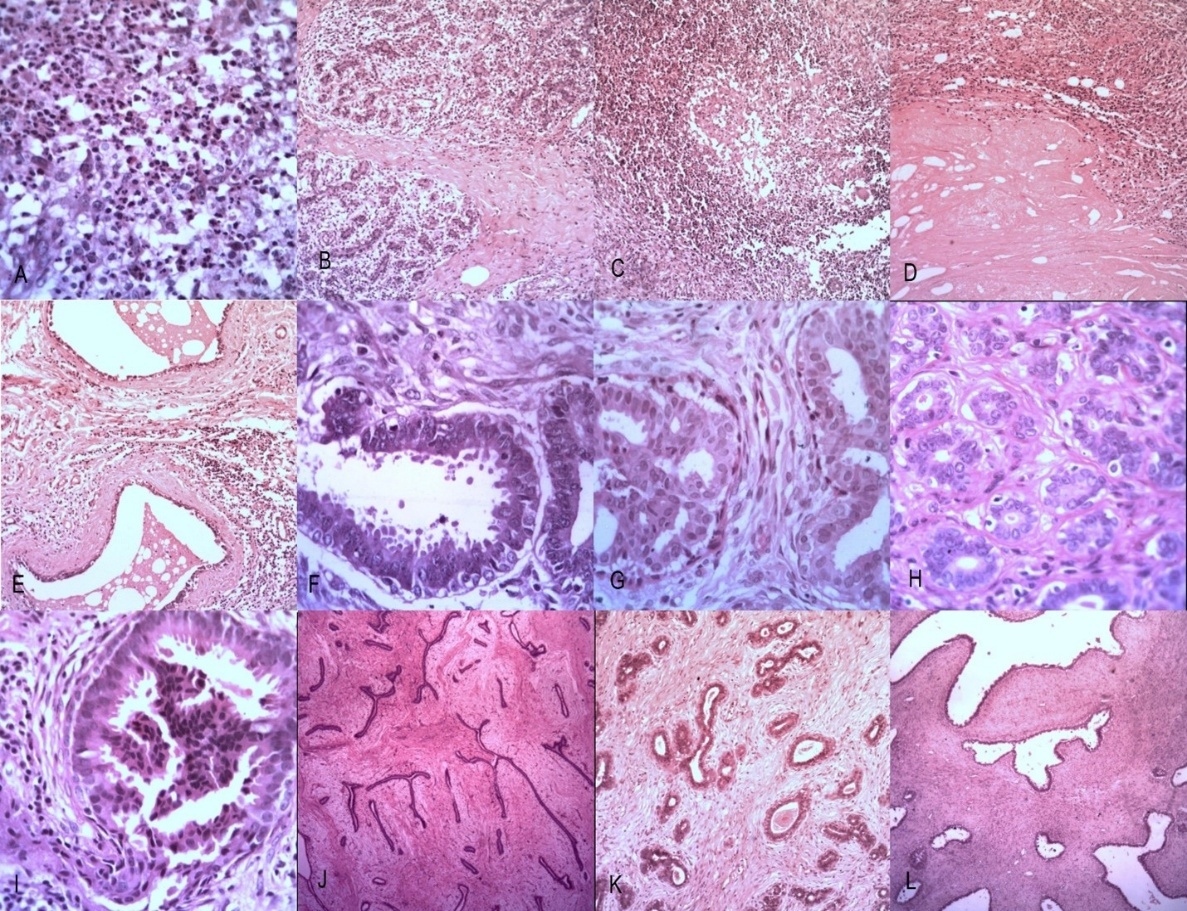
Inflammatory and Non-Proliferative Lesions (Table 1, Figure 1- A to F)
Of the 21 female inflammatory cases, acute mastitis was the most frequent (n = 10), followed by chronic mastitis (n = 5), granulomatous mastitis (n = 5), and fat necrosis (n = 1). Non-proliferative breast pathology (fibrocystic disease) was diagnosed in 9 female patients; the majority (n = 7) were aged 31–50 years, with single cases noted in the 11–20 and 21–30 age cohorts.
Proliferative Lesions and Fibroepithelial Tumors (Table 1, Figure 1- G to L)
Proliferative breast disease without atypia was identified in 8 cases, dominated by usual ductal hyperplasia (UDH, n = 6), followed by solitary cases of intraductal papilloma and tubular adenoma. UDH cases peaked in the 21–30 age group (n = 3). The single tubular adenoma occurred in the 11–20 age cohort, whereas the intraductal papilloma occurred in the 41–50 age range. Atypical ductal hyperplasia (ADH) was rare, detected in a single 36-year-old female, displaying characteristic cribriform spaces with monomorphic, regularly spaced cells forming rigid bars and micro papillae.
Fibroepithelial lesions comprised a substantial portion of the cohort. Fibroadenoma was highly prevalent (n = 50; 35% of all female breast lesions), with 76% of cases presenting before age 30. Phyllodes tumors were less frequent (n = 5; 3.5% of all female breast lesions) and peaked in the 21–30 age cohort.
| Pathological Diagnosis | 11–20 Years | 21–30 Years | 31–40 Years | 41–50 Years | Total Cases (n) | (%) |
| Inflammatory Lesions | ||||||
| Acute Mastitis | 1 | 1 | 4 | 4 | 10 | 10.64% |
| Chronic Mastitis | 0 | 1 | 2 | 2 | 5 | 5.32% |
| Granulomatous Mastitis | 0 | 4 | 1 | 0 | 5 | 5.32% |
| Fat Necrosis | 1 | 0 | 0 | 0 | 1 | 1.06% |
| Non-Proliferative Lesions | ||||||
| Fibrocystic Disease | 1 | 1 | 4 | 3 | 9 | 9.57% |
| Proliferative Lesions (Without Atypia) | ||||||
| Usual Ductal Hyperplasia (UDH) | 1 | 3 | 2 | 0 | 6 | 6.38% |
| Intraductal Papilloma | 0 | 0 | 0 | 1 | 1 | 1.06% |
| Tubular Adenoma | 1 | 0 | 0 | 0 | 1 | 1.06% |
| Proliferative Lesions (With Atypia) | ||||||
| Atypical Ductal Hyperplasia (ADH) | 0 | 0 | 1 | 0 | 1 | 1.06% |
| Fibroepithelial Lesions | ||||||
| Fibroadenoma | 18 | 20 | 7 | 5 | 50 | 53.19% |
| Phyllodes Tumor | 1 | 3 | 1 | 0 | 5 | 5.32% |
| Total Group Category (N) | 24 | 33 | 22 | 15 | 94 | 100.00% |
Malignant Lesions and Histological Grading (Table 2, Figure 2)
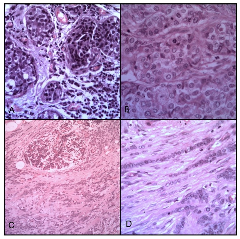
Among the malignancies, Ductal Carcinoma in Situ (DCIS) was identified in 5 patients, demonstrating a dual peak in the 31–40 and 51–60 age cohorts (n = 2 each). Invasive Carcinoma-No Special Type (IDC-NST) was the most frequent malignancy (n = 44), peaking significantly in the sixth decade (n = 16). A single case of Invasive Lobular Carcinoma (ILC) was reported in a 50-year-old female.
Histological grading of the 44 IDC-NST cases revealed that the majority were moderately differentiated/Grade II (n = 19; 43%), followed by well-differentiated/Grade I (n = 15; 34%) and poorly differentiated/Grade III (n = 10; 23%).
Immunohistochemical (IHC) Profiling and Molecular Subtyping (Table 2, Figure 3)
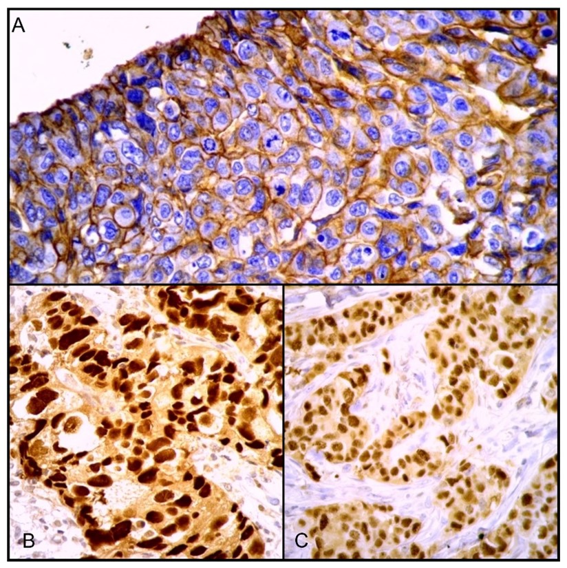
IHC was performed on 15 of the 44 invasive cases (34.1%). ER and PR positivity were each identified in 12 cases (80%), with 3 cases being double-negative; strong nuclear localization was confirmed microscopically. HER2/neu was positive in 7 cases (47%)demonstrating strong, circumferential membrane stainingand negative in 8 cases (53%).
The 15 cases were classified into the following molecular subtypes:
-
Luminal A (ER+/PR+/HER2-): 33.3% (n = 5)
-
Luminal B/HER2+ (ER+/PR+/HER2+): 26.7% (n = 4)
-
Luminal B/HER2- (ER+/PR+/HER2-): 13.3% (n=2)
-
HER2-enriched (ER-/PR-/HER2+): 20.0% (n = 3)
-
Triple-Negative (TNBC; ER-/PR-/HER2-): 6.7% (n = 1)
Chi-square analysis demonstrated a highly significant association between molecular subtype and histological grade (χ²= 25.29, df = 8, p = 0.00139), with HER2-rich and TNBC subtypes exclusively displaying poorly differentiated (Grade III) morphology. Furthermore, significant correlations were confirmed between molecular subtyping and ER/PR status (χ²= 10.83, df = 4, p = 0.0285), as well as HER2/neu status (χ²= 15.00, df = 4, p = 0.0047).
| Malignant Parameter Category | Sub-Classification | 31-40 Years | 41-50 Years | 51-60 Years | 61-70 Years | 71-80 Years | Total Cases (n) | (%) |
| Histological Tumor Type | Ductal Carcinoma in Situ (DCIS) | 2 | 1 | 2 | 0 | 0 | 5 | 10.00% |
| Invasive Ductal Carcinoma (IDC-NST) | 9 | 10 | 16 | 7 | 2 | 44 | 88.00% | |
| Invasive Lobular Carcinoma (ILC) | 0 | 1 | 0 | 0 | 0 | 1 | 2.00% | |
| Nottingham/Bloom-Richardson Grade* | Grade I (Well Differentiated) | — | — | — | — | — | 15 | 34.10% |
| Grade II (Moderately Differentiated) | — | — | — | — | — | 19 | 43.20% | |
| Grade III (Poorly Differentiated) | — | — | — | — | — | 10 | 22.70% | |
| Immunohistochemical Molecular Subtype** | Luminal A (n=5) | — | — | — | — | — | 4 (G-I) | 33.30% |
| Luminal B / HER2- (n=2) | — | — | — | — | — | 2 (G-II) | 13.30% | |
| Luminal B / HER2+ (n=4) | — | — | — | — | — | 4 (G-II) | 26.70% | |
| HER2-Rich / Non-Luminal (n=3) | — | — | — | — | — | 3 (G-III) | 20.00% | |
| Triple Negative / TNBC (n=1) | — | — | — | — | — | 1 (G-III) | 6.70% | |
| Total Malignant Cohort (N) | 13 | 12 | 18 | 7 | 2 | 50 | 100.00% |
*Histological Grading: Grading percentages strictly reflect the 44 invasive ductal carcinoma (IDC-NST) cases evaluated under the Nottingham modification system. **Molecular Subtyping: IHC profiling was performed on a validated subset of 15 invasive carcinoma cases. Subtype breakdown displays exact cross-tabulated tumor grades inside parentheses (e.g., G-I, G-II, G-III) to maintain your core findings showing HER2-rich and TNBC variants exclusively tracking with Grade III morphology.
Male Breast Pathology
All 6 male breast specimens were histopathologically diagnosed as gynecomastia. The highest incidence was observed in the third decade (n = 3), followed by the fourth (n = 2) and second (n = 1) decades of life.
Discussion
Breast lesions encompass a broad spectrum of clinical conditions, ranging from benign to malignant, across all stages of adult life. While the primary driver for clinical presentation is often the fear of malignancy, the widespread adoption of mammographic screening has significantly increased the detection of both symptomatic and asymptomatic breast diseases over the last decade.
Clinicopathological Spectrum and Inflammatory lesions of breast
Integrating local data within domestic and international literature is vital for mapping regional disease patterns. In our cohort, inflammatory lesions comprised 14.6% (n=21) of female specimens (n=144). This baseline frequency aligns with established data from the Indian subcontinent and Asian nations, closely mirroring reports from Pakistan (Siddiqui MS et al., 14%) [10] and India (Sharma K et al., 24%) [11], while remaining higher than findings from Vadodara, Gujarat (Thakral A et al., 5%) [12] and Saudi Arabia (Mansoor I et al., 11%) [13].
Subtype analysis demonstrates that our localized findings conform closely to broader Asian trends (2–9% for acute mastitis, 0.5–5% for chronic/granulomatous mastitis, and 0.5–2% for fat necrosis):
-
Acute Mastitis: Comprising 7% (n=10) of our cohort, this matches standard regional ranges and correlates well with findings from Bangladesh (Rahman MA et al., 9%) [14] and Pakistan (Aslam HM et al., 6%) [15], though it sits higher than the 2% reported in Saudi Arabia.
-
Chronic Mastitis: This diagnosis accounted for 3.5% (n=5) of cases, falling squarely within the international average and replicating rates documented in Saudi Arabia (3%), Bangladesh (4%), and Indian series by Sulhyan KR et al. (4%) [16] and Sharma K et al. (4%) [11].
-
Granulomatous Mastitis: Identified in 3.5% (n=5) of specimens, our prevalence is consistent with patterns in developing health ecosystems. It reflects a slightly higher local concentration than seen in Saudi Arabia (1%) and Maharashtra, India (0.5%), while identically matching data from Dinajpur, Bangladesh (3%).
-
Fat Necrosis: Observed in 1% (n=1) of our cohort, this remains a rare but expected entity that perfectly mirrors the 1% incidence reported by Mansoor I et al.,[13] Rahman MA et al.,[14] Sulhyan KR et al.,[16] and Sharma K et al.,[11]
These strong correlations show that despite the Kachchh region's unique geographic position, its inflammatory breast pathology profile mirrors the broader baseline established across India and neighbouring Asian territories. This uniformity indicates shared regional timelines in patient clinical presentations and similar diagnostic thresholds for non-neoplastic breast conditions.
Clinicopathological Spectrum and Benign Lesions (Table 3)
The pathological landscape of breast disease in the Kachchh region reflects a diverse spectrum of benign, inflammatory, and malignant conditions. In this study, benign lesions constituted 50.7% of female cases (n = 73/144), reinforcing the global consensus that most palpable or radiologically detected breast masses are non-malignant. Fibroepithelial and cystic pathologies dominated this category, with fibroadenoma being the most frequent diagnosis (35%, n = 50), followed by fibrocystic disease (6%, n = 9) and phyllodes tumor (4%, n = 5). These results align closely with established data from India and other Asian regions, where the reported incidence ranges are 17–69% for fibroadenoma, 2–18% for fibrocystic disease, and 0.3–4% for phyllodes tumors.
Consistent with literature from India and Bangladesh, these benign entities predominantly affected women in their reproductive years (11–40 years), with fibroadenoma and phyllodes tumors peaking in the third decade, and fibrocystic disease peaking in the fourth decade.
Intraductal proliferative lesions were less frequent but epidemiologically consistent with Asian cohorts. Usual Ductal Hyperplasia (UDH) was identified in 6 cases (4%), peaking between 11 and 40 years, while solitary cases of intraductal papilloma, tubular adenoma, and Atypical Ductal Hyperplasia (ADH) were observed. These numbers fall within the previously cited regional ranges of 0.4–3% for UDH, 0.4–2.5% for papilloma, 0.2–1.2% for tubular adenoma, and 0.3–1.8% for ADH.
Male breast disease was restricted to gynecomastia (4%, n = 6), showing a peak incidence in the third decade without an identifiable aetiology. This falls securely within the 1–11% prevalence range reported across various Asian and Indian cohorts.
| Study | Year | Country | FA cases & (%) | Fibrocystic disease cases & (%) | PT cases & (%) | UDH cases & (%) | Papilloma cases & (%) | TA cases & (%) | ADH cases & (%) | Gynecomastia cases & (%) | Total cases & (%) |
| Foreign Studies | |||||||||||
| Mansoor I et al. [13] | 2001 | Saudi Arabia | 254 (27%) | 126 (13%) | 0 | 13 (1.4%) | 0 | 2 (0.2%) | 0 | N/A | 542 (57%) |
| Aslam HM et al.,[15] | 2013 | Pakistan | 181 (69%) | 7 (3%) | 3 (1%) | 1 (0.4%) | 1 (0.4%) | 0 | 0 | 10 (4%) | 201 (76%) |
| Rahman MA et al.,[17] | 2014 | Bangladesh | 129 (41%) | 17 (5%) | 1 (0.3%) | 7 (2.2%) | 8 (2.5%) | 1 (0.3%) | 0 | 3 (1%) | 182 (57%) |
| Raza AK et al., [22] | 2017 | Bangladesh | 90 (40%) | 42 (18%) | 0 | N/A | N/A | N/A | N/A | N/A | 138 (61%) |
| Siddiqui MS et al.,[10] | 2018 | Pakistan | 556 (17%) | 484 (15%) | 46 (1%) | 32 (1.3%) | 34 (1.0%) | 5 (0.2%) | 4 (0.1%) | N/A | 1423 (43%) |
| Indian Studies | |||||||||||
| Shanthi V et al.,[23] | 2011 | India | 51 (51%) | 6 (6%) | 4 (4%) | N/A | N/A | N/A | N/A | N/A | 76 (76%) |
| Bagale P et al.,[24] | 2013 | India | 151 (31%) | 55 (11%) | 7 (1%) | N/A | N/A | N/A | N/A | 11 (2%) | 295 (60%) |
| Pudale S et al.,[5] | 2015 | India | N/A | N/A | N/A | 0 | 6 (1.1%) | 3 (0.6%) | 0 | 12 (2%) | N/A |
| Thakral A et al.,[12] | 2016 | India | 103 (30%) | 8 (2%) | 4 (1%) | 0 | 3 (0.9%) | 2 (0.6%) | 1 (0.3%) | 6 (2%) | 138 (41%) |
| Sulhyan KR et al.,[16] | 2017 | India | 60 (37%) | 6 (4%) | 1 (1%) | 0 | 2 (1.2%) | 2 (1.2%) | 0 | 4 (2%) | 76 (47%) |
| Nandam MR et al.,[25] | 2017 | India | 78 (59%) | 0 | 2 (2%) | N/A | N/A | N/A | N/A | 14 (11%) | 94 (71%) |
| Reddy MM et al.,[26] | 2017 | India | 85 (51%) | 18 (11%) | 6 (4%) | 5 (3.0%) | 1 (0.6%) | 0 | 3 (1.8%) | N/A | 131 (78%) |
| Sharma K et al.,[11] | 2018 | India | 62 (36%) | 6 (4%) | 2 (1%) | 0 | 2 (1.0%) | 2 (1.2%) | 0 | 6 (4%) | 76 (45%) |
| Present study | 2018 | India | 50 (35%) | 9 (6%) | 5 (4%) | 6 (4.0%) | 1 (0.7%) | 1 (0.7%) | 1 (0.7%) | 6 (4%) | 73 (51%) |
FA = Fibroadenoma, PT = Phyllodes tumor, UDH = Usual ductal hyperplasia, TA = Tubular adenoma, ADH = Atypical ductal hyperplasia.
Malignant Neoplasms and Histological Grading (Table 4)
Malignant tumors were identified in 35% (n = 50) of cases, comprising Invasive Ductal Carcinoma (IDC-NST; 31%, n = 44), Ductal Carcinoma in Situ (DCIS; 3%, n = 5), and Invasive Lobular Carcinoma (ILC; 0.7%, n = 1). This distribution is consistent with international and domestic studies, which report incidences of 8–44% for IDC, 0.3–3% for DCIS, and 0.6–3% for lobular carcinoma. Malignancies spanned ages 31 to 80, peaking in the 51–60 age group for IDC, which corresponds to the known peak among pre- and postmenopausal women in South Asia.
Among the IDC cases, Grade II (moderately differentiated) morphology was most prevalent (43%), followed by Grade I (34%) and Grade III (23%). This high proportion of intermediate-to-high grade tumors (Grades II and III) mirrors data from India, Pakistan, and Western nations, underscoring a persistent trend of delayed clinical presentation. Conversely, the presence of Grade I tumors suggests a positive shift toward early clinical intervention, likely aided by expanding diagnostic awareness.
| Study | Year | Country | DCIS cases & (%) | IDC cases & (%) | Lobular Carcinoma cases & (%) | Grade 1 IDC cases & (%) | Grade 2 IDC cases & (%) | Grade 3 IDC cases & (%) |
| Foreign Studies | ||||||||
| Onitilo AA et al.,[6] | 2009 | USA | N/A | N/A | N/A | 240 (21%) | 435 (38%) | 407 (36%) |
| Mansoor I et al.,[13] | 2001 | Saudi Arabia | 21 (2%) | 250(26%) | 17 (2%) | N/A | N/A | N/A |
| Aslam HM et al.,[15] | 2013 | Pakistan | 0 | 30 (11%) | 0 | N/A | N/A | N/A |
| Ali EM et al.,[27] | 2014 | Egypt | N/A | N/A | N/A | 13 (5%) | 179 (75%) | 47 (20%) |
| Rahman MA et al.,[14] | 2014 | Bangladesh | 2 (0.6%) | 68 (22%) | 2 (0.6%) | N/A | N/A | N/A |
| Raza AK et al.,[22] | 2017 | Bangladesh | 6 (3%) | 18 (8%) | 0 | N/A | N/A | N/A |
| Siddiqui MS et al.,[10] | 2018 | Pakistan | 21 (1%) | 1224(37%) | 30 (1%) | 111 (11%) | 577 (59%) | 287 (29%) |
| Indian Studies | ||||||||
| Shanthi V et al.,[23] | 2011 | India | 0 | 22 (22%) | 2 (2%) | 4 (25%) | 7 (44%) | 5 (31%) |
| Ambroise M et al.,[28] | 2011 | India | N/A | N/A | N/A | 0 (9%) | 184 (57%) | 107 (33%) |
| Arya RC et al.,[29] | 2015 | India | 1 (0.3%) | 82 (26%) | 9 (3%) | N/A | N/A | N/A |
| Doval DC et al.,[30] | 2015 | India | N/A | N/A | N/A | 114 (9%) | 600 (47%) | 570 (44%) |
| Thakral A et al.,[12] | 2016 | India | 5 (2%) | 150 (44%) | 0 | N/A | N/A | N/A |
| Sulhyan KR et al.,[16] | 2017 | India | 2 (1.2%) | 43 (27%) | 0 | 8 (24%) | 24 (71%) | 2 (6%) |
| Nandam MR et al.,[25] | 2017 | India | 4 (3%) | 22 (17%) | 2 (2%) | 2 (9%) | 16 (77%) | 4 (18%) |
| Reddy MM et al.,[26] | 2017 | India | 4 (2%) | 14 (8%) | 1 (0.6%) | N/A | N/A | N/A |
| Sharma K et al.,[11] | 2018 | India | 3 (2%) | 43 (25%) | 0 | 10 (23%) | 20 (47%) | 13 (30%) |
| Present study | 2018 | India | 5 (3%) | 44 (31%) | 1 (0.7%) | 15 (34%) | 19 (43%) | 10 (23%) |
Methodological Context, Limitations, and Future Trends
During the study period, the immunohistochemistry (IHC) laboratory was in its implementation phase at our institution. To guarantee optimal diagnostic accuracy, a stringent internal validation protocol was followed, meaning only cases yielding highly unequivocal staining patterns for ER, PR, and HER2/neu were selected for final molecular analysis. While this approach preserved sample validation and data integrity, it limited the final IHC sample size, preventing broad comparative statistical testing with large, multi-centre clinical trials.
On the global stage, breast oncology has shifted toward precision diagnostics, incorporating next-generation sequencing, liquid biopsies (ctDNA), and advanced multigene expression profiling to individualize care [7,17,18]. The androgen receptor (AR), expressed in a substantial proportion of breast cancer cases, has emerged as a potential biomarker and therapeutic target. In breast cancer, AR exhibits diverse functions across subtypes, often interacting with other hormone receptors, thereby influencing tumor progression and treatment responses [19]. Concurrently, deep learning models applied to digital pathology and radiological imaging are being explored to predict surrogate receptor status and risk-stratify lesions non-invasively [8,20]. The future of breast cancer detection will likely involve a multi-modal approach, combining AI-driven imaging, advanced biosensors, and wearable technologies to create a more efficient, non-invasive, and personalized diagnostic framework [21].
In Conclusion
This study provides a comprehensive histopathological profile of breast lesions in the Kachchh region, emphasizing the critical role of morphology and molecular subtyping in clinical diagnosis. Our findings reveal that while benign lesions, particularly fibroadenoma, are the most frequent breast pathology in younger women (aged 11–30 years), malignant tumors–predominantly Infiltrating Ductal Carcinoma (NST)—show a rising incidence in the fifth and sixth decades of life.
A significant highlight of this research was the successful integration of IHC marker analysis (ER, PR, and HER2/neu) despite the nascent stage of the laboratory setup. By maintaining a conservative inclusion criterion focused on unequivocal staining patterns, we established that the Luminal (HER2-) profile is the most common molecular subtype in our localized cohort.
The statistically significant correlation found between molecular subtypes and histological grades (p = 0.00139) reinforces the prognostic value of these markers. Higher histological grades were consistently associated with more aggressive molecular profiles, such as HER2-enriched and Triple Negative subtypes.
Although advanced genomic and AI-driven workflows represent the future of oncology, they remain cost-prohibitive and structurally unavailable in many developing health ecosystems. In resource-constrained settings, routine histopathological evaluation combined with targeted IHC remains the most practical, reliable, and foundational diagnostic approach—forming the baseline of breast cancer care and guiding therapeutic algorithms where advanced testing is unavailable.
Declarations
Acknowledgements
We are thankful to the Dean- Dr Gurudas Khilnani, the Head of Pathology Department – Dr. D. N. Lanjewar and the entire Department of Pathology of Gujarat Adani Institute of Medical Sciences and all our patients.
Conflict of interest
The authors declare that they have no conflicts of interest.
Funding/ financial support
Not applicable
Contributors
Dr. Arohi P. Parekh, Assistant Professor Department of Pathology, Government Medical College, Sir T. Hospital, Bhavnagar, 364001, Gujarat, India.
Dr. Shweta A. Kanabar, Senior resident Department of Pathology, Government Medical College, Sir T. Hospital, Bhavnagar, 364001, Gujarat, India.
Dr. Riti P. Dixit, Professor Department of Pathology, Shri M. P. Shah Medical College and Guru Govind Singh Hospital, Jamnagar, 361001, Gujarat, India.
All authors have read and agreed to the published version of the manuscript.
Ethical Clearance
Ethics approval and consent to participate the research involving human participants was reviewed and approved by the Institute Research Ethics Committee of Gujarat Adani Institute of Medical Sciences (Approval no.: GAIMS/IEC/29/Res.Proj/2016).
Trial details
Not applicable