Introduction
Fascial defects involving the umbilicus are a prevalent occurrence, accounting for approximately 6% to 14% of all abdominal wall hernias in adults [1]. Typically, these defects manifest as small, symptomatic anomalies that necessitate prompt repair upon detection [2]. Surgical intervention represents the sole definitive treatment modality for adults with hernias, often involving the reinforcement of the abdominal wall with a prosthetic mesh. The mesh can be positioned through various techniques, such as Onlay, Inlay, Sublay, or Underlay placement. Notably, Onlay and Inlay mesh placements are associated with a heightened risk of mesh infection, given their proximity to the subcutaneous tissue. Sublay mesh placement entails a more extensive dissection process and extended operative time compared to the underlay approach. Underlay (intraperitoneal) meshplasty for umbilical hernia can be executed through either an open or laparoscopic method. The laparoscopic approach carries certain disadvantages, including post-operative ileus and increased postoperative pain scores, leading to prolonged hospital stays and heightened overall morbidity. Consequently, smaller hernias (with defects smaller than 3 cm) are conventionally addressed through open surgery, due to its cost-effectiveness and comparable surgical outcomes when compared to laparoscopy [3].
In developing countries, effective strategies for repairing umbilical hernias are imperative to enable patients to swiftly resume their daily activities, reduce hospitalization durations, and accommodate a large patient population, particularly in tertiary care centres with high demand for emergency patient services. Ventral Umbilical Patch repair presents a promising solution to these challenges, although its efficacy remains relatively unexplored. Notably, there is a paucity of studies conducted concerning this specific approach. Therefore, the intent is to undertake a comprehensive study to address this gap in the current literature.
Materials and Methods
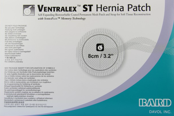
The VentralexTM ST Hernia Patch, invented in 2006, this patch is a self-expanding, non-absorbable, circular bilayer prosthesis made of an outer polypropylene monofilament mesh and an inner ePTFE surface (expanded polytetrafluroethylene). The solid, white ePTFE layer has sub-micronal porosity (<1), which minimises tissue attachment, and this part of the prosthesis can be applied safely against the bowel [4]. The patch has a memory recoil ring, which allows it to pop open, lay fat, and maintain its shape within the peritoneal cavity. The outer polypropylene layer of the mesh is designed to form straps, which facilitate placement, positioning and fixation of the device. The straps along with the patient’s own abdominal pressure keep the mesh in place [5]. The mesh used in this study is a large mesh of 8 x 8cm (3.2 x 3.2inch) which is available on schedule as per the Hospital administrative policy permit with no extra cost to the patient (Figure 1).
This study constituted a retrospective cohort analysis based on data extracted from a prospective database encompassing all patients who underwent repair for umbilical hernias in a tertiary care centre in India, between January 2022 and June 2023. A total of 112 patients underwent surgical intervention for umbilical and paraumbilical hernias, employing this specific patch.
Inclusion and Exclusion criteria
Inclusion criteria encompassed patients with umbilical and paraumbilical hernias with a diameter of less than 3cm, regardless of gender or age. Conversely, exclusion criteria encompassed pregnant individuals, patients with cirrhosis and ascites due to chronic liver disease, those undergoing emergency procedures for obstructed or strangulated hernias, cases of recurrent hernias, and instances of incisional hernias.
Patients were admitted one day prior to surgery following pre-anesthetic assessment and clearance on an outpatient basis. All patients received a single dose of intravenous antibiotics (1.2 g Inj. AmoxyClav) just prior to incision, based on the choice of regional or general anesthesia determined by the patient's fitness level. The surgical procedure involved a small infra-umbilical incision resembling a smile, with subsequent dissection of subcutaneous fat from the hernia sac. The sac was then dissected and opened to inspect its contents. A finger was introduced into the defect to clear the surrounding peritoneum, and the Ventralex™ hernia patch was placed within the peritoneal cavity. This placement was facilitated by grasping the outer borders with forceps or a blunt clamp, ensuring a minimum overlap of 2-3 cm on all sides (Figure 2).

Following the introduction, the positioning straps were delicately manipulated to ensure the patch adhered smoothly to the abdominal wall. As the positioning straps were drawn upwards, a finger or peanut sponge was introduced into the defect, creating a barrier between the anterior portion of the patch and the peritoneum. A methodical sweep was performed around the patch to verify that it lay flat, with special attention to ensuring no entrapment of bowel or omentum between the patch and the abdominal wall. To secure the mesh against the abdominal wall, the positioning straps were sutured to the margins of the fascia defect using polypropylene sutures on each side, followed by removal of any excess straps. In all cases, the anterior fascia was closed over the mesh using Polypropylene sutures to minimize the risk of mesh infection. Closure of both subcutaneous tissue and skin was accomplished using Polyglactin and Polyamide sutures, respectively. The surrounding subcutaneous tissue and dermis were infiltrated with a local anesthetic such as lignocaine or bupivacaine. Postoperatively, patients received 1 g of Inj. Paracetamol within the first hour immediately following the operation, and were subsequently transitioned to oral analgesic tablets. Discharge typically occurred on the following day, with scheduled follow-up appointments at 3 days, 3 weeks, 3 months, and 6 months in the outpatient department. Any occurrences at suture sites were documented and addressed as necessary.
Patient demographics, body mass index, operation duration, postoperative analgesic consumption, duration of hospitalization, postoperative complications, and instances of recurrence were systematically recorded.
Results
A total of 112 patients were operated with Ventral Umbilical Patch repair, of which 70 were male and 42 were female, with a mean age at presentation of 54.5 years (range 30-86). 35 patients were obese (BMI >30 kg/m2) with a mean BMI of 31 kg/m2 (Table 1).
| Males | Females | Total |
| 70 | 42 | 112 |
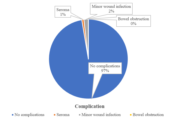
Median operative time was 33 min (range 25–60). 30 patients required general anaesthesia, 65 spinal anaesthesia and 17 regional anaesthesia. Patients had a short hospital stay post-surgery. 106 patients were discharged within 48 hours of surgery, out of which 68 patients were discharged within 24 hours of surgery and 38 patients in the next 24 hours. Only 6 patients had a prolonged hospital stay for more than 48 hours due to social reasons. Half of the patients resumed their normal daily activities within 3 days (Table 2).
| Duration | Number of patients |
| Less than 48 hrs. Within 24 hrs. 24-48 hrs. | 106 68 38 |
| More than 48 hrs. | 6 |
| Total | 112 |
Concerning postoperative analgesia, all the patients received 1 g of Inj. Paracetamol within an hour immediately post-op. 72 patients did not require any further analgesia. 40 patients required moderate postoperative analgesia (1 g of Inj. Paracetamol 8 hourly). None required opioids or another analgesic.
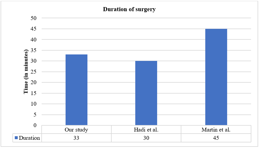
Patients were followed up for a duration of 6 months where we observed was one seroma and two minor wound infections (Figure 3). Skin stitches were removed to drain the seroma and the infections were treated successfully with antibiotics. No recurrence was observed in the follow up period.
Discussion
Adult umbilical hernias are a common surgical condition mainly encountered in the fifth and sixth decades of life. Open repair of umbilical hernias is considered the standard procedure for most surgeons. The mean duration of surgery in our study was 33 min. These results are comparable [6,7] if not better than other published series, even though most operations were performed by residents helped by senior surgeons (Figure 4).
The median hospitalization duration in our investigation was one day. Based on these findings, we posit that this prosthesis is highly suitable for day care surgical procedures. Additionally, it facilitates a rapid turnover rate, enabling early surgery following the initial outpatient department (OPD) visit. This translates to a reduced waiting period for the procedure compared to more developed healthcare systems, such as in the United States. Furthermore, it leads to shorter hospital stays and earlier return to work.
Our experience with the composite patch demonstrates an exceptionally low complication rate of 2.6%. Specifically, we encountered one case of seroma (0.89%) and two instances of minor wound infections (1.78%). These findings align favourably with existing literature, where the mean infection rate typically falls within the range of 2–4% [6,7]. The application of this technique yields several advantages, provided it is employed judiciously based on specific indications.
An intrinsic benefit of intraperitoneal mesh placement lies in the avoidance of extraperitoneal or prefascial dissection, consequently mitigating the risk of complications and obviating the need for wound drains. Additionally, the utilization of microporous ePTFE mesh in an intraperitoneal position diminishes tissue adherence, thereby reducing the likelihood of adhesion formation and the development of intestinal fistulas [8]. Notably, our series, consistent with the literature, did not report any instances of small bowel obstruction subsequent to Ventral Umbilical patch repair. Moreover, we observed no recurrences in our cohort. To further minimize the risk of recurrences, we ensured a minimum mesh overlap of 2-2.5cm in all directions.
Conclusion
We conclude that use of Ventral Umbilical patch in the repair of small (<3cm) umbilical and paraumbilical hernias by open method is a safe and efficient method, as it requires less duration of operation, less postoperative analgesia and faster recovery, shorter hospital stay, minimal post-operative complications, and no recurrence rate during 6 months of follow-up. As the patch was available on Scheduled items in our hospital, it proved to be cost-effective, with early resumption of work for the patient. From healthcare personnel point of view, the method is easy and reproducible with quick patient discharge at a Tertiary Health centre where ward occupancy for other emergency patients prevails.
Declarations
Ethical Approval and Consent to Participate
All procedures performed in this case series were conducted in accordance with institutional ethical standards and the principles of the Declaration of Helsinki. Ethical approval was obtained from the appropriate institutional review board where required. Written informed consent was obtained from all patients or their legal guardians prior to the procedures.
Consent for Publication
Written informed consent for publication of clinical details and images was obtained from the patients or their legal guardians. All identifying information has been anonymized to protect patient confidentiality.
Availability of Supporting Data
The data supporting the findings of this study are available from the corresponding author upon reasonable request, subject to institutional and ethical regulations.
Competing Interests
The authors declare that they have no competing interests related to this work.
Funding
This study received no specific grant from any funding agency in the public, commercial, or not-for-profit sectors.
Authors’ Contributions
All authors contributed substantially to the conception, data acquisition, analysis, drafting, and critical revision of the manuscript. All authors have read and approved the final version of the manuscript.