Introduction
Tracheal intubation is placement of a flexible plastic tube into the trachea to maintain an open airway, to facilitate ventilation of the lung and prevention of aspiration in critically ill or anesthetized patients. Direct laryngoscopy and passage of an endotracheal tube are noxious stimuli produces adverse hemodynamic responses, due to reflex sympathetic discharge caused by epipharyngeal and laryngopharyngeal stimulation. Increases plasma catecholamines concentration leads to hypertension, tachycardia and arrhythmia [1]. The magnitude of hemodynamic response is greater with increasing force and duration of laryngoscopy and endotracheal intubation [2]. The elevation of blood pressure typically starts within 5 sec of laryngoscopy, reached pick in 1-2 min and returns to control level within 5 min [3]. Transient hypertension and tachycardia are probably of no consequence in healthy individuals but either or both may be hazardous to those with hypertension, myocardial insufficiency and cerebrovascular disease. At least in such individuals there is a necessity to blunt this response. Reid and Brace in 1940 were the first to report the circulatory responses to laryngeal and tracheal stimulation in an anesthetized man [4]. Dexmedetomidine, a highly selective alfa-2 adrenergic agonist has sedative, anxiolytic, sympatholytic and analgesic effect. In addition, Dexmedetomidine has been shown to decrease perioperative catecholamines concentration and promote hemodynamic and adrenergic stability, as well as it decreases induction doses of intravenous (IV) anaesthetic and also decrease intra operative opioid and volatile anaesthetic requirements for maintenance of anaesthesia [5,6]. The purpose of our study was to investigate and compare the effects of two different doses of Dexmedetomidine on controlling hemodynamic response to induction of anaesthesia and tracheal intubation.
Material and Methods
The study protocol was approved by institutional ethical committee and written informed consent was obtained from all the patients. Sixty normotensive patients (ASA Grade I or II, age between 19-60 years, weight between 40-80 kg, Mallampati Grade I or II) were randomly allocated in two groups (30 patients each). Gr A received inj. Dexmedetomidine 1µg/kg in 10ml normal saline, IV over 10 min. While Gr B received inj. Dexmedetomidine 0.5µg/kg in normal saline, IV over 5 min.
In all the patients, IV line was secured and routine monitor like pulse oximeter (SpO2), non-invasive blood pressure (BP) and electrocardiogram were attached. Vital data like B.P. (SBP, DBP), heart rate (HR) and SpO2 were recorded as pre-induction parameters (basal) and pre-medication inj. Glycopyrrolate 0.2mg IV was given. According to group, inj. dexmedetomidine was given and patients were pre-oxygenated with 100% oxygen via facemask. Anaesthesia was induced with inj. thiopentone 5 mg/kg IV. Laryngoscopy and tracheal intubation were done after 3 min of inj. Vecuronium o.1mg/kg. Anaesthesia was maintained using 66% nitrous oxide, 33% oxygen, inj. Vecuronium and Sevoflurane 1%. Measurements of HR, SBP, DBP, and SpO2 were performed as basal, 60 sec after inj. Dexmedetomidine (t1), 60 sec after induction (t2), during laryngoscopy and intubation (t3), 60 sec after intubation (t4), 2 min after intubation (t5) and 5 min after intubation (t6).
Data were analysed with unpaired independent sample t-test to measure difference between the groups. P>0.05 was consider as not significant, P<0.05 was considered as significant and p<0.01 was considered as highly significant. The results were presented as means and Standard Deviation.
Observation and Results
Demographic profile
| Characteristics | Group A | Group B | P value | Signifi-cance | |
| Total no. of patients | 30 | 30 | |||
| Weight (kg) | 53.66±6.28 | 53.83±6.39 | 0.36 | N.S. | |
| Age (years) | 30.66±11.20 | 28.26±9.13 | 0.91 | N.S. | |
| Sex | Male | 15(50%) | 16(60%) | ||
| Female | 15(50%) | 14(40%) |
Demographic data of patients in both groups were comparable and not significant (N.S.) (P value> 0.05).
Hemodynamic changes
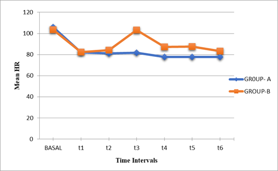
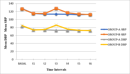
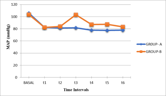
Statistical evaluation between groups shows that mean HR at pre-induction, t1 and t2 were statistically not significant (P value> 0.05). While mean HR at t3 (during laryngoscopy and intubation), t4, t5 and t6 were significantly higher in Gr B as compare to Gr A (P value<0.01) (Table II, Graph-I). At t3 and t4 there were increase in mean SBP, DBP and MAP in Gr B as compare to Gr A, which was statistically highly significant (P<0.01). The mean SBP, DBP and MAP at pre-induction, t1, t2, t5 and t6 were comparable in both groups (P>0.05) (Table III, Graph II, III). There were no significant changes in SpO2 in both groups (p>0.05) (Table IV).
None of patients in either group had any cardiovascular or respiratory side effects due to dexmedetomidine.
| Time | Pre-Induction (Basal) | t1 | t2 | t3 | t4 | t5 | t6 |
| Group- A | 105.9±10.28 | 82.17±5.58 | 81.20±7.51 | 81.77±8.68 | 77.70±8.63 | 77.60±8.71 | 77.77±8.59 |
| Group- B | 103.66±4.73 | 82.37±3.65 | 84.07±4.25 | 103.33±5.17 | 87.27±3.35 | 87.53±3.40 | 83.13±2.40 |
| P- Value | 0.28 | 0.87 | 0.074 | 0.000 | 0.000 | 0.000 | 0.002 |
| Signi-Finance | N.S. | N.S. | N.S. | H.S. | H.S. | H.S. | H.S. |
P value>0.05-statistically N.S, P value<0.05- statistically significant (S), P value<0.01- statistically highly significant (H.S.), Mean± SD.
| Time | MEAN SBP | S.G. | Mean DBP | S.G. | MAP | S.G. | |||
| Group A | Group B | Group A | Group B | Group A | Group B | ||||
| BASAL | 127.3±5.59 | 126.5±3.85 | N.S. | 83.46±2.09 | 85.00±3.80 | N.S. | 98.08±2.82 | 98.93±3.81 | N.S. |
| t1 | 115.00±3.95 | 114.87±3.62 | N.S. | 74.40±3.87 | 73.80±3.16 | N.S. | 87.93±3.83 | 87.49±3.18 | N.S. |
| t2 | 113.60±4.73 | 115.13±3.26 | N.S. | 74.27±4.32 | 75.00±3.95 | N.S. | 87.38±4.12 | 88.38±3.48 | N.S. |
| t3 | 113.53±4.25 | 127.47±2.34 | H.S. | 72.87±4.19 | 85.80±3.37 | H.S. | 86.42±3.52 | 99.69±2.85 | H.S. |
| t4 | 113.07±3.85 | 116.83±3.91 | H.S. | 73.07±4.29 | 76.13±2.77 | H.S. | 86.40±3.58 | 89.70±2.95 | H.S. |
| t5 | 112.53±4.06 | 113.40±3.64 | N.S. | 72.73±3.61 | 72.87±3.35 | N.S. | 86.00±3.53 | 86.38±3.37 | N.S. |
| t6 | 113.07±4.57 | 112.40±3.54 | N.S. | 72.53±3.19 | 72.27±3.00 | N.S. | 86.04±3.52 | 85.64±3.11 | N.S. |
S.G.- Significance
| Time | BASAL | t1 | t2 | t3 | t4 | t5 | t6 |
| Group-A | 99.03±0.18 | 99.13±0.34 | 99.07±0.36 | 99.13±0.43 | 99.23±0.43 | 99.27±0.45 | 99.10±0.30 |
| Group-B | 98.96±0.18 | 99.06±0.25 | 99.03±0.18 | 99.03±0.18 | 99.17±0.37 | 99.17±0.37 | 99.20±0.40 |
| P -Value | 0.162 | 0.398 | 0.656 | 0.250 | 0.527 | 0.356 | 0.286 |
| Signi-Finance | N.S. | N.S. | N.S. | N.S. | N.S. | N.S. | N.S. |
Discussion
Most of general anaesthetic procedures in modern anaesthetic practice are carried out with endotracheal intubation. Tracheal intubation and direct laryngoscopy are considered as the most critical event during administration of general anaesthesia as they provoke transient but marked sympathoadrenal response as hypertension and tachycardia [7]. These responses are transitory variable and may not be significant in otherwise normal individuals. But in patient with cardiovascular compromise like hypertension, IHD, Cerebrovascular disease and in patient with intracranial aneurysms, even these transient changes in haemodynamics can result in potentially harmful effects like left ventricular failure, pulmonary oedema, myocardial ischemia, ventricular dys-arrhythmias and cerebral haemorrhage [3,8]. This is by far the most important indication for attenuation of haemodynamic response to laryngoscopy and tracheal intubation during general anaesthesia.
Many methods like use of inhalational anaesthetic agents, lidocaine, opioids, direct acting vasodilator, calcium channel blockers, and β-blockers have been tried by various authors for blunting haemodynamic response to laryngoscopy and tracheal intubation [9-16]. But all such manoeuvres had their own limitations. For example, with opioids respiratory depression and chest wall rigidity were potential problems, use of halothane was associated with dys-arrhythmias, calcium channel blocker produced reflex tachycardia, direct acting vasodilator needed invasive haemodynamic monitoring and lidocaine did not give consistent result in blunting the haemodynamic responses to laryngoscopy and intubation [17-20]. Beta blockers are also one group of pharmacological agents employed for blunting haemodynamic response to laryngoscopy and intubation but they blunt HR response better than BP response [16].
The -2 Adrenoreceptor are involved in regulating the autonomic and cardiovascular systems, which are located on blood vessels, where they mediate vasoconstriction and on sympathetic terminals, they inhibit norepinephrine release. The -2 receptors are also located within the central nervous system (CNS) and their activation leads to sedation, a reduction of tonic levels of sympathetic outflow and an augmentation of cardiac-vagal activity. This can result in a decrease in HR and cardiac output. The use of α-2 agonists in the perioperative period has been associated with reduced anaesthetic requirements and attenuated HR and BP responses to stressful events. In addition, α-2 receptors within the spinal cord modulate pain pathways, providing some degree of analgesia [21-22]. The analgesic, sedative, anxiolytic, sympatholytic and blunting of exaggerated haemodynamic responses by administration of dexmedetomidine are being extensively studied and are mainly mediated by the activation of alpha-2 receptors located in the post-synaptic terminals in the CNS, which causes decreased neuronal activity and augmentation of the vagal activity [23]. Another α -2 agonist Clonidine, is also used by various authors to blunt the haemodynamic response for laryngoscopy and intubation [24].
Recently, the use of dexmedetomidine has been dramatically increased. This highly selective α-2 agonist has a set of unique effects that include titratable sedation, sympatholysis and analgesia without significant respiratory depression [25].
The present study was undertaken to know two different doses of dexmedetomidine (0.5µg/kg and 1µg/kg), in attenuation of haemodynamic response to laryngoscopy and tracheal intubation during general anaesthesia.
In our study after giving dexmedetomidine the mean HR, SBP, DBP and MAP were decreased in both groups. In Gr B, we have observed increases in mean SBP, DBP and MAP as compare to Gr A at t3 (during laryngoscopy and intubation) and t4, which was statistically highly significant. (p<0.01) At pre-induction, t2, t4, t5 and t6 mean SBP, DBP and MAP were comparable in between both groups (p>0.05). There were increases in mean HR in Gr B as compare to Gr A at t3 (during laryngoscopy and intubation), t4, t5 and t6 which was statistically highly significant (p<0.01), while mean HR at pre-induction, t1 and t2 were comparable in between groups (p>0.05). There was no significant change in Spo2 in both groups at all intervals (p>0.05).
In a similar study done by A. Esra Sağıroğlu et al., they found that after giving dexmedetomidine 1 μg.kg-1 (Gr I) and 0.5 μg.kg-1 (Gr II), SAP, DAP, MAP and HR levels were significantly lower at 60 sec after induction and 5 min after intubation than baseline levels [26]. But in Gr II, these levels were significantly higher at 60 sec after tracheal intubation (t3) while in Gr I, SAP, DAP, MAP were significantly lower in at t3 (p<0.01). They conclude that dexmedetomidine 1 μg.kg-1 is effective to suppress haemodynamic responses to tracheal intubation but dexmedetomidine 0.5 μg.kg-1 hasn’t the same effect. In this study any hypotension or bradycardia were not observed and any medical intervention was not required. Significant respiratory depression, apnoea, muscle rigidity or decrease in SpO2 were also not seen in any patient.
In study done by Ferdi Menda et al. they demonstrated that in dexmedetomidine 1µg/kg group, SAP, DAP and MAP were lower at all times in comparison to baseline values [27]. While in the placebo group, SAP, DAP and MAP decreased after the induction of general anaesthesia and five min after the intubation compared to baseline values. They also demonstrated that after induction of general anaesthesia, the drop in HR was higher in dexmedetomidine 1µg/kg group than placebo group. Whereas, 1 min after intubation HR increased significantly in placebo group, while it decreased in dexmedetomidine group.
Tanyoung Pipanmekaporn et al. demonstrated that during intubation and 10 min afterward (T1-T10), the mean HR, SBP, DBP and MAP in the control Gr were significantly higher than those in the dexmedetomidine 0.7µg/kg Gr throughout the study period except at T1 [28].
In similar study by Varshali M. Keniya et al., the increase in SBP after intubation was 40% in control Gr as compared to 8% in dexmedetomidine 1µg/kg Gr (P=0.00) [29]. While the increase in DBP after intubation was 25% in control Gr as compared to 11% in dexmedetomidine 1µg/kg Gr (P=0.001). They also found that 1 µg/kg of dexmedetomidine Gr received more treatments for bradycardia than patients in the control Gr. Arpita Laha et al. demonstrated that pretreatment with dexmedetomidine 1µg/kg attenuated but did not totally abolish cardiovascular and catecholamine responses to tracheal intubation after induction of anaesthesia [30]. They found increase in HR, SBP and DBP after intubation and at 1, 2, 3 and 5 min in both dexmedetomidine and control Gr, but this rise was significantly less with dexmedetomidine. Bajawa S J S et al. found that the dose of 1 µg/kg of dexmedetomidine attenuate but did not completely obtund the haemodynamic responses to laryngoscopy and tracheal intubation [31]. In a similar study done by Jeong Han Lee et al., they observed that in dexmedetomidine 1 μg/kg Gr, the increase in SBP and DBP due to tracheal intubation were significantly lower than that of control Gr [32].
Scheinin et al reported that the use of α-2 agonist leads to bradycardia [33]. Basar et al had also reported that the incidence of bradycardia after single dose of 0.5µg/kg of dexmedetomidine was about 5% [34].
In our study, there was no significant changes in Spo2 in both groups at all intervals. Similar to our study, Ebert et al. didn’t observe any apnoea, airway obstruction or hypoxemia with bolus doses of dexmedetomidine [11]. They reported that depression of respiration may be seen due to deep sedation, for the reason that α-2 adrenergic agonists don’t have active role on the respiration centres. In contrary to our study, Belleville et al found that dexmedetomidine, given as bolus dose of 1-2 µgkg-1, intravenously within two minutes, causes irregular ventilation and apnoea episodes [21].
Limitations
There were three important limitations regarding this study.
-
Not assessed the quality of intubation.
-
We had not measure the plasma catecholamines levels.
-
We have not studied extubation response, postoperative sedation and hemodynamic variations.
Abbreviations
TEMP: Temperature
HR: Heart Rate
BP: Blood Pressure
SBP: Systolic Blood Pressure
DBP: Diastolic Blood Pressure
MAP: Mean Arterial Pressure
SPO2: Peripheral Oxygen Saturation
RS: Respiratory System
CVS: Cardio Vascular System
CNS: Central Nervous System
MPG: Mallampatti Grading
OC: Oral Cavity
HB: Haemoglobin
RBS: Random Blood Sugar
CXR: Chest X-Ray
ECG: Electrocardiogram
IV: Intravenous
WT: Weight
ASA grade: American Society of Anaesthesiologists Grading
mean ±SD: Mean± Standard Deviation
NS: non-significant
S: Significant
HS: Highly significant
Min: Minute
Sec: Second
Declarations
Conflict of interest
Authors declares no conflicts of interest.
Funding/ financial support
No funding sources.
Ethical Clearance
The study was approved by the Institutional Ethics Committee.
Data Availability
All data available on corresponding author upon responsible request.