Introduction
Traumatic brachial plexus injuries are clinically significant because of their potential to cause permanent neurological deficits, functional disability, and chronic neuropathic pain [1]. These injuries predominantly affect neonates and young adults [1]. In the neonatal population, they are most commonly associated with shoulder dystocia during vaginal delivery, whereas in adults they primarily result from blunt trauma secondary to motor vehicle accidents, particularly motorcycle collisions, and falls from height. Penetrating trauma accounts for a smaller proportion of cases [2].
From a clinical perspective, brachial plexus injuries are classified according to the severity, anatomical location, and mechanism of injury [1][4]. Based on injury severity, they range from neurapraxia, characterized by focal demyelination without axonal disruption, to neurotmesis, which represents complete nerve transection [1]. Anatomically, these injuries are categorized as preganglionic, involving the spinal nerve roots proximal to the dorsal root ganglion, or postganglionic, affecting neural structures distal to the dorsal root ganglion. This distinction is of paramount importance because preganglionic injuries carry a substantially poorer prognosis due to the inability to achieve direct surgical repair [4].
The mechanism of injury also provides valuable diagnostic and prognostic information. Blunt trauma typically results from traction forces that may lead to nerve root avulsion, whereas penetrating injuries produce focal nerve lacerations. The position of the upper extremity at the time of trauma influences the level of injury: forced shoulder abduction is more commonly associated with upper plexus injuries (C5–C6), whereas arm flexion is more likely to involve the lower plexus (C8-T1) [4].
Clinically, brachial plexus injuries are grouped into three major syndromes: upper brachial plexus palsy (Erb palsy), lower brachial plexus palsy (Dejerine–Klumpke palsy), and complete brachial plexus palsy. The latter represents the most severe presentation and is most commonly associated with extensive nerve root avulsion or complete plexus disruption.
Accurate evaluation requires a thorough clinical history, comprehensive physical examination, and early imaging assessment to determine the extent of neural injury and to prevent irreversible complications such as muscle denervation and atrophy. Treatment is determined by the severity of the lesion. Low-grade injuries are generally managed conservatively with rehabilitation and physical therapy, whereas high-grade injuries, particularly postganglionic lesions, often require surgical intervention, including primary nerve repair, nerve grafting, or nerve transfer procedures [6].
Case Presentation
A 21-year-old right-handed woman with a history of occasional active smoking and previous left ankle osteosynthesis presented to the emergency department following a high-energy motorcycle accident. The patient sustained blunt polytrauma associated with transient loss of consciousness. Upon regaining consciousness, she exhibited complete monoplegia and anesthesia of the left upper extremity, accompanied by severe neuropathic pain predominantly involving the palmar aspect of the hand.
Initial physical examination revealed complete paralysis of the left upper limb with profound sensory loss involving the C5–T1 dermatomes. Examination of the trapezius muscle demonstrated absence of active contraction, initially attributed to pain-related limitation secondary to associated injuries of the shoulder girdle. Cervical computed tomography (CT) demonstrated displaced comminuted fractures of the left transverse processes, transverse foramina, and anterior and posterior tubercles extending from C4 to C7, as well as widening of the atlanto-odontoid joint. Conventional radiographs confirmed concomitant fractures of the left clavicle and scapula.
Magnetic resonance imaging (MRI) demonstrated multiple cerebrospinal fluid–intensity cystic collections consistent with post-traumatic pseudomeningoceles (Figure 1), located within the left neural foramina from C5 through T1, with caudal extension to the T2 level (Figure 2). These findings were associated with nonvisualization of the corresponding nerve roots, highly suggestive of nerve root avulsion. The left C5, C6, C7, C8, and T1 preganglionic nerve roots were not identified, with complete loss of the normal anatomical continuity of the ipsilateral brachial plexus (Figure 3).
Increased signal intensity on fluid-sensitive sequences was observed within the muscles of the left shoulder girdle, consistent with acute denervation edema. The trunks, divisions, cords, and terminal branches of the contralateral brachial plexus demonstrated normal morphology and signal intensity. No abnormalities were identified within the interscalene triangle, costoclavicular space, or retropectoralis minor space. No additional soft-tissue collections or masses were detected. Several cervical lymph nodes with reactive morphology were observed, without imaging features suggestive of pathological lymphadenopathy.
High-resolution MR neurography with three-dimensional reconstructions was subsequently performed for detailed assessment of the peripheral nervous system. The examination demonstrated multiple cerebrospinal fluid–intensity cystic lesions adjacent to the preganglionic dorsal nerve roots (C5-T1) and extending to the infraganglionic segments (C4-T2) (Figure 1), consistent with post-traumatic pseudomeningoceles. Complete absence of root continuity and nonvisualization of the proximal nerve root stumps from C5 through T1 (Figure 2) confirmed the diagnosis of complete preganglionic and postganglionic avulsion of the left brachial plexus. Associated imaging findings included acute denervation changes and trophic muscle replacement involving the left shoulder girdle musculature. Follow-up electromyography confirmed complete denervation without evidence of reinnervation or preserved functional continuity.
During follow-up in January 2026, the patient sustained a fall while using public transportation, resulting in worsening left shoulder pain. Repeat imaging excluded new major neural or osseous injuries but demonstrated acromioclavicular joint dislocation with widening of the joint space and associated periarticular soft-tissue edema.
Given the complete nerve root avulsion demonstrated by MR neurography, direct neurorrhaphy and autologous nerve grafting were deemed unfeasible because of the absence of viable proximal cervical nerve root stumps. The patient was therefore referred to the Hand Surgery and Orthopedic Trauma Unit for surgical planning, where reconstruction with extraplexal nerve transfers and pedicled muscle transfer procedures was indicated.
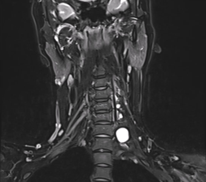
A well-defined hyperintense cystic lesion is identified in the left supraclavicular region, adjacent to the expected course of the brachial plexus, with signal intensity similar to cerebrospinal fluid, consistent with a post-traumatic pseudomeningocele
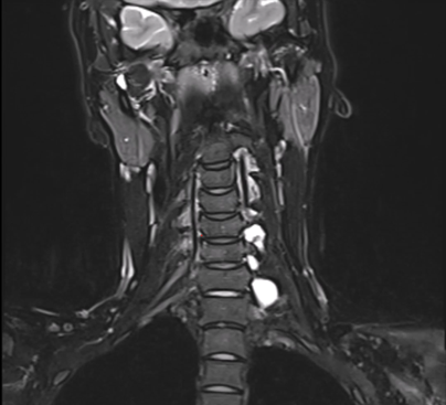
Multiple cerebrospinal fluid–intensity pseudomeningoceles are demonstrated extending from the left C5 to T1 neural foramina, with caudal extension toward T2. The corresponding nerve roots are not visualized, consistent with multilevel preganglionic nerve root avulsion
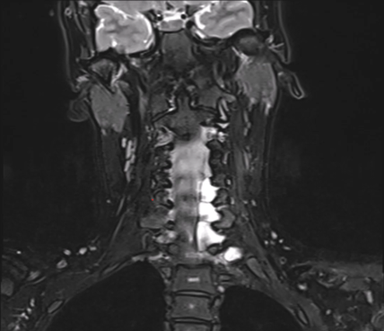
Multiple left-sided cerebrospinal fluid–intensity pseudo meningoceles extending from C5 through T1 are associated with nonvisualization of the corresponding nerve roots, indicating complete preganglionic brachial plexus avulsion. Associated denervation edema of the left shoulder girdle musculature is also evident.
Discussion
Traumatic brachial plexus injuries represent one of the most challenging entities in musculoskeletal radiology and neuroradiology because of the intricate anatomy of the brachial plexus and the need for precise localization of neural injury. Although clinical examination and electrodiagnostic studies remain essential components of the diagnostic workup, magnetic resonance imaging (MRI) has become the cornerstone of evaluation by providing direct visualization of the nerve roots, trunks, and adjacent soft tissues. This comprehensive assessment enables differentiation between potentially repairable injuries and those with limited potential for spontaneous recovery, thereby directly influencing surgical planning and functional prognosis [1][3][5].
In adults, most brachial plexus injuries result from high-energy trauma, particularly motorcycle accidents, in which traction forces applied to the neck and shoulder produce a spectrum of injuries ranging from nerve stretching to complete nerve root avulsion. The present case illustrates this classic mechanism, as the patient sustained high-energy blunt trauma associated with cervical spine and shoulder girdle fractures, followed by immediate complete monoplegia and anesthesia of the left upper extremity. Correlation between the mechanism of injury, the initial neurological deficit, and the imaging findings is fundamental for the early recognition of severe preganglionic brachial plexus injury [2][4].
One of the primary objectives of imaging evaluation is to distinguish preganglionic from postganglionic injuries. This distinction has major clinical implications because nerve root avulsion represents complete separation of the nerve root from the spinal cord, precluding direct nerve repair or primary nerve grafting. In contrast, postganglionic injuries preserve proximal neural continuity and may be amenable to a variety of reconstructive techniques (Table 1). In this setting, MRI offers significant advantages over other imaging modalities by simultaneously assessing nerve root integrity, brachial plexus continuity, and secondary muscle denervation changes [1][4][5].
| Seddon | Sunderland | Lesion | MRI Findings |
| Neuropraxia | I | Demyelination | T2 hyperintensity 24 h after trauma without muscle denervation |
| Axonotmesis | II | Axonal disruption | T2 hyperintensity with prominent fascicles, with or without neuroma |
| III | Axonal and endoneurial disruption | Muscle denervation | |
| IV | Axonal, endoneurial and perineurial disruption | Heterogeneous signal with neuroma in continuity; muscle denervation | |
| Neurotmesis | V | Complete nerve disruption | Nerve discontinuity and terminal neuroma; muscle denervation |
In the present case, the identification of multiple pseudomeningoceles extending from C5 through T1 represented one of the most significant imaging findings. These cerebrospinal fluid collections result from dural disruption secondary to nerve root avulsion and, although not entirely pathognomonic, are highly associated with traumatic preganglionic injuries when accompanied by nonvisualization of the corresponding nerve roots. The multilevel distribution observed in this patient, together with caudal extension to the T2 level, reflected a high-energy traction mechanism and strongly supported the diagnosis of multiple nerve root avulsions [1][3][5].
In addition to demonstrating multiple pseudomeningoceles, MRI revealed complete nonvisualization of the left C5, C6, C7, C8, and T1 nerve roots. This finding represents one of the most reliable direct imaging signs of preganglionic nerve root avulsion and has high specificity when identified using high-resolution MR neurography protocols. Recent advances in MRI, including fat-suppressed T2-weighted sequences, isotropic three-dimensional acquisitions, and multiplanar reconstructions, have substantially improved diagnostic sensitivity by providing more accurate anatomical depiction of the brachial plexus and facilitating preoperative planning [1][5].
Another noteworthy finding in this case was the presence of denervation edema involving the left shoulder girdle musculature. These imaging changes represent an early pathophysiological response to interruption of neural input and appear as diffuse hyperintensity on fluid-sensitive sequences, preceding fatty infiltration and irreversible muscle atrophy. Identification of these abnormalities complements the assessment of nerve root integrity and provides valuable indirect evidence of injury severity, particularly when correlated with complete neurological deficit and electrodiagnostic findings [1][3][5].
MR neurography has significantly advanced the evaluation of traumatic brachial plexus injuries by enabling direct visualization of neural architecture with substantially higher spatial resolution than conventional MRI sequences. Three-dimensional fat-suppressed T2-weighted acquisitions combined with multiplanar reconstructions and maximum intensity projection (MIP) images facilitate assessment of the continuity of the nerve roots, trunks, and fascicles, thereby increasing sensitivity for detecting nerve root avulsions, neuromas, and traction-related injuries. In the present case, MR neurography confirmed the complete absence of continuity of the C5-T1 nerve roots, providing unequivocal anatomical confirmation of complete preganglionic injury and complementing the findings observed on conventional MRI [1][5].
Although MRI is currently the imaging modality of choice for evaluating the brachial plexus, comprehensive assessment of polytrauma patients requires a multimodality approach. Computed tomography plays a pivotal role in identifying fractures of the transverse processes, transverse foramina, and other cervical osseous injuries that increase the likelihood of associated nerve root injury. Likewise, electromyography provides functional information regarding the degree of denervation and the potential for reinnervation, although electrodiagnostic abnormalities generally become evident only several weeks after trauma. In the present patient, the concordance among the clinical findings, computed tomography, MRI, and electromyographic evaluation established the diagnosis of complete left brachial plexus avulsion with a high degree of diagnostic confidence [2][4][6].
From the radiologist's perspective, recognizing both the direct and indirect imaging signs of nerve root avulsion has immediate therapeutic implications. The combination of multiple pseudomeningoceles, nonvisualization of the corresponding nerve roots, and denervation-related muscle changes is highly suggestive of complete preganglionic injury, allowing differentiation of patients who are candidates for reconstruction with extraplexal nerve transfers from those in whom conventional nerve repair techniques may still be feasible. Consequently, the radiology report should systematically describe the level of injury, neural continuity, the presence of pseudomeningoceles, and associated muscle denervation, thereby providing essential information for multidisciplinary surgical planning [1][4][5].
Conclusion
Traumatic brachial plexus injuries require accurate imaging assessment because of their major prognostic and therapeutic implications. High-resolution magnetic resonance imaging, complemented by MR neurography, is the imaging modality of choice for localizing the site of injury, distinguishing preganglionic from postganglionic involvement, and identifying the direct and indirect signs of nerve root avulsion. This case highlights the pivotal role of MRI in the comprehensive evaluation of complete traumatic brachial plexus injury, providing essential information for surgical planning and multidisciplinary patient management.
Declarations
Conflicts of Interest
The authors declare that they have no conflicts of interest
Ethical Considerations
As this is a review article, no ethical approval was needed.
Funding Statement
None
Consent to participate
Not applicable as this is a review article
Acknowledgments
None