Introduction
Osteoarticular infections can affect any joint. The intra-articular administration of injectable medications represents a recognized risk factor [1]. This infection is termed septic arthritis, which may lead to joint destruction as well as short- or long-term complications [2].
Joint fluid aspiration is a valuable diagnostic technique for septic arthritis, yielding synovial fluid for biochemical analysis, direct Gram stain, and culture. Additional diagnostic utility includes leukocyte count with a predominance of polymorphonuclear neutrophils, reduced glucose levels, and elevated protein values (>3 mg/dL) [3].
Empiric antibiotic therapy must be initiated as promptly as possible. Although obtaining culture samples is critical for diagnosis, if the patient's clinical condition is unstable or critical, antibiotic therapy should be started immediately without awaiting sample collection [2].
In addition to antibiotic therapy, surgical debridement is essential. In this patient, the indications for surgical intervention were the presence of an intra-articular fluid collection, a large chest wall collection along the midaxillary line communicating with the glenohumeral joint, and acute osteomielitis [3].
Antibiotic therapy depends on pathogen epidemiology, with treatment response monitored through laboratory studies until results allow directing targeted therapy against the definitive etiologic agent [2]. A review conducted by Huang Y, et al., using a database from Taiwan published in 2020 reported that septic monoarthritis has an increasing incidence [4]. Incidence varies from one country to another and is influenced by ethnicity, age, and socioeconomic status [5].
Septic arthritis has a prevalence of 6 per 100,000 cases, which increases among patients with rheumatoid arthritis. It carries a mortality rate of up to 15% in patients with single-joint involvement (monoarticular) and increases up to 50% if more than one joint is affected (polyarthritis) [6]. The most significant risk factor is the placement of prosthetic material, followed by minimally invasive procedures such as intra-articular injections, arthroscopies, and bone punctures for various indications. Conditions causing immunosuppression include advanced age, diabetes, HIV, intravenous drug use, steroids, and immunosuppressive medications [7].
In adults, the most common organisms are Staphylococcus aureus, coagulase-negative Staphylococcus, Streptococcus, Pseudomonas, and other Gram-negative bacteria [8]. Sepsis is a clinical condition characterized by organ complications with a high mortality rate [9]. The onset of sepsis is driven by systemic vasodilation, which leads to decreased organ blood flow and cellular hypoxia [10].
At the hepatic level, sepsis causes alterations in bile metabolism with a cholestatic pattern [10]. This pattern manifests as jaundice due to an increase in direct bilirubin, along with liver enzyme alterations [10]. A mild elevation of alkaline phosphatase has also been described [10]. The elevation of bilirubin is explained by hepatocyte injury caused by circulating endogenous toxins, the inflammatory process, hypoxia, hepatitis, decreased hepatic perfusion, ischemia, as well as metabolic and genetic alterations [11]. Jaundice presents in patients with serum bilirubin levels exceeding 2.5 mg/dL [11].
Hepatic impairment serves as a marker of severity and mortality in septic patients with cholestasis, enabling timely diagnosis and treatment [12]. The objective of this case report was to describe a case of sepsis secondary to intra-articular shoulder injection in a middle-aged adult patient, including its clinical evolution and management. Additionally, it aimed to emphasize the importance of doctor-patient communication when making clinical management decisions.
Case presentation
A 55-year-old male patient, resident of a rural area, married, mechanic, retired military personnel, blood type O Rh-positive. Additionally, he denied previous blood transfusions and stated he would accept them if necessary. He had a recently diagnosed lumbar hernia and partial rupture of the right shoulder rotator cuff (approximately one month prior to hospitalization). He reported no significant surgical or family history. He also reported an allergy to nonsteroidal anti-inflammatory drugs (NSAIDs), which was confirmed with an allergy test during hospitalization.
The patient presented to the emergency department of the Specialty Hospital from the outpatient clinic (February 2024) due to generalized jaundice, epigastric abdominal pain accompanied by vomiting, and unquantified fever predominantly at night, with an onset of 3 days prior to admission. He reported several previous visits to the emergency department of the same health institution (specialty hospital) on multiple occasions over approximately 5 weeks prior to admission. The reason for consultation was pain and functional limitation of the right shoulder and lumbosacral region.
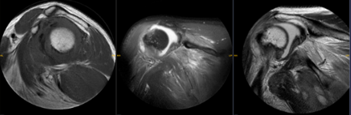
He was diagnosed ultrasonographically (Figure 1) with partial rupture of the rotator cuff muscles, subacromial and subdeltoid bursitis, as well as tendinitis and tenosynovitis of the long head of the biceps tendon of the right shoulder. This was confirmed by a magnetic resonance imaging (MRI) study (Figure 2). Following diagnosis, a single intra-articular glenohumeral injection was performed. He received outpatient treatment based on tapentadol, tramadol, celecoxib, etoricoxib, and ketorolac, with mild improvement of the clinical picture.
The patient showed no neurological deterioration, was afebrile, and presented with an icteric tinge (Kramer grade 2), an equivocal Murphy's sign, and no signs of peritoneal irritation. The upper limbs were asymmetrical, with a dropped right shoulder and restricted range of motion. Positive Apley, Jobe, and Neer signs were observed. The neurovascular examination was unremarkable, except for moderate edema.
Laboratory tests revealed leukocytosis with neutrophilia, hyperbilirubinemia predominantly driven by direct bilirubin, and elevated GGT and alkaline phosphatase levels. Due to the laboratory findings described in Table 1. Evaluations by General Surgery and Gastroenterology were warranted, both of which ruled out surgical pathology and biliary tract disorders via magnetic resonance cholangiopancreatography and ultrasonography
| Parameter | Admission (Feb 2024) | Discharge (Jun 2024) | Trend |
| White blood cells (×10³/µL) | 23.96 | 5.89 | ↓ Improved |
| Hemoglobin (g/dL) | 13.1 | 10.9 | ↓ |
| Platelets (×10³/µL) | 234 | 240 | Stable |
| Glucose (mg/dL) | 146.35 | 105.92 | ↓ Improved |
| Urea (mg/dL) | 58.10 | 27.88 | ↓ Improved |
| Creatinine (mg/dL) | 1.00 | 0.73 | ↓ Improved |
| AST (U/L) | 52.4 | 16.7 | ↓ Normalized |
| ALT (U/L) | 38.2 | 16.2 | ↓ Normalized |
| C-reactive protein (mg/L) | 28.76 | 1.12 | ↓ Markedly improved |
During subsequent clinical history taking, the presence of indoor pets was reported; therefore, the patient was admitted to Internal Medicine to investigate a potential zoonotic etiology (he keeps 4 dogs and 1 cat indoors, all with complete vaccination schedules). Arthrocentesis of the right glenohumeral joint was performed, yielding purulent synovial fluid. Serial surgical debridements were performed on five occasions at the right glenohumeral joint by Orthopedics and Cardiothoracic Surgery, with placement of a vacuum-assisted closure (VAC) system for 18 days due to joint collection and a chest wall abscess, without complications.
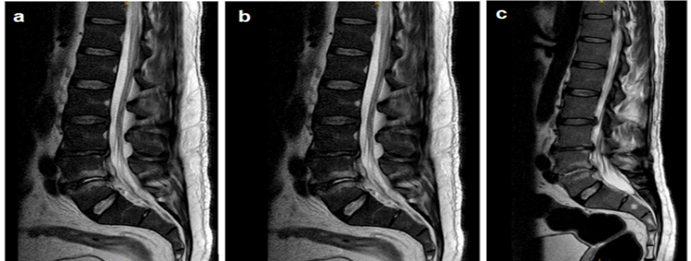
From admission, the patient reported severe lumbosacral pain, requiring pain management therapy with opioids via continuous infusion pump. A lumbar spine magnetic resonance imaging (MRI) study was performed, which reported spondylodiscitis and an apparent collection at the L4-L5 level (Figure 2-a). Due to the findings in the lumbar region, he was kept under observation by Spine Orthopedics and Neurosurgery due to suspicion of disseminated infection, pending therapeutic response to medical management.
The patient received intravenous antibiotic therapy consisting of piperacillin/tazobactam 4.5 g every 6 hours (PIP/TAZ) combined with vancomycin 1 g every 12 hours for 11 days. Cultures isolated oxacillin-susceptible Staphylococcus aureus, leading to a regimen change to intravenous oxacillin 2 g every 8 hours for the remainder of his hospital stay. Follow-up lumbar spine MRIs were performed to monitor the imaging finding suggestive of a collection at L4-L5 (Figures 2-a, 2-b, 2-c), which persisted until discharge. Neurosurgery decided to manage the case with outpatient imaging evaluations upon completion of the proposed antibiotic regimen. The surgical intervention on the shoulder warranted follow-up by Orthopedics. Antibiotic management was directed by Internal Medicine with subsequent follow-up visits.
Upon hospital discharge, completion of an outpatient antibiotic regimen for a total duration of 3 to 6 months was recommended. The prescribed treatment was clindamycin 300 mg every 8 hours until outpatient consultation. Outpatient follow-up visits were scheduled with all consulting specialties. During follow-up, C-reactive protein (CRP) levels were similar to those at discharge (Table 2); therefore, the dosing interval was shortened, prescribing clindamycin 300 mg every 6 hours with a planned follow-up in one month.
| Parameter | Feb 26, 2024 | Mar 15, 2024 | Change / Outcome |
| Leukocytes | 5.89 | 6.50 | Stable |
| Neutrophils | 3.99 | 4.05 | Stable |
| Lymphocytes | 1.35 | 1.84 | Improved |
| Hemoglobin | 10.90 | 12.90 | Improved |
| Hematocrit | 33.40 | 40.70 | Improved |
| Platelets | 240.00 | 220.00 | Stable |
| Glucose | 105.92 | 88.72 | Improved |
| Urea | 27.88 | 32.89 | Stable |
| Creatinine | 0.73 | 0.80 | Stable |
| Total Bilirubin | 0.87 | 0.95 | Stable |
| Direct Bilirubin | 0.65 | 0.49 | Improved |
| Indirect Bilirubin | 0.22 | 0.46 | Stable |
| AST (U/L) | 16.67 | 17.30 | Stable |
| ALT (U/L) | 16.24 | 15.80 | Stable |
| CRP | 1.12 | 0.37 | Improved |
ALT: alanine aminotransferase; AST: aspartate aminotransferase; CRP: C-reactive protein.
Discussion
Sepsis is defined as organ dysfunction with a high potential to be life-threatening as a consequence of the host response to an infectious process [13]. Sepsis of osteoarticular origin depends on the patient's age and the specific joint affected. In adult patients, it depends on medical history, immune status, and drug use, among other factors [8]. Hyperbilirubinemia may represent a manifestation of the septic process. A hyperbilirubinemic state with mild elevation above normal values can be beneficial due to the antioxidant, anti-inflammatory, immunomodulatory, and anti-toxicity properties of bilirubin. However, when these values increase above 3 mg/dL, damage from oxidative stress, inflammation, and apoptosis is enhanced [14].
In this case report, the history of joint puncture caused an infection leading to pyogenic arthritis, which subsequently progressed to sepsis. The clinical picture was caused by oxacillin-susceptible S. aureus, which was managed in an inpatient setting. Broad-spectrum antibiotics were initially administered; following culture results, targeted therapy was initiated and continued as outpatient treatment after hospital discharge.
Vassallo and McBride, in their respective studies published in 2020, stated that the knee is the most frequently affected joint1 [5]. The second most common joint affected varies depending on the population and underlying risk factors. The shoulder joint accounts for between 5% and 10% of single-joint septic arthritis cases [15].
Diagnosis is made through clinical manifestations, laboratory testing, imaging studies, and direct inspection. The clinical picture is characterized by inflammatory signs accompanied by functional limitation [16]. Laboratory studies reveal leukocytosis, neutrophilia, and elevated acute-phase reactants (C-reactive protein, erythrocyte sedimentation rate, elevated procalcitonin). Blood cultures should be performed, in addition to analyzing synovial fluid obtained via arthrocentesis, arthroscopy, or during surgical debridement [2].
Alterations in liver markers during sepsis can be attributed to medication use, hemolysis, inflammatory processes, or altered bilirubin metabolism [12]. Prompt diagnosis and treatment of sepsis serve to preserve patient well-being. Furthermore, hyperbilirubinemia in septic patients can act as an indicator of complications, a marker of severity, and a prognostic factor [11]. Imaging studies are useful for delineating the extent of infection, as well as muscular and osseous involvement. The most commonly utilized imaging modalities include plain radiography, musculoskeletal ultrasonography, computed tomography, and magnetic resonance imaging, depending on the anatomical location of the lesion or suspicion of dissemination [16].
Infection caused by methicillin-resistant S. aureus carries a higher rate of complications. The most frequent complications reported in the Mexican Clinical Practice Guideline for the Diagnosis and Treatment of Septic Arthritis in Children and Adults are deep vein thrombosis, osteomyelitis, pulmonary embolism, and multiple organ failure. Joint-specific complications also exist; at the hip level, gait abnormalities, chronic pain, limb length discrepancy, and loss of the femoral head and neck may occur [17]. In septic arthritis, drainage of the intra-articular collection is essential. Additionally, surgical debridement should be performed using a suction drainage system if available at the healthcare facility. Revision procedures and additional debridements should be carried out if clinical or laboratory signs of localized or disseminated infection persist [18].
Initially, immobilization of the affected joint is implemented to manage pain [19]. Joint activity and movement should subsequently be resumed, as prolonged joint immobilization can lead to structural damage. Early mobilization of the affected limb promotes proper synovial fluid distribution, thereby protecting the joint capsule and cartilage [17]. The initiation of physical therapy allows for the recovery of mobility, muscle strength, and muscle tone [18]. Surgical drainage may be delayed for up to 24 hours only if the patient has undergone prior invasive or minimally invasive surgical treatment with irrigation, if empirical antibiotic therapy has already been initiated, or in the absence of a specialist available to perform the procedure [2].
The selection of empiric antibiotic therapy depends on local resistance patterns, patient risk factors, and comorbidities. There is no clear consensus regarding antibiotic administration in this condition; however, current literature consistently recommends clinical and laboratory reassessment to adjust or switch antibiotic regimens [13].
An indicator for modifying antibiotic therapy is the culture result, which identifies the specific antimicrobial needed to target the causative pathogen18. Jeong Joo et al. conducted a retrospective review in 2023 including 137 patients who met the inclusion criteria. With a follow-up period exceeding two years, they observed a relapse rate of 9.5% within the first 30 days following treatment completion, which was significantly higher among patients who received antibiotic therapy for four weeks or less [20].
The total duration of antibiotic therapy depends on infection severity, the causative microorganism, and the clinical and laboratory response demonstrated by the patient during outpatient follow-up. Table 3 shows the frequency of bacterial pathogens isolated from cultures in the Orthopedics and Traumatology service, based on the microbiology database of the Specialty Hospital of the Armed Forces HE-1. Escherichia coli, Staphylococcus aureus, and Enterococcus faecalis were identified as the primary isolated microorganisms, collectively accounting for more than 50% of positive cultures submitted by this department.
| Microorganism | Frequency (n) | Percentage (%) |
| Escherichia coli | 77 | 17.70% |
| Staphylococcus aureus | 75 | 17.24% |
| Enterococcus faecalis | 68 | 15.63% |
| Pseudomonas aeruginosa | 54 | 12.41% |
| Staphylococcus epidermidis | 51 | 11.72% |
| Klebsiella pneumoniae | 28 | 6.44% |
| Enterobacter cloacae | 24 | 5.52% |
| Candida albicans | 18 | 4.14% |
| Others | 40 | 9.20% |
| Total | 435 | 100.00% |
Conclusion
Invasive intra-articular interventions represent a critical risk factor for pyogenic joint infection and subsequent systemic dissemination. This case underscores that a meticulous medical history specifically identifying prior minimally invasive procedures is essential to prevent diagnostic delays, avoid misattribution to intra-abdominal or biliary etiologies despite cholestatic laboratory presentations, and initiate timely targeted surgical and antimicrobial therapy.
The successful resolution of complex septic monoarthritis complicated by extra-articular collections and secondary spondylodiscitis relies on prompt joint debridement, dynamic imaging monitoring, and prolonged targeted antibiotic therapy. Furthermore, persistent or fluctuating acute-phase reactants such as CRP during outpatient surveillance necessitate agile antimicrobial dose adjustments and close multidisciplinary follow-up to prevent clinical relapse.
Declarations
Conflicts of Interest
The authors declare that they have no conflicts of interest
Ethical Considerations
As this is a review article, no ethical approval was needed.
Funding Statement
None
Consent to participate
Not applicable as this is a review article
Acknowledgments
None