Introduction
Mayer–Rokitansky–Küster–Hauser Syndrome (MRKH) is a congenital anomaly in the embryonic development of the genital tract, presenting with primary amenorrhoea in the presence of phenotypically female secondary sexual characteristics [1]. The Mayer-Rokitansky-Küster-Hauser (MRKH) spectrum refers to congenital anomalies of the female genital tract characterized by uterovaginal agenesis with normal secondary sexual characteristics [2]. It has a prevalence of approximately 1 in 4,500–5,000 female births. The condition is classified into Type I (isolated uterovaginal agenesis) and Type II (associated anomalies, commonly renal and skeletal) [3]. MRI is considered the imaging modality of choice to delineate uterine remnants, vaginal agenesis, and associated anomalies [4]. This case series presents five patients with diverse phenotypic presentations of MRKH spectrum disorders, emphasizing the importance of MRI in diagnosis and management.
This case series was conducted at Basaweshwar hospital in Karnataka, India. Patients presenting with primary amenorrhea were evaluated clinically and referred for MRI. MRI protocol included multiplanar, multi-sequence scans (T1, T2, SPAIR, DWI). Parameters assessed included uterine morphology, cervix and vaginal canal, ovarian morphology, renal anomalies, and associated pelvic findings [3].
Case 1
15 years, Indian residing in Kalaburagi, Karnataka with chief complaints of primary amenorrhea with normal female phenotype, no significant past history or similar complains in family. The patient did take medical hormonal therapy previously, but did not attain menarche and was suggested MRI Abdomen & Pelvis to rule out congenital malformations of mullerian tract.
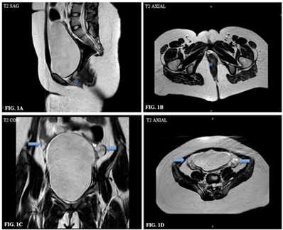
Impression: Type I MRKH.
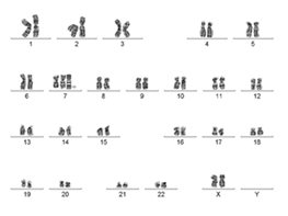
The patient was subjected to genetic karyotyping to confirm our findings.
Case 2
20 years old Indian residing in Kalaburagi, Karnataka with chief complaints of primary amenorrhea with normal female phenotype, no significant past history or similar complains in family. The patient did take medical hormonal therapy previously, but did not attain menarche and was suggested MRI Abdomen & Pelvis to rule out congenital malformations of mullerian tract.
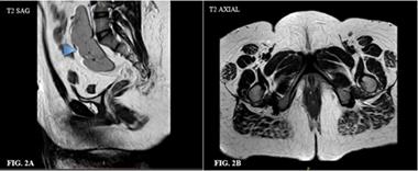
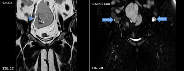
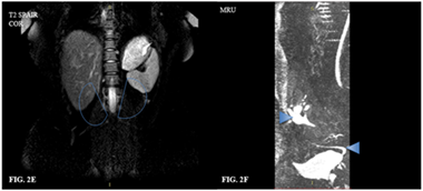
Impression: Type II MRKH with renal anomaly.
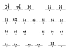
The patient was subjected to genetic karyotyping to confirm our findings.
Case 3
19 years, Indian residing in Kalaburagi, Karnataka with chief complaints of primary amenorrhea with normal female phenotype, no significant past history or similar complains in family. The patient did take medical hormonal therapy previously, but did not attain menarche and was suggested MRI Abdomen & Pelvis to rule out congenital malformations of mullerian tract.
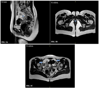
Impression: Type I MRKH.
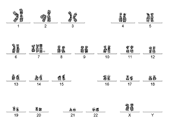
The patient was subjected to genetic karyotyping to confirm our findings.
Case 4
17 years, Indian residing in Kalaburagi, Karnataka with chief complaints of primary amenorrhea with normal female phenotype, no significant past history or similar complains in family. The patient did take medical hormonal therapy previously, but did not attain menarche and was suggested MRI Abdomen & Pelvis to rule out congenital malformations of mullerian tract.
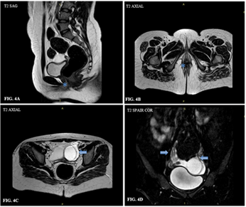
Impression: Type I MRKH with left simple ovarian cyst.
The patient was subjected to genetic karyotyping to confirm our findings.
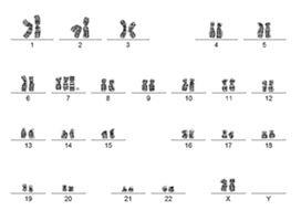
Case 5
18 years, Indian residing in Kalaburagi, Karnataka with chief complaints of primary amenorrhea with normal female phenotype, no significant past history or similar complains in family. The patient did take medical hormonal therapy previously, but did not attain menarche and was suggested MRI Abdomen & Pelvis to rule out congenital malformations of mullerian tract.
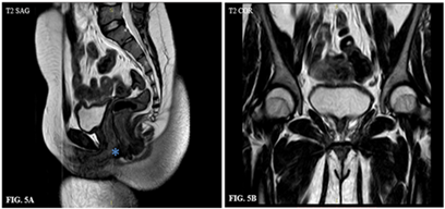
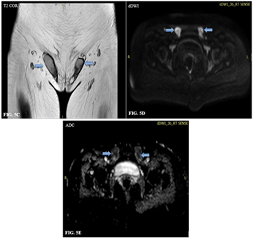
Impression: Congenital Androgen Insensitivity Syndrome (AIS).
The patient was subjected to genetic karyotyping to confirm our findings.
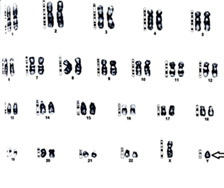
| Complaints | Age (years) | Phenotype | MRI Findings | Impression | Karyotype |
| Primary Amenorrhea | 15 | Female with well - developed secondary sexual characteristics. | Hypoplastic uterus, absent upper 2/3 vagina, normal ovaries | Type I MRKH | 46 XX |
| Primary Amenorrhea | 20 | Female with well - developed secondary sexual characteristics. | Uterus, cervix, vagina agenesis; cross-fused ectopic kidney | Type II MRKH | 46 XX |
| Primary Amenorrhea | 19 | Female with well - developed secondary sexual characteristics. | Hypoplastic uterus, normal ovaries | Type I MRKH | 46 XX |
| Primary Amenorrhea | 17 | Female with well - developed secondary sexual characteristics. | Fibrotic uterine remnant, absent cervix/proximal vagina, ovarian cyst | Type I MRKH with simple ovarian cyst | 46 XX |
| Primary Amenorrhea | 18 | Female with well - developed breasts but absent pubic hair. | Absent uterus/ovaries, bilateral undescended testes | Congenital Androgen insensitivity syndrome | 46 XY |
Discussion
The MRKH syndrome was first described by Mayer in 1829 and by Rokitansky in 1838 while Hauser and Schreiner in 1961 described the distinguishing features of MRKH syndrome from androgen insensitivity syndrome [5]. Ultrasonography is often the initial imaging modality for the diagnosis of MRKH syndrome. However, small uterine remnants, ectopic ovaries or intrauterine endometrium may not be thoroughly assessed by US. Moreover, a partially developed small rudimentary uterine remnant may be misinterpreted as a small tubular prepubertal uterus [6]. Principal findings of the present study can be summarized as follows: 1) Typical Müllerian remnants in MRKH syndrome consisted of three structural components, bilateral uterine buds, fibrous band-like structures, and midline triangular soft tissue, and 2) Bilateral uterine buds were connected by the fibrous band-like structures, which converged at the midline triangular soft tissue lying above the bladder dome [7]. Associations of MRKH syndrome are GRES (Genital, renal and ear syndrome) and MURCS (Mullerian, Renal and Cervical Somite dysplasia) [8]. This case series demonstrates the heterogeneity of MRKH spectrum disorders. MRI reliably differentiates Type I and Type II MRKH and detects associated renal anomalies. Renal anomalies such as ectopic kidney are common in Type II MRKH. type-II MRKH syndrome is associated with ovarian cancers and cardiac malformations. Hence, differentiation between MRKH syndrome and androgen insensitivity syndrome are essential for treatment planning of such patients. In present study we didn’t find any ovarian and cardiac anomalies [9]. MRI imaging modalities are also known to have the same effectiveness when compared to a combination of clinical examinations and ultrasound examinations [10]. The first finding that suggests the diagnosis of MRKH syndrome is failure to monitor menarche during puberty in spite of nor-mal development of secondary sex characteristics. Diagnosis would be clear after using radiologic modalities and karyo-type analysis [11]. Misdiagnosis is also common where doctors make further observations and do not provide early therapy so that there is a delayed diagnosis. Women with MRKH syndrome have never had menstruation or pregnancy. Even though ovarian cell donors and in vitro fertilization can be carried out, the patient may still feel depressed. In patients with vaginal agenesis, surgery is also needed so that they can have sexual intercourse with partners [12]. Ovarian function is usually preserved, though ovarian cysts may be incidental findings. AIS may mimic MRKH, underscoring the importance of imaging in differential diagnosis. MRI aids in surgical planning, patient counselling, and long-term management. Psychosocial counselling and multidisciplinary management, including gynecology, radiology, endocrinology, and psychiatry, are essential.
Conclusion
MRKH spectrum disorders present variably, ranging from isolated uterovaginal agenesis to complex associations with renal anomalies and conditions mimicking MRKH, such as AIS. MRI is indispensable in diagnosis, classification, and management planning. Timely diagnosis allows counselling, fertility planning, and psychosocial support.
Declarations
Ethical Approval and Consent to Participate
All procedures performed in this case series were conducted in accordance with institutional ethical standards and the principles of the Declaration of Helsinki. Ethical approval was obtained from the appropriate institutional review board where required. Written informed consent was obtained from all patients or their legal guardians prior to the procedures.
Consent for Publication
Written informed consent for publication of clinical details and images was obtained from the patients or their legal guardians. All identifying information has been anonymized to protect patient confidentiality.
Availability of Supporting Data
The data supporting the findings of this study are available from the corresponding author upon reasonable request, subject to institutional and ethical regulations.
Competing Interests
The authors declare that they have no competing interests related to this work.
Funding
This study received no specific grant from any funding agency in the public, commercial, or not-for-profit sectors.
Authors’ Contributions
All authors contributed substantially to the conception, data acquisition, analysis, drafting, and critical revision of the manuscript. All authors have read and approved the final version of the manuscript.