Introduction
Rheumatoid arthritis (RA) is a chronic, systemic autoimmune inflammatory disorder characterized by persistent synovitis, progressive joint destruction, and varying degrees of extra-articular involvement [1]. The disease predominantly affects the small and medium-sized joints, resulting in cartilage damage, bone erosion, pain, functional impairment, and reduced quality of life [1,2]. Among the musculoskeletal manifestations of RA, foot involvement is common and may present as pain, swelling, stiffness, deformity, and disability, significantly affecting mobility and daily activities [3-5].
Foot pathology in RA arises primarily from synovial inflammation and subsequent destruction of articular structures [3,4]. The forefoot is most frequently affected, particularly the metatarsophalangeal (MTP) joints, where chronic synovitis and tendon involvement lead to joint instability, contractures, subluxations, and deformities [3,4,6] Progressive weight-bearing stress on inflamed joints may result in hallux valgus, hallux rigidus, claw toe, mallet toe, and splay toe deformities. Midfoot involvement commonly manifests as pes planus due to collapse of the longitudinal arch, whereas hindfoot and ankle involvement may lead to subtalar instability, calcaneal varus or valgus deformities, restricted ankle dorsiflexion, and impaired gait [4,6,7]. These structural changes contribute substantially to pain, disability, and deterioration in functional status [5,8].
Assessment of disease burden in RA traditionally relies on composite indices such as the Disease Activity Score-28 (DAS28) [9]. However, the DAS28 does not include evaluation of foot and ankle joints, potentially leading to under-recognition of ongoing disease activity in these regions [10]. Consequently, patients who are otherwise classified as being in clinical remission may continue to experience significant foot-related symptoms and functional limitations [5,10]. Instruments such as the Health Assessment Questionnaire (HAQ) and the Foot Function Index (FFI) provide valuable insights into the functional impact of foot involvement and its effect on quality of life [11,12].
Despite the high prevalence of foot-related symptoms among patients with RA, routine clinical examination often overlooks detailed assessment of the feet [13]. Furthermore, there is a relative paucity of data from Indian populations regarding the prevalence, clinical profile, functional implications, and outcomes of foot involvement in RA, particularly in tertiary care settings. Early identification and appropriate management of foot involvement are essential to prevent irreversible deformities, preserve function, and improve patient outcomes [13,14].
In view of these considerations, the present prospective observational study was undertaken to determine the proportion of foot involvement among patients with rheumatoid arthritis attending a tertiary care centre, to evaluate their clinical and laboratory characteristics, to assess the functional impact of foot involvement using standardized assessment tools, and to study clinical outcomes following treatment over a six-month follow-up period.
Materials and Methods
This prospective observational study was conducted in the Rheumatology Outpatient Department of a tertiary care centre over a period of 18 months. The study endpoint involved data collection at the time of initial patient recruitment and after six months of follow-up. Prior to enrolment, written informed consent was obtained from all participants, and approval for the study was secured from the Institutional Ethics Committee.
A detailed clinical history was obtained from all patients, followed by a comprehensive physical examination. Demographic details, clinical findings, laboratory investigations, and radiological parameters were systematically recorded in a predesigned data collection form. As part of the study protocol, all participants underwent radiographic evaluation (X-ray) and ultrasonography (USG) of the affected foot joints without any additional cost. Disease activity and functional status were assessed using validated instruments, including the Disease Activity Score using 28 joints with Erythrocyte Sedimentation Rate (DAS28-ESR), the Health Assessment Questionnaire (HAQ), and the Foot Function Index (FFI) [9,11,12,15].
Patients were selected based on predefined eligibility criteria. Individuals aged 18–75 years diagnosed with rheumatoid arthritis according to the American College of Rheumatology/European League Against Rheumatism (ACR/EULAR) classification criteria, with a score of 6 or more and involvement of two or more foot joints, were included in the study [16]. Only those willing to provide written informed consent were enrolled.
Patients with overlap syndromes despite fulfilling ACR/EULAR criteria for rheumatoid arthritis were excluded from the study. Pregnant women and lactating mothers were also excluded because disease activity in rheumatoid arthritis is known to fluctuate during pregnancy and the postpartum period, potentially influencing study outcomes. Additionally, patients with foot pain or deformity resulting from trauma, previous surgery, or congenital abnormalities were excluded. Individuals who were non-compliant with prescribed medications or follow-up schedules were also excluded.
Based on the study by Borman et al., the prevalence of foot involvement in rheumatoid arthritis was assumed to be 89% [10]. Considering a confidence level of 95% (Z = 1.96), a margin of error of 5%, and a study power of 80%, the calculated sample size was 151 participants.
The collected data were entered into a database and analyzed using appropriate statistical methods. Continuous variables were expressed as mean ± standard deviation or median with interquartile range, while categorical variables were presented as frequencies and percentages. Analysis of Variance (ANOVA) was used to compare means across groups where applicable. A p-value of less than 0.05 was considered statistically significant.
Results
A total of 151 patients with rheumatoid arthritis (RA) fulfilling the 2010 ACR/EULAR classification criteria were enrolled in this prospective observational study. Among them, 128 patients demonstrated clinical and/or radiological evidence of foot involvement, yielding a prevalence of 84.77%.
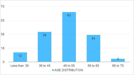
The mean age of the study population was 49.13 ± 9.2 years, with the majority (42%) of patients belonging to the 46–55-year age group (Figure 1) Females constituted 60.93% of the study population, resulting in a male-to-female ratio of 1:1.56. The duration of symptoms was most commonly between 1 and 5 years (45.70%), followed by less than 1 year (31.78%) as seen in Table 1.
| Duration of onset of symptoms | Number of subjects | Percentage |
| Less than 1year | 48 | 31.78% |
| 1 to 5 years | 69 | 45.70% |
| 5 to 10 years | 22 | 14.57% |
| More than 10 years | 12 | 7.95% |
| Total | 151 | 100.00% |
Analysis of body mass index (BMI) revealed a significant association with disease severity. Lower BMI was associated with higher disease activity as measured by DAS28-ESR, HAQ, and Foot Function Index (FFI) scores. The association remained statistically significant on Kruskal–Wallis testing (p < 0.001), while FFI demonstrated a significant inverse correlation with BMI (Spearman's rho = –0.164, p = 0.044).
Among laboratory parameters, significant changes over six
months were observed in total leukocyte count, platelet count, SGOT, blood urea nitrogen, potassium, and C-reactive protein levels (Table 2&3). CRP levels showed a significant reduction from 20.74 ± 21.89 mg/L at baseline to 13.54 ± 16.39 mg/L at six months (p < 0.001). Rheumatoid factor was positive in 92.05% of patients, while anti-CCP antibodies were positive in 86.09%. Both rheumatoid factor and anti-CCP positivity were observed in 78.14% of participants (Table 2).
| Haematological parameters | Mean ± SD at presentation | Mean ± SD after 6 months | p value |
| Hb | 11.10 ± 1.42 | 11.22 ±1.09 | 0.153 |
| TLC | 7256.56 ±1450.93 | 7944.90 ±1181.43 | 0.001 |
| Platelet count | 229344.37 ±71234.45 | 216556.30 ± 41393.18 | 0.012 |
| Total bilirubin | non-icteric | non-icteric | — |
| Direct bilirubin | non-icteric | non-icteric | — |
| SGOT | 28.54 ±9.89 | 30.76 ±8.51 | 0.001 |
| SGPT | 24.06 ±9.99 | 24.69 ±7.79 | 0.202 |
| Total protein | 7.45 ±0.44 | 7.48 ±0.25 | 0.795 |
| Albumin | 3.97 ±0.35 | 3.97 ±0.28 | 0.212 |
| Serological parameters | Mean ±SD at presentation | Mean ± SD after 6 months | p value |
| BUN | 12.84 ±3.61 | 11.31 ±3.36 | 0.001 |
| Serum creatinine | 1.09 ±0.22 | 1.16 ±0.66 | 0.506 |
| Sodium | 136.97 ±3.18 | 136.85 ±2.38 | 0.960 |
| Potassium | 3.81 ±0.37 | 3.92 ±0.30 | 0.008 |
| Chloride | 102.16 ±4.12 | 101.52 ±3.51 | 0.337 |
| ESR | 40.43 ±18.27 | 38.77 ±15.12 | 0.335 |
| C-reactive protein (CRP) | 20.74 ±21.89 | 13.54 ±16.39 | 0.001 |
Clinical examination demonstrated significant improvement in joint tenderness and swelling after six months of follow-up. Joint tenderness decreased from 85.43% to 58.28% (p < 0.001), while joint swelling decreased from 34.43% to 11.25% (p < 0.001). The prevalence of joint deformity remained unchanged at 8.60%.
| Physical finding | At presentation | At 6 months | p value | ||
| Present (n) | Absent (n) | Present (n) | Absent (n) | ||
| Joint Tenderness | 128 | 22 | 88 | 63 | 0.0001 |
| Joint Swelling | 52 | 99 | 17 | 134 | 0.0001 |
| Joint Deformity | 13 | 138 | 13 | 138 | 1.00 |
The first metatarsophalangeal (MTP) joint was the most commonly affected foot joint, with tenderness observed in 54.30% of left first MTP joints and 52.98% of right first MTP joints. Swelling was most frequently noted in the right first MTP joint. Foot deformities were identified in 13 patients (8.60%), with bilateral hallux valgus being the most common deformity, followed by hallux rigidus, mallet toe, pes planus, and hammer toe deformities (Table 5).
Radiographic evaluation revealed reduced joint space in 43.7% of patients, periarticular osteopenia in 27.81%, and erosions in 11.25%. The first MTP joints were the most frequently affected sites, particularly with respect to reduced joint space narrowing and erosive changes. No significant radiographic progression was observed during the six-month follow-up period (Table 5).
| Radiographic findings | At presentation | At 6 months | ||
| Present (n) | Absent (n) | Present (n) | Absent (n) | |
| Periarticular Osteopenia (X-ray) | 42 | 109 | 42 | 109 |
| Reduced joint space (X-ray) | 66 | 85 | 66 | 85 |
| Erosions (X-ray) | 17 | 134 | 17 | 134 |
| Synovial Effusion (USG) | 15 | 42 | 10 | 47 |
| Synovial Hypertrophy (USG) | 19 | 38 | 25 | 32 |
Ultrasonography was performed in 57 patients without definitive radiographic abnormalities. Among these, 34 patients demonstrated evidence of joint involvement. Synovial hypertrophy was more common than synovial effusion and was most frequently observed in the first tarsometatarsal (TMT) joint. Synovial effusion was most commonly identified in the first MTP joint (Table 5).
Analysis of foot joint involvement demonstrated that the right first MTP joint (55.62%) and left first MTP joint (54.96%) were the most frequently involved joints, followed by the right first TMT joint (26.5%) and right fifth MTP joint (23.84%). Metatarsophalangeal joints represented the most commonly affected anatomical region, followed by TMT joints, ankle joints, and proximal interphalangeal joints. No distal interphalangeal or calcaneocuboid joint involvement was observed.
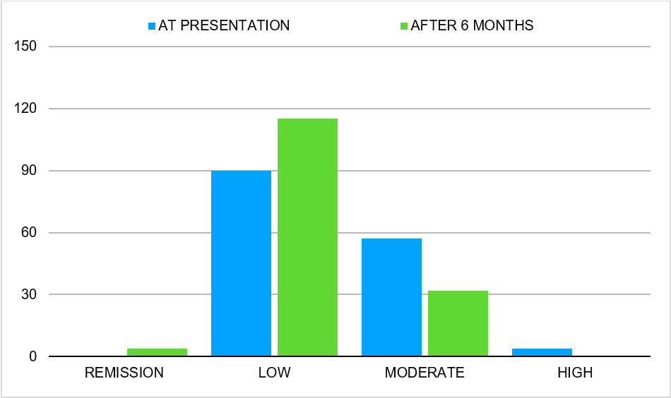
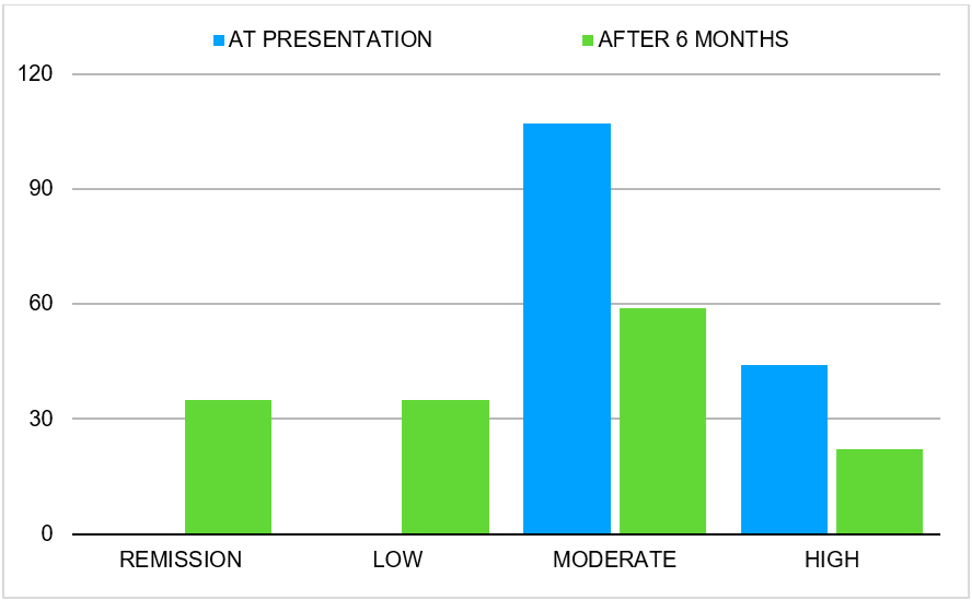
Disease activity and functional status improved significantly over the six-month follow-up period. Mean DAS28-ESR scores decreased from 4.63 ± 1.09 to 3.61 ± 1.12 (p < 0.001), HAQ scores decreased from 3.27 ± 2.21 to 1.97 ± 1.90 (p < 0.001), and FFI scores decreased from 34.53 ± 26.53 to 23.94 ± 22.74 (p < 0.001). At baseline, all patients had moderate or high disease activity, whereas after six months, 23.18% achieved remission and an additional 23.18% had low disease activity.
Drug-specific analysis demonstrated a significant association between methotrexate use and improvement in DAS28-ESR, HAQ, and FFI scores (all p < 0.05). In contrast, hydroxychloroquine and tofacitinib did not demonstrate statistically significant associations with disease activity or functional outcomes during the follow-up period. (Table 6, and Figure 2,3)
| Scores | Mean ±SD at presentation | Mean ± SD after 6 months | p value |
| DAS28-ESR Score | 4.63 ±1.09 | 3.61 ±1.12 | 0.0001 |
| HAQ Score | 3.27 ±2.21 | 1.97 ±1.90 | 0.0001 |
| Foot Functionality Index | 34.53 ±26.53 | 23.94±22.74 | 0.0001 |
Discussion
Rheumatoid arthritis (RA) is a chronic systemic autoimmune disease characterized by persistent synovial inflammation, progressive joint destruction, and functional disability [1]. Although involvement of the small joints of the hands has traditionally received considerable attention, foot involvement remains an under-recognized yet clinically significant manifestation of the disease [3,4]. The present prospective observational study was undertaken to evaluate the prevalence, clinical characteristics, functional impact, and outcomes of foot involvement in patients with rheumatoid arthritis attending a tertiary care centre.
The mean age of the study population was 49.13 ± 9.2 years, with the majority of patients belonging to the 46–55-year age group. A female predominance was observed, with females accounting for 60.93% of the study population. These findings are consistent with the established epidemiology of RA, which demonstrates a higher prevalence among middle-aged women. Similar demographic characteristics have been reported by Papadopoulos et al. and Shrivastava et al., supporting the representativeness of our study population [17,18].
A major finding of the present study was the high prevalence of foot involvement, observed in 84.77% of patients. This finding closely parallels the prevalence reported by Borman et al., who documented foot-related symptoms in 89% of patients with rheumatoid arthritis [10]. The high prevalence observed emphasizes that foot involvement is not merely an ancillary manifestation but rather a core component of disease burden in RA. Despite this, routine rheumatologic assessment frequently underestimates foot pathology because commonly used disease activity indices such as DAS28 do not include foot and ankle joints [9,10].
An important observation in our study was the association between lower body mass index and greater disease severity, as reflected by DAS28-ESR, HAQ, and Foot Function Index scores. This finding contrasts with reports suggesting poorer outcomes among obese patients with rheumatoid arthritis [19]. The observed inverse relationship may reflect the influence of chronic inflammation, rheumatoid cachexia, reduced nutritional reserves, and advanced disease burden in patients with lower BMI. Further studies are required to clarify the complex relationship between nutritional status and disease activity in RA.
Inflammatory markers demonstrated a significant reduction in CRP levels over the six-month follow-up period, indicating effective suppression of systemic inflammation with treatment. Although ESR also showed a numerical decline, the change was not statistically significant. The high prevalence of rheumatoid factor positivity (92.05%) and anti-CCP antibody positivity (86.09%) observed in our study reflects a predominantly seropositive disease population, which is generally associated with more aggressive disease and increased risk of structural joint damage [20].
Clinical examination revealed that tenderness was the most common physical finding, followed by swelling and deformity. Significant reductions in tenderness and swelling were observed after six months of treatment, demonstrating improvement in inflammatory activity. However, deformities remained unchanged during follow-up, highlighting the irreversible nature of established structural damage once deformities have developed. Bilateral hallux valgus was the most common deformity identified, followed by hallux rigidus, mallet toe, pes planus, and hammer toe deformities. These findings are consistent with the known pathophysiology of RA, where chronic synovitis and ligamentous laxity contribute to progressive forefoot deformity [21].
The forefoot was the most frequently affected region of the foot, with the first metatarsophalangeal joint being the single most commonly involved joint. This observation is consistent with previous studies demonstrating a predilection of rheumatoid arthritis for the metatarsophalangeal joints [3,22]. In contrast, hindfoot and ankle involvement were less common. The predominance of forefoot disease is clinically important because involvement of the first MTP joint substantially affects gait mechanics, weight-bearing capacity, and overall mobility [23].
Radiographic evaluation demonstrated reduced joint space as the most frequent abnormality, followed by periarticular osteopenia and erosions. The first metatarsophalangeal joints were the most commonly affected sites. These findings reflect the progressive cartilage loss and structural damage associated with chronic synovitis [24]. Importantly, radiographic abnormalities remained largely unchanged during the six-month follow-up period, suggesting that structural changes require longer periods to demonstrate measurable progression or improvement.
Ultrasonography proved valuable in detecting inflammatory changes among patients without radiographic abnormalities. Synovial hypertrophy was more common than synovial effusion and was predominantly observed in the first tarsometatarsal joint. These findings support the growing role of musculoskeletal ultrasonography as a sensitive modality for identifying early inflammatory changes before the appearance of radiographic damage [25]. The detection of subclinical disease activity may facilitate earlier intervention and potentially prevent progression to irreversible deformity.
Assessment of disease severity demonstrated significant improvement over six months, as evidenced by reductions in DAS28-ESR, HAQ, and Foot Function Index scores. Furthermore, a substantial proportion of patients transitioned from moderate or high disease activity to remission or low disease activity. Improvement in FFI scores indicates better foot function and suggests that successful control of inflammatory activity translates into meaningful improvements in mobility and quality of life [8,26].
Among the disease-modifying antirheumatic drugs evaluated, methotrexate demonstrated a significant association with improvement in disease activity and functional outcomes. In contrast, hydroxychloroquine and tofacitinib did not show statistically significant associations with outcome measures. However, these observations should be interpreted cautiously because treatment allocation was not randomized and may have been influenced by baseline disease severity, physician preference, treatment adherence, and concomitant therapies.
The findings of the present study underscore the substantial burden of foot involvement in rheumatoid arthritis and highlight the need for routine foot assessment as part of comprehensive disease evaluation. Incorporation of foot-specific clinical examination, functional assessment tools, and imaging modalities into routine rheumatology practice may improve early detection of disease activity, facilitate timely intervention, and reduce long-term disability. Greater attention to foot health is essential for optimizing patient outcomes and improving quality of life in individuals living with rheumatoid arthritis [10,13].
Strengths and Limitations
The strengths of this study include its prospective design, standardized clinical assessment, and combined use of radiography and ultrasonography. However, the study was limited by its single-centre setting, relatively short follow-up duration, and non-randomized treatment allocation, which may limit the generalizability of the findings.
Conclusion
Foot involvement is a common and clinically significant manifestation of rheumatoid arthritis, affecting the majority of patients and contributing substantially to pain, functional impairment, and reduced quality of life [10,13]. The present study demonstrated a high prevalence of foot joint involvement, with the forefoot, particularly the first metatarsophalangeal joint, being the most frequently affected site [3,22].
Clinical examination, when complemented by imaging modalities, provides a comprehensive assessment of foot disease. While conventional radiography remains useful for identifying structural damage, ultrasonography proved valuable in detecting early inflammatory changes, including synovial hypertrophy and effusion, even in the absence of radiographic abnormalities [25]. These findings highlight the importance of incorporating imaging into routine evaluation for early diagnosis and timely therapeutic intervention.
Significant improvements in disease activity, functional status, and foot-related disability were observed following treatment, emphasizing the benefits of early initiation and regular optimization of disease-modifying antirheumatic drugs. Nevertheless, established deformities persisted despite treatment, underscoring the importance of early recognition before irreversible structural damage occurs [21].
Regular assessment of foot involvement should form an integral component of the routine management of patients with rheumatoid arthritis. A multidisciplinary approach involving rheumatologists, physiotherapists, occupational therapists, and podiatric care services can facilitate early detection, appropriate intervention, prevention of disability, and preservation of mobility. Such an approach has the potential to improve functional outcomes, maintain independence, and enhance the overall quality of life of patients living with rheumatoid arthritis [8,13,26].
Declarations
Ethical Approval and Consent to Participate
All procedures performed in this prospective observational study were conducted in accordance with institutional ethical standards and the principles of the Declaration of Helsinki. Ethical approval was obtained from the appropriate institutional review board where required. Written informed consent was obtained from all patients or their legal guardians prior to the procedures.
Consent for Publication
Written informed consent for publication of clinical details was obtained from the patients or their legal guardians. All identifying information has been anonymized to protect patient confidentiality.
Availability of Supporting Data
The data supporting the findings of this study are available from the corresponding author upon reasonable request, subject to institutional and ethical regulations.
Competing Interests
The authors declare that they have no competing interests related to this work.
Funding
This study received no specific grant from any funding agency in the public, commercial, or not-for-profit sectors.
Authors’ Contributions
All authors contributed substantially to the conception, data acquisition, analysis, drafting, and critical revision of the manuscript. All authors have read and approved the final version of the manuscript.