Introduction
Invasive mechanical ventilation (IMV) is an indispensable life-support modality for critically ill patients with respiratory failure. Although it is lifesaving, prolonged dependence on mechanical ventilation is associated with significant complications, making timely liberation from ventilatory support a major therapeutic goal in intensive care units (ICUs). The decision to extubate requires a careful balance, as premature extubation increases the likelihood of reintubation, whereas delayed weaning exposes patients to ventilator-associated complications, including ventilator-associated pneumonia, ventilator-induced lung injury, diaphragmatic dysfunction, prolonged ICU stay, and increased mortality [1-2].
Successful weaning is generally defined as the ability to sustain spontaneous breathing without invasive ventilatory support for at least 48 hours after extubation. Conversely, weaning failure includes failure of the spontaneous breathing trial (SBT), need for reintubation or non-invasive ventilatory support within 48 hours, or death during this period [3]. Despite standardized weaning protocols and daily assessment of extubation readiness, extubation failure continues to occur in approximately 15–20% of mechanically ventilated patients, emphasizing the need for more reliable predictors of weaning success [4].
Several physiological indices have been proposed to assess readiness for weaning, including minute ventilation, maximal inspiratory pressure, breathing frequency, airway occlusion pressure, the compliance-rate-oxygenation-pressure (CROP) index, and the Rapid Shallow Breathing Index (RSBI) [5]. Among these, the RSBI, calculated as respiratory rate divided by tidal volume, remains the most widely accepted bedside predictor. An RSBI of <105 breaths/min/L has demonstrated high sensitivity but only moderate specificity, indicating that reliance on this parameter alone may lead to inaccurate prediction of extubation outcomes [5].
A major limitation of conventional weaning indices is that they evaluate overall respiratory performance without directly assessing diaphragmatic function, despite the diaphragm being the principal muscle of respiration. Ventilator-induced diaphragmatic dysfunction is increasingly recognized as an important contributor to weaning and extubation failure [6]. Traditional methods for evaluating diaphragmatic function, such as fluoroscopy, trans-diaphragmatic pressure measurement, and phrenic nerve stimulation, are either invasive, technically demanding, or impractical for routine bedside use [7].
Bedside diaphragm ultrasonography has emerged as a simple, non-invasive, reproducible, and radiation-free technique for real-time assessment of diaphragmatic function. Ultrasound-derived parameters, particularly the Excursion-Time Index (ETI), provide objective evaluation of diaphragmatic mechanics and may complement conventional weaning criteria by identifying patients with occult diaphragmatic dysfunction [8]. Furthermore, integrating diaphragm ultrasonography into routine weaning assessment has the potential to reduce failed extubation, decrease reintubation rates, and shorten ICU stay [9-10]. However, evidence regarding the incremental value of ETI over conventional weaning parameters remains limited.
Therefore, the present study was undertaken to compare the predictive performance of ultrasonography-guided diaphragm Excursion-Time Index (ETI) combined with conventional weaning criteria versus conventional weaning criteria alone for predicting successful extubation in critically ill ICU patients.
Materials and Methods
Study Design and Ethical Approval
This prospective, parallel-group, randomized comparative study was conducted in the Department of Anaesthesiology and Critical Care, Sri Guru Ram Das Institute of Medical Sciences & Research, Amritsar, Punjab, India, from July 2024 to December 2025. The study was approved by the Institutional Research and Ethics Committee, and written informed consent was obtained from the legally authorized representatives of all participants. The study adhered to the principles of the Declaration of Helsinki.
Study Population
Adult patients (≥18 years) receiving invasive mechanical ventilation (MV) and considered clinically ready for weaning were screened for inclusion.
Inclusion criteria: Patients aged ≥18 years on invasive MV fulfilling readiness-to-wean criteria.
Exclusion criteria: Pregnancy, neuromuscular disorders, diaphragmatic paralysis (diaphragmatic excursion <10 mm), pneumothorax, massive pleural effusion, and tracheostomy.
Sample Size and Randomization
A total of 80 patients were enrolled and randomized in a 1:1 ratio into two groups (40 patients each) using a computer-generated randomization sequence. Allocation concealment was ensured through sequentially numbered, opaque, sealed envelopes opened immediately before the weaning process.
Study Groups
Group A (Conventional Group): Extubation based on conventional weaning criteria.
Group B (USG-ETI Group): Extubation based on conventional weaning criteria supplemented with diaphragm ultrasonography-guided Excursion-Time Index (ETI).
Study Protocol
Baseline demographic data, indication for mechanical ventilation, duration of ventilation, and APACHE II score were recorded at ICU admission. Patients were ventilated in Assist-Control mode and assessed daily for readiness to wean.
Conventional weaning criteria included:
-
PaO₂ >60 mmHg
-
PaCO₂ <50 mmHg
-
FiO₂ ≤0.5
-
PaO₂/FiO₂ >300
-
PEEP ≤5 cmH₂O
-
Respiratory rate <35 breaths/min
-
Tidal volume >5 mL/kg
-
Vital capacity >10 mL/kg
-
Minute ventilation 4–10 L/min
-
Hemodynamic stability
-
Afebrile state
-
Absence of significant metabolic or electrolyte abnormalities
Eligible patients underwent a 30-minute spontaneous breathing trial (SBT) using pressure support ventilation (pressure support 5 cmH₂O and PEEP 5 cmH₂O). The Rapid Shallow Breathing Index (RSBI) was calculated as:
RSBI = Respiratory Rate (breaths/min) ÷ Tidal Volume (L)
Patients with RSBI <105 breaths/min/L were considered suitable for extubation.
Diaphragm Ultrasonography
In Group B, diaphragm ultrasonography was performed using a 2–10 MHz convex transducer (GE Wipro, P21 M-mode probe) with patients in the semi-recumbent position (20°–40°). The right hemidiaphragm was examined via the subcostal approach using the liver as an acoustic window. Diaphragmatic excursion (DE) and inspiratory time (Ti) were measured over three consecutive tidal breaths, and the average value was recorded.
The Excursion-Time Index (ETI) was calculated as:
ETI = Diaphragmatic Excursion (cm) × Inspiratory Time (s)
Ultrasound assessment was performed at ICU admission, after successful SBT, and 4–24 hours following extubation. Patients in Group B were extubated only after fulfilling conventional weaning criteria, demonstrating RSBI <105 breaths/min/L and DE >10 mm.
Patient Monitoring
Arterial blood gas analysis was performed at initiation of mechanical ventilation, after SBT, and 2 hours after extubation. Patients were monitored for vital signs and respiratory status for 48 hours following extubation.
Outcome Measures
Primary outcome: To evaluate whether diaphragm ultrasonography-guided ETI improves prediction of successful extubation when added to conventional weaning criteria.
Secondary outcomes: Comparison of extubation success, reintubation, need for non-invasive ventilation (NIV), mortality within 48 hours, association between APACHE II score and duration of mechanical ventilation, and changes in diaphragmatic excursion after extubation.
Successful extubation was defined as spontaneous breathing without invasive or non-invasive ventilatory support for 48 hours, whereas extubation failure was defined as reintubation, NIV requirement, or death within 48 hours.
Statistical Analysis
Data were analyzed using IBM SPSS Statistics version 26.0. Continuous variables were tested for normality using the Shapiro–Wilk test and expressed as mean ± SD, while categorical variables were expressed as frequencies and percentages. Intergroup comparisons were performed using the independent Student's t-test and Chi-square/Fisher's exact test, as appropriate. Pearson's correlation assessed the relationship between APACHE II score and duration of mechanical ventilation. A two-sided p-value <0.05 was considered statistically significant.
Results
A total of 105 patients receiving invasive mechanical ventilation were assessed for eligibility. Of these, 25 patients were excluded, including 20 who did not meet the inclusion criteria and 5 who declined to participate. The remaining 80 eligible patients were randomized in a 1:1 ratio into two groups. Group A (n=40) underwent extubation based on conventional weaning criteria, whereas Group B (n=40) was extubated using conventional weaning criteria supplemented with ultrasonography-guided diaphragm Excursion-Time Index (ETI). No participants discontinued the intervention or were lost to follow-up in either group. Consequently, all 80 randomized patients (40 in each group) completed the study and were included in the final analysis of the primary outcome (Figure 1).
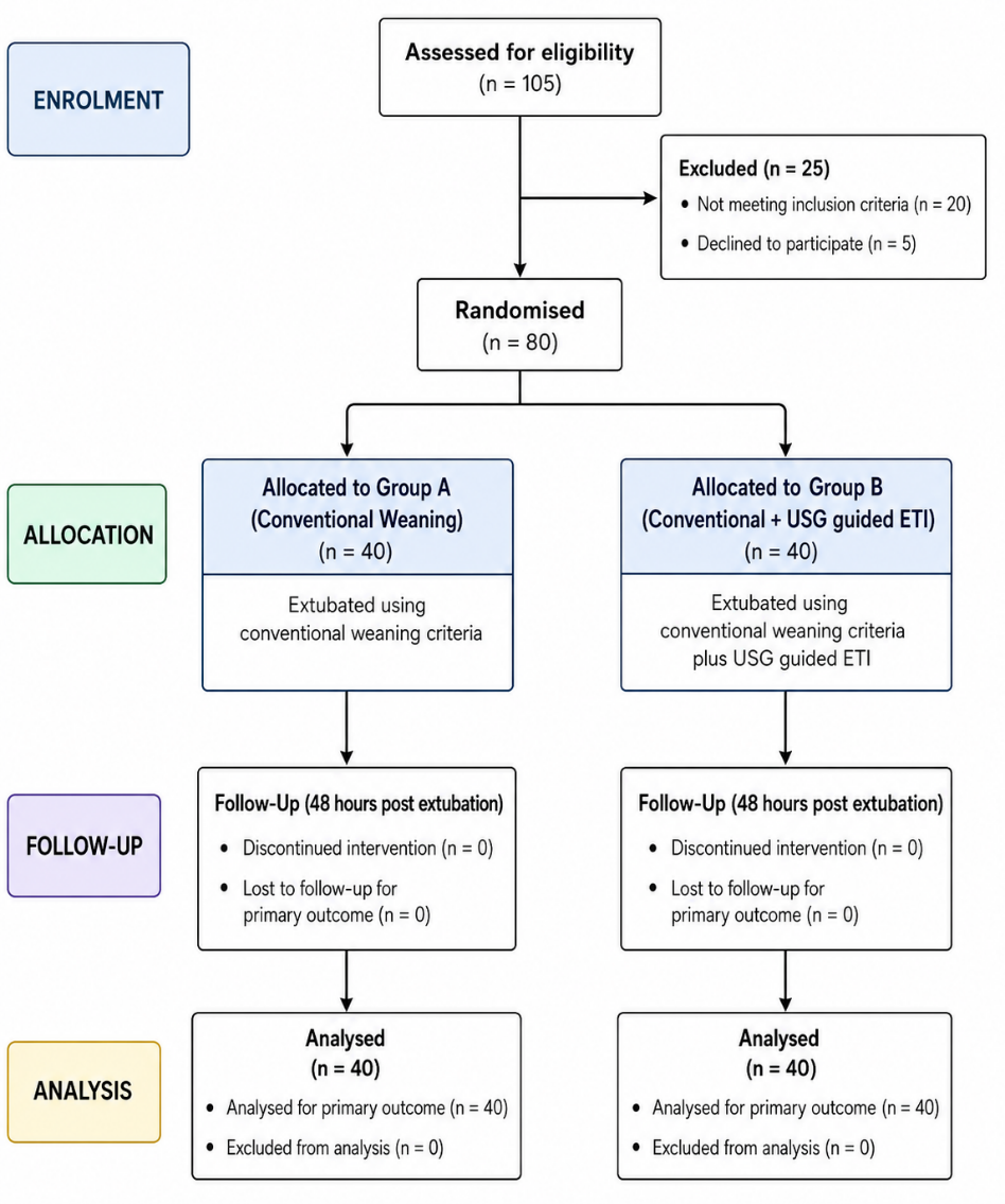
Table 1 compares baseline demographic, clinical, and diaphragmatic parameters between patients with successful and failed extubation. Most baseline variables, including age, Glasgow Coma Scale, respiratory rate, diaphragm excursion, inspiratory time, expiratory time, diaphragmatic RSBI, and diaphragm thickness fraction, were comparable between the two groups (all p>0.05). However, patients with extubation failure had significantly higher APACHE II scores (22.70 ± 5.13 vs. 16.98 ± 4.66; p<0.001) and RSBI (68.82 ± 20.27 vs. 41.67 ± 11.87; p<0.001), indicating greater illness severity and poorer respiratory reserve at baseline.
| Variable | Success (n=53) | Failure (n=27) | P-value |
| Age (years) | 49.89 ± 17.34 | 55.04 ± 17.08 | 0.252 |
| Baseline GCS | 6.40 ± 2.06 | 6.67 ± 1.90 | 0.548 |
| Respiratory Rate | 18.87 ± 6.39 | 18.56 ± 5.98 | 0.890 |
| Diaphragm Excursion (cm) | 1.63 ± 0.42 | 1.51 ± 0.39 | 0.164 |
| Inspiratory Time (sec) | 1.18 ± 0.29 | 1.03 ± 0.31 | 0.081 |
| Expiratory Time (sec) | 2.26 ± 0.66 | 2.01 ± 0.71 | 0.097 |
| D-RSBI | 1.21 ± 0.66 | 1.12 ± 0.52 | 0.722 |
| Thickness Fraction (%) | 37.95 ± 14.08 | 31.38 ± 17.27 | 0.112 |
| APACHE II Score | 16.98 ± 4.66 | 22.70 ± 5.13 | <0.001 |
| RSBI | 41.67 ± 11.87 | 68.82 ± 20.27 | <0.001 |
After 30 minutes of the spontaneous breathing trial, patients with successful extubation demonstrated significantly lower respiratory and heart rates, higher pH and bicarbonate levels, lower lactate concentrations, greater diaphragm excursion, longer inspiratory and expiratory times, lower diaphragmatic RSBI, and higher diaphragm thickness fraction than those with extubation failure (all p<0.05). In contrast, PaO₂ and PaCO₂ did not differ significantly between the two groups (p>0.05), indicating that diaphragmatic function and respiratory mechanics were better predictors of extubation outcome than gas exchange parameters alone (Table 2).
| Variable | Success | Failure | P-value |
| Respiratory Rate | 19.32 ± 3.84 | 22.37 ± 6.31 | 0.019 |
| Heart Rate | 96.1 ± 13.7 | 103.6 ± 15.8 | 0.041 |
| pH | 7.409 ± 0.054 | 7.375 ± 0.065 | 0.014 |
| PaO₂ | 94.8 ± 16.5 | 86.9 ± 18.4 | 0.071 |
| PaCO₂ | 38.8 ± 7.4 | 41.9 ± 8.8 | 0.118 |
| HCO₃ | 22.90 ± 4.80 | 19.82 ± 3.19 | 0.001 |
| Lactate | 1.86 ± 0.74 | 2.52 ± 0.96 | 0.003 |
| Diaphragm Excursion | 2.02 ± 0.30 | 1.61 ± 0.45 | 0.017 |
| Inspiratory Time | 1.35 ± 0.25 | 0.76 ± 0.09 | <0.001 |
| Expiratory Time | 2.71 ± 0.61 | 1.23 ± 0.41 | <0.001 |
| D-RSBI | 0.98 ± 0.31 | 1.69 ± 0.48 | <0.001 |
| Thickness Fraction | 47.73 ± 15.51 | 28.45 ± 15.34 | 0.003 |
Following extubation, patients with successful outcomes had significantly lower respiratory rate, systolic and diastolic blood pressure, lower lactate levels, higher pH and bicarbonate concentrations, greater diaphragm excursion, longer inspiratory and expiratory times, lower diaphragmatic RSBI, and higher diaphragm thickness fraction than those with extubation failure (all p<0.05). These findings indicate that preserved diaphragmatic function and stable physiological parameters were strongly associated with successful extubation, whereas impaired diaphragmatic performance was linked to extubation failure (Table 3).
| Variable | Success | Failure | P-value |
| Respiratory Rate | 18.8 ± 3.5 | 24.5 ± 5.90 | <0.001 |
| SBP | 130.45 ± 20.37 | 139.78 ± 20.17 | 0.039 |
| DBP | 77.00 ± 11.97 | 85.04 ± 11.67 | 0.005 |
| pH | 7.416 ± 0.053 | 7.372 ± 0.084 | 0.018 |
| HCO₃ | 22.95 ± 4.21 | 19.94 ± 4.03 | 0.007 |
| Lactate | 1.71 ± 0.63 | 2.63 ± 0.88 | <0.001 |
| Diaphragm Excursion | 1.91 ± 0.31 | 1.32 ± 0.39 | 0.001 |
| Inspiratory Time | 1.26 ± 0.33 | 0.68 ± 0.11 | <0.001 |
| Expiratory Time | 2.36 ± 0.55 | 0.89 ± 0.34 | <0.001 |
| D-RSBI | 1.20 ± 0.35 | 1.86 ± 0.55 | 0.002 |
| Thickness Fraction | 45.7 ± 14.2 | 24.8 ± 13.6 | <0.001 |
Multivariable logistic regression identified APACHE II score, RSBI, diaphragm excursion, and diaphragm thickness fraction as independent predictors of extubation outcome. Higher APACHE II scores and RSBI were associated with increased odds of extubation failure, whereas greater diaphragm excursion and higher diaphragm thickness fraction significantly reduced the risk of failure. These findings highlight that both illness severity and diaphragmatic function independently influence the likelihood of successful extubation (Table 4).
| Variable | Adjusted OR | 95% CI | P-value |
| APACHE II Score | 1.31 | 1.12–1.57 | <0.001 |
| RSBI | 1.12 | 1.05–1.21 | <0.001 |
| Diaphragm Excursion | 0.28 | 0.12–0.65 | 0.002 |
| Thickness Fraction | 0.94 | 0.91–0.98 | 0.004 |
ROC analysis demonstrated excellent predictive performance of both conventional and diaphragm ultrasonography-derived parameters for extubation outcome. RSBI showed the highest diagnostic accuracy (AUC = 0.91), followed by diaphragm thickness fraction (AUC = 0.89), diaphragm excursion (AUC = 0.87), and APACHE II score (AUC = 0.86). The identified optimal cut-off values provided high sensitivity (81.5–88.9%) and specificity (79.2–84.9%), indicating that these parameters reliably discriminate between successful extubation and extubation failure (Table 5).
| Variable | AUC | Sensitivity | Specificity | Optimal Cut-off |
| RSBI | 0.91 | 88.9% | 84.9% | >55 |
| APACHE II | 0.86 | 81.5% | 79.2% | >19 |
| Diaphragm Excursion | 0.87 | 85.2% | 81.1% | <1.7 cm |
| Thickness Fraction | 0.89 | 87.0% | 83.0% | <35% |
Discussion
Liberation from invasive mechanical ventilation remains a critical milestone in the management of critically ill patients. Despite the widespread use of standardized weaning protocols, extubation failure continues to occur in 10–20% of mechanically ventilated patients and is associated with prolonged ICU stay, ventilator-associated complications, and increased mortality. Conventional weaning indices, particularly the Rapid Shallow Breathing Index (RSBI), assess global respiratory mechanics but do not directly evaluate diaphragmatic performance, which has emerged as a key determinant of successful ventilator liberation. Therefore, bedside diaphragm ultrasonography has gained increasing attention as an adjunctive tool for predicting extubation outcomes.
In the present study, 53 (66.3%) patients achieved successful extubation, whereas 27 (33.7%) experienced extubation failure. Baseline demographic characteristics, including age, Glasgow Coma Scale, respiratory rate, diaphragm excursion, inspiratory time, expiratory time, diaphragmatic RSBI (D-RSBI), and diaphragm thickness fraction, were comparable between the two groups, indicating that both groups had similar baseline characteristics. However, patients with extubation failure had significantly higher APACHE II scores and RSBI than those with successful extubation (p<0.001), suggesting that disease severity and respiratory mechanics remain important determinants of weaning outcome. Similar observations have been reported by Burns et al.,[11] demonstrated that increasing APACHE II scores are associated with prolonged ventilatory support and a greater likelihood of weaning failure. Likewise, the predictive utility of RSBI has been consistently validated since its original description by Yang and Tobin [17], although its specificity remains suboptimal when used alone.
Following the spontaneous breathing trial (SBT), patients with successful extubation demonstrated significantly lower respiratory and heart rates together with better acid-base status, as evidenced by higher pH and bicarbonate levels and lower serum lactate concentrations. These findings indicate superior cardiopulmonary adaptation during spontaneous breathing. Similar physiological responses have been described by Cohen et al.,[12] and Amir et al.,[13] reported that metabolic acidosis and elevated lactate are associated with respiratory muscle fatigue and increased risk of extubation failure.
A major finding of the present study was the significant difference in diaphragmatic ultrasound parameters between the success and failure groups. During SBT, patients who were successfully extubated had significantly greater diaphragm excursion, longer inspiratory and expiratory times, lower diaphragmatic RSBI, and higher diaphragm thickness fraction. These differences persisted after extubation, indicating sustained preservation of diaphragmatic function in successfully liberated patients. These observations support previous reports [2,14,15] who demonstrated that preserved diaphragmatic excursion and thickening fraction reflect adequate diaphragmatic contractility and are strongly associated with successful weaning from mechanical ventilation.
Multivariable logistic regression further demonstrated that APACHE II score, RSBI, diaphragm excursion, and diaphragm thickness fraction were independent predictors of extubation outcome. Higher APACHE II scores and RSBI independently increased the likelihood of extubation failure, whereas greater diaphragm excursion and higher thickness fraction significantly reduced this risk. These findings emphasize that diaphragmatic function contributes independently to extubation success beyond conventional clinical and respiratory variables. Similar results have been reported by Umbrello et al.,[16] and Dres et al.,[17] who identified diaphragm ultrasonographic parameters as independent predictors of weaning success after adjustment for disease severity and respiratory indices.
Receiver operating characteristic (ROC) analysis further confirmed the excellent diagnostic performance of both conventional and ultrasound-derived parameters. RSBI demonstrated the highest discriminative ability (AUC 0.91), followed by diaphragm thickness fraction (AUC 0.89), diaphragm excursion (AUC 0.87), and APACHE II score (AUC 0.86). The optimal cut-off values identified in the present study (>55 for RSBI, <1.7 cm for diaphragm excursion, and <35% for diaphragm thickness fraction) provided good sensitivity and specificity for predicting extubation failure. These findings are comparable with those reported by Parada-Gereda et al.,[18] whose meta-analysis demonstrated pooled AUC values between 0.80 and 0.90 for diaphragm ultrasonography in predicting successful extubation, and by Alam et al.,[14] reported similar diagnostic accuracy for diaphragm excursion and thickening fraction during spontaneous breathing trials.
Overall, the present study demonstrates that while conventional indices such as RSBI remain valuable predictors of extubation readiness, incorporation of bedside diaphragm ultrasonography substantially enhances assessment of respiratory muscle function. Objective evaluation of diaphragm excursion and thickness fraction provides additional prognostic information that cannot be obtained from conventional respiratory parameters alone. Integration of these ultrasound-derived measurements into routine weaning protocols may facilitate more accurate identification of patients ready for extubation, reduce extubation failure, and improve overall ICU outcomes.
This study has several limitations. It was conducted at a single tertiary care center with a relatively small sample size, which may limit the generalizability of the findings. Diaphragm ultrasonography is operator-dependent and may be subject to inter-observer variability. Assessment was limited to the right hemidiaphragm because of superior acoustic visualization. Additionally, extubation outcomes were evaluated only up to 48 hours, and longer-term clinical outcomes were not assessed. Larger multicenter studies are warranted to validate these findings and establish standardized ultrasonographic cut-off values for predicting successful extubation.
Conclusion
Bedside diaphragm ultrasonography is a practical and reliable adjunct to conventional weaning assessment. In the present study, diaphragm excursion and thickness fraction independently predicted extubation outcome and demonstrated excellent diagnostic performance. Although RSBI remained the strongest conventional predictor, combining diaphragmatic ultrasonographic parameters with established weaning criteria improved identification of patients ready for extubation. Incorporation of diaphragm ultrasonography into routine ICU practice may reduce extubation failure, minimize reintubation, and optimize ventilator liberation.
Abbreviations
ABG: Arterial Blood Gas
APACHE II: Acute Physiology and Chronic Health Evaluation II
AUC: Area Under the Curve
DBP: Diastolic Blood Pressure
D-RSBI: Diaphragmatic Rapid Shallow Breathing Index
DE: Diaphragm Excursion
ETI: Excursion-Time Index
FiO₂: Fraction of Inspired Oxygen
GCS: Glasgow Coma Scale
HCO₃⁻: Bicarbonate
ICU: Intensive Care Unit
IMV: Invasive Mechanical Ventilation
MV: Mechanical Ventilation
NIV: Non-invasive Ventilation
PaCO₂: Partial Pressure of Carbon Dioxide
PaO₂: Partial Pressure of Oxygen
PEEP: Positive End-Expiratory Pressure
PSV: Pressure Support Ventilation
RR: Respiratory Rate
RSBI: Rapid Shallow Breathing Index
SBP: Systolic Blood Pressure
SBT: Spontaneous Breathing Trial
Declarations
Ethical Approval and Consent to Participate
The study protocol was approved by the Institutional Research and Ethics Committee of Sri Guru Ram Das Institute of Medical Sciences & Research, Amritsar, Punjab, India (SGRD/IEC/2024-315 & CTRI/2024/10/075857). Written informed consent was obtained from the legally authorized representatives of all participants before enrolment. The study was conducted in accordance with the ethical principles of the Declaration of Helsinki.
Consent for Publication
Not applicable.
Availability of Data and Materials
The datasets generated and/or analyzed during the current study are available from the corresponding author upon reasonable request.
Competing Interests
The authors declare that they have no competing interests.
Funding
The authors received no external funding for this study.
Authors' Contributions
Dr. Arshdeep Kaur conceived and designed the study, collected the clinical data, conducted the literature review, interpreted the findings, and drafted the initial manuscript.
Dr. Jonny Dhawan contributed to patient recruitment, supervised the clinical conduct of the study, and assisted in data interpretation.
Dr. Sunil Chawla critically revised the manuscript for important intellectual content and approved the final version for publication.
Dr. Ruchi Gupta contributed to formal validation of the study, reviewed the manuscript for scientific accuracy, and approved the final manuscript.
Dr. Prabhjot Kaur Gill performed the statistical analysis, contributed to manuscript preparation, critically reviewed the final draft, coordinated the manuscript submission process.