Introduction
The thyroid gland is an essential component of the human body that plays an important role in metabolism. Dysfunction of the thyroid gland invariably affects human health and life quality. Disorders of the thyroid gland are one of the most common anomalies. According to the World Health Organization, more than 190 million people have iodine deficiency disorders. Understanding of the wide spectrum of thyroid disorders is of great importance, and most of these lesions are treatable by medical or surgical management. Thyroid diseases, including carcinoma, hyperthyroidism, and local compression, require surgical management [1]. Another common clinical problem of the thyroid gland is thyroid nodules; it is important to distinguish between benign and malignant nodules.
Fine needle aspiration (FNA) cytology is a useful tool widely accepted for the diagnostic assessment of thyroid nodules. This method distinguishes between benign and malignant nodules with approximately 95% accuracy. It is easy to perform and free of hazards [2]. Information provided by the FNA guides the management of patients with thyroid nodules by identifying if patients require a surgical resection or if no further interventions are needed [3]. A successful implementation of this method substantially reduces mortality and morbidity [4]. Hence, FNA is an important test for assessing patients with thyroid nodules [3]. For successful reporting of the FNA, proper communication among the medical team, including pathologists, clinicians, radiologists, and surgeons, along with cytohistological correlation, is necessary. Having a consistent diagnostic terminology is also essential. To achieve uniformity in the diagnostic terminology, morphologic criteria, and risk of malignancy for reporting of thyroid FNA, the National Cancer Institute (NCI) organised NCI Thyroid Fine Needle Aspiration State of the Science Conference in 2007, which proposed a 6-tier system, entitled as The Bethesda System for Reporting Thyroid Cytopathology (TBSRTC) [5].
This study was performed to find the incidence of different neoplastic and non-neoplastic thyroid lesions among the people of Odisha, to determine the utility and diagnostic accuracy of FNA for thyroid lesions using TBSRTC, and to correlate it with histopathological findings.
Methods
Study design
This was a prospective, hospital-based, single-centered study, conducted at Department of Pathology, Government Medical College and Hospital, Sundargarh, Odisha., from July 2024 to June 2025. All procedures were conducted in accordance with the International Council for Harmonization Good Clinical Practice and the Declaration of Helsinki. Ethical approval for this study was obtained from the Institutional Ethics Committee, Fakir Mohan Medical College and Hospital, Balasore, approval no: 44/IEC dated 25.11.2020. Informed consent forms were obtained from the patients before participation.
Patient population
Patients with cases of thyroid swelling aged 10 to 80 yrs were included in the study. Those with neck swelling other than thyroid were excluded from the study.
Data collection
Data were collected from patients presenting with thyroid swelling who underwent fine needle aspiration cytology (FNAC). Records of patients undergoing FNAC during the study period were reviewed, and relevant data, including age, sex, FNAC findings, and histopathological diagnosis, were collected from case records and original slides.
Fine-needle aspiration and cytomorphological evaluation
FNAC was performed under aseptic precautions using a 23–24-gauge needle. Both air-dried and wet-fixed smears were prepared and stained with Diff-Quick, Hematoxylin-Eosin, and Papanicolaou stains.
Cytological reporting was carried out independently by two pathologists within 24 hours, in accordance with TBSRTC. Histopathological correlation was done wherever surgical specimens were available.
Statistical analysis
All data were compiled in Microsoft Excel. Descriptive statistics were used to calculate frequencies, percentages, and mean, standard deviation (SD) for continuous variables.
Results
A total of 216 patients with different types of thyroid dysfunction were recruited. Out of these, 13.88% were male, and 86.11% were female. The majority of patients were in the age group 21-30 years (24.73% females, 16.67% males), followed by 31-40 years (24.19% were females and 13.33% male) (Table 1).
| Age group | Gender | |
| Female [n=186] | Male [n=30] | |
| 10- 20 | 22 (11.82) | 2 (6.67) |
| 21- 30 | 46 (24.73) | 5 (16.67) |
| 31- 40 | 45 (24.19) | 4 (13.33) |
| 41- 50 | 21 (11.29) | 5 (16.67) |
| 51- 60 | 22 (11.82) | 6 (20.00) |
| >60 | 30 (16.13) | 8 (26.67) |
The majority of the patients (81.48%) had benign lesions (Category II), and 11.11% were non-diagnostic (Category I). In category III, 2.78% cases were observed (Table 2).
| Bethesda Category | Cytology cases [N=216] |
| I Non-diagnostic | 24 (11.11) |
| II Benign | 176 (81.48) |
| III Atypia of undetermined significance | 6 (2.7) |
| IV Follicular neoplasm | 2 (0.93) |
| V Suspicious for malignancy | 5 (2.31) |
| VI Malignant | 3 (1.39) |
| Data represented as n (%) |
A total of 102 cases were analysed by histopathology, of which 93 (91.17%) were confirmed as benign and 9 (8.82%) as malignant. A thyroid lesion reported as TBSRTC category III on FNAC cytology demonstrated features of multinodular goiter on subsequent histopathological examination (Figure 1).
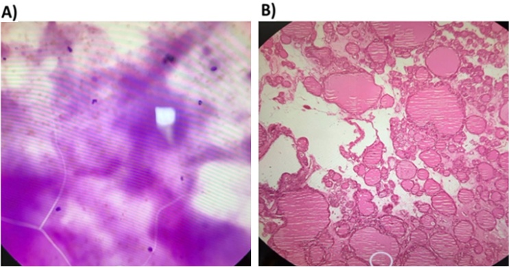
TBSRTC category III on cytology [A) Hematoxylin and Eosin, high power view] proved to be Multinodular goiter on histopathology [ B) Hematoxylin and Eosin, low power view].
The majority of malignant cases had a papillary carcinoma (55.55%) (Figure 2).
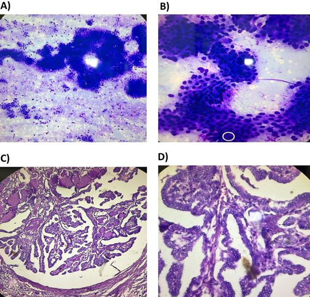
TBSRTC category VI on cytology, A) Hematoxylin and Eosin, scanner view, and B) low power view proved to be papillary carcinoma of thyroid on histopathology, C) Hematoxylin and Eosin, low power view, and D) high power view.
Among the benign cases on histopathology, the majority (93.55%) had colloid goiter, followed by the follicular adenoma (4.30%) (Table 3).
| Histology | Cases [N=102] |
| Benign | 93 (91.17) |
| Colloid goiter | 87 (93.55) |
| Follicular adenoma | 4 (4.30) |
| Hurthle cell adenoma | 2 (2.15) |
| Malignant | 9(8.82) |
| Papillary carcinoma | 5 (55.55) |
| Adenoid cystic carcinoma | 1 (11.11) |
| Follicular carcinoma | 1 (11.11) |
| Anaplastic carcinoma | 1(11.11) |
| Lymphoma | 1(11.11) |
| Data represented as n (%) |
Discussion
The present study evaluated the cytological spectrum of thyroid lesions and correlated the FNAC findings with histopathological diagnosis wherever available.
Patients included in the study were in the age group of 10 to 80 years, and the majority were females (86.11%). Another study also included patients in the age group of 0 to 80 years and almost 88 % females [6]. The majority of thyroid lesions were observed in the age group 21-40 years (48.92%). Similar results were observed in another study where the maximum patients with lesions were in the age group of 20-39 years (47.52%), indicating a higher prevalence among middle-aged adults [7].
The majority of cases belonged to the benign category (Bethesda category II), accounting for 81.48% of the total cases. Similar results were observed in other studies where benign lesions constituted the major proportion (81-85%) of thyroid FNAC [7-9]. This highlights the predominance of non-neoplastic thyroid lesions in routine clinical practice. In the current study, a total of 102 cases were analysed by histopathology, of which 91.17% were confirmed as benign and 8.82% as malignant. In a parallel study, among 171 patients who underwent surgery, 87.72% were having benign lesions, and 12.28% were having malignant lesions [10]. Among the benign cases on histopathology, the majority (993.55%) had colloid goiter, followed by the follicular adenoma (4.30%). Another study by Nandedkar et al. also reported that colloid goiter (57.07%) was more prevalent in benign lesions [10]. The strong concordance between cytological and histopathological findings in the present study further highlights the diagnostic reliability of FNAC in evaluating thyroid lesions.
In this study, a thyroid lesion reported as TBSRTC category III on FNAC cytology demonstrated features of multinodular goiter, and category VI demonstrated papillary carcinoma on subsequent histopathological examination, which was comparable to that reported in a parallel study [11]. Overall, the Bethesda System remains an essential tool in thyroid cytopathology by providing malignancy risk estimates for each diagnostic category, thereby assisting clinicians in determining appropriate follow-up, surgical management, and the extent of therapeutic intervention required [7]. Regular standardization and histopathological correlation can further improve diagnostic accuracy and reinforce the role of FNAC in thyroid lesion evaluation.
Limitations
The present study has certain limitations. In developing countries such as India, especially in rural and outreach settings where implementation of standard guidelines may be difficult, dependence on FNAC alone can sometimes result in either over-treatment or under-treatment of patients. Being a single-center study with a relatively limited sample size, the findings may not be entirely generalizable to the wider population. Histopathological correlation was not available for all FNAC cases, which may limit the comprehensive assessment of diagnostic accuracy. In addition, follow-up data and long-term clinical outcomes were not evaluated.
Conclusion
Most thyroid lesions in the present study were benign, with colloid goiter being the predominant lesion. The FNAC, along with the Bethesda System, proved to be a reliable, minimally invasive, and effective tool for the evaluation and management of thyroid lesions, with good histopathological correlation.
Declarations
Ethical clearance
Study was approved by the Institutional Ethics Committee, Fakir Mohan Medical College and Hospital, Balasore, approval no: 44/IEC dated 25.11.2020
Conflict of interest
None.
Funding/ financial support
Nil
Contributors
Behera S: conceptualization, methodology, investigation, data curation, formal analysis, visualization, writing-review original draft; Subhadarsini S: Conceptualization, methodology, validation, supervision, project administration, writing-review and editing; Sahu S: investigation, data curation, validation, resources, writing- review and editing. All authors read and approved the final manuscript and agree to be accountable for all aspects of the work.
Acknowledgements
None