Introduction
Appendiceal mucinous neoplasm may have disastrous effects if rupture and cause dissemination of disease into the abdomen resulting in pseudomixoma peritonei, which necessitates more aggressive treatment [1]. Occurrence of Mucinous appendiceal neoplasms is rare and adenocarcinoma usually is diagnosed in elderly individuals around the age of 60 to 70 years [1,2]. There are different variety of appendiceal mucinous neoplasm which comprises of serrated polyps, hyperplastic polyps, and low-grade appendiceal mucinous neoplasms (LAMN), high grade appendiceal mucinous neoplasm (HAMN), and mucinous adenocarcinomas [3]. Though the majority of Mucinous neoplasms of the appendix are usually present without any symptom and diagnosis, some patients may have a manifestation as a sign and symptom of acute appendicitis [1]. Other symptoms could be abdominal pain, a palpable mass, gastrointestinal obstruction or gastrointestinal bleeding [4]. Mucinous neoplasm of the appendix in women can frequently be misdiagnosed with ovarian cancer [5]. This case report has been framed in accordance with the CARE guidelines.
Case report
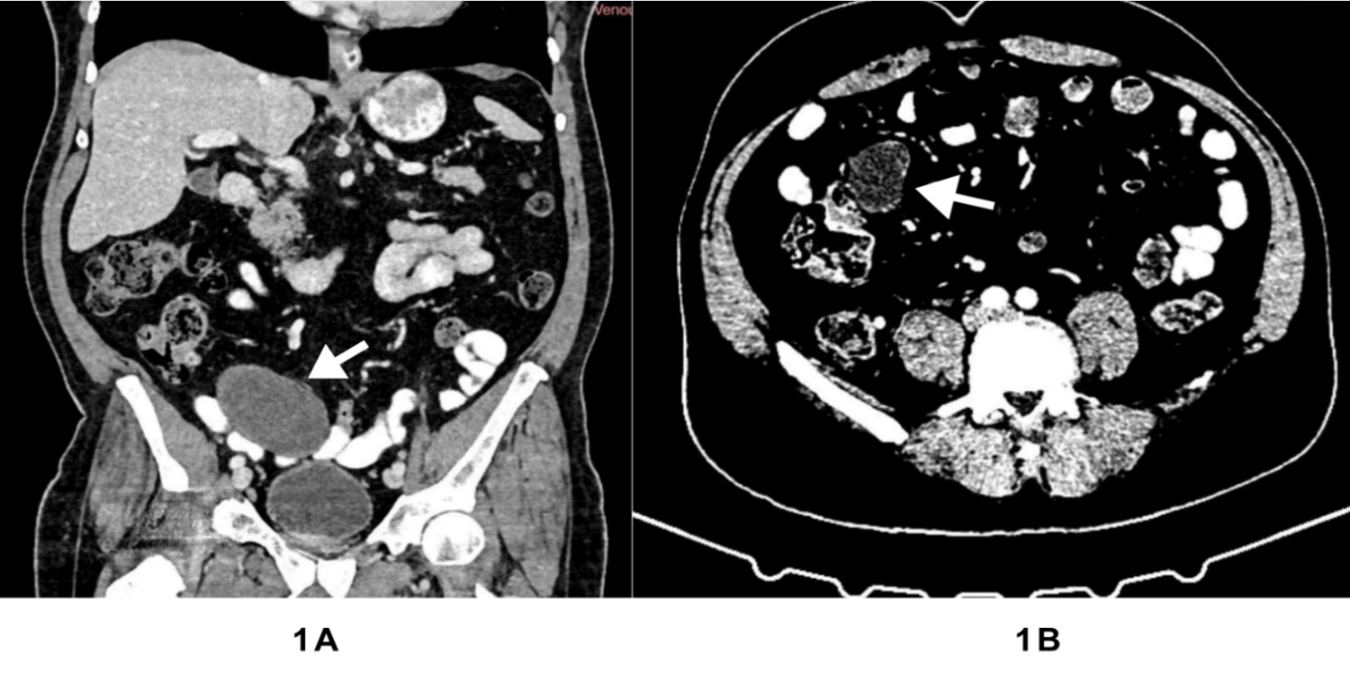
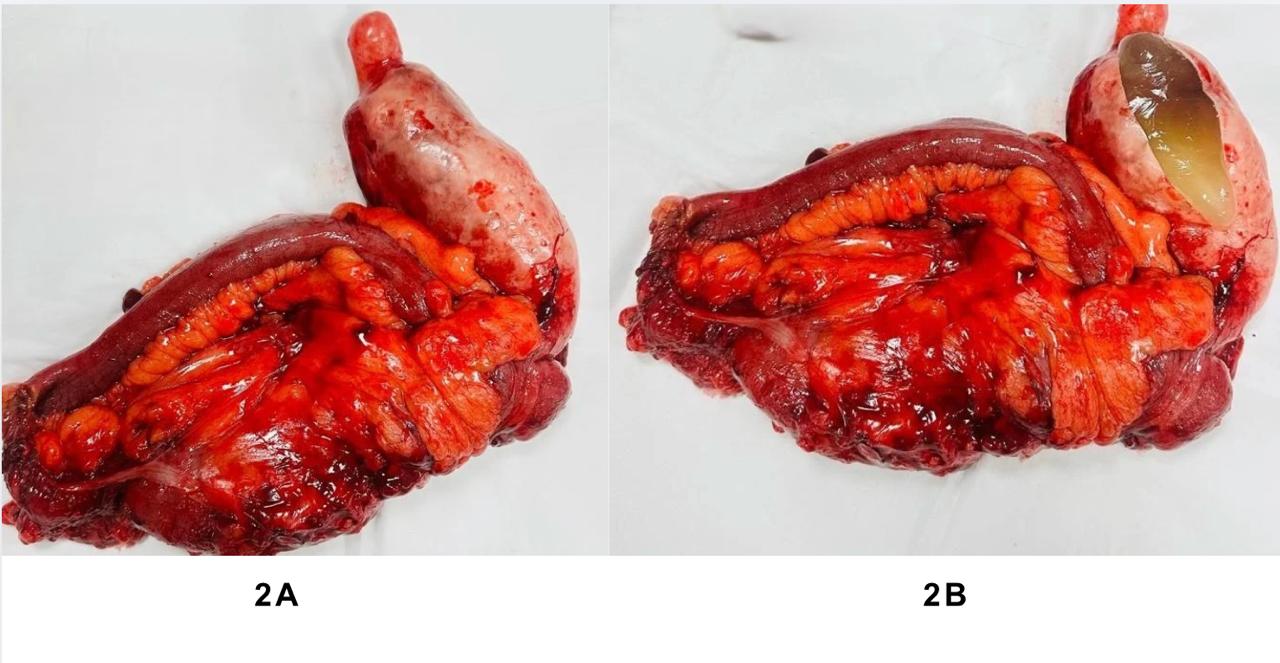
A 75-year-old male patient presented with unintentional weight loss of more than 10% over 3 months and it was associated with vague abdominal pain located mainly in the right iliac fossa. On further enquiry, the patient was on a non-steroidal anti-inflammatory medication for arthritis. He had no previous history of any abdominal operations. On physical examination, a non-tender mass was detected in the right iliac fossa measuring 6cm by 8cm and no obvious abnormality was found on rectal examination. Initial assessment was considered carcinoma of colon and offered Colonoscopy. However, no abnormality was noticed on Colonoscopy. Computer tomography (CT) abdomen performed and revealed a large, dilated mucus filled appendix which measured 170mm in length and 50mm in transverse diameter (Figure 1). The wall is slightly thickened with minimal contrast enhancement after intra venous contrast. No caecal masses were demonstrated and concluded with an appendiceal mucocele. Patients underwent laparoscopic procedure. An enlarged distended appendix was found (Figure 2A) and there were not any mucinous deposits in the peritoneum or any signs of metastasis. After mobilization of ascending colon, right hemicolectomy was performed extracorporeally after extension of incision about 5 cm through the umbilical port and the bowel was anastomosed. The cut section of the appendix specimen had shown mucinous jelly (Figure 2B). In the postoperative course, the patient recovered well. Histology reported the features of high grade appendiceal mucinous neoplasm with no stromal invasion and there was no metastasis of lymph nodes (0/11). On further follow up after 6 months there was no complaint.
Discussion
Majority of Mucinous neoplasms of the appendix are usually present without any symptom and diagnosis usually is made incidentally after a histological report of appendectomy specimens and found near 1 % of the appendectomy specimens [1]. Computer tomography (CT) or magnetic resonance (MRI) imaging is the important modalities to detect the mucinous neoplasm and stage primary adenocarcinomas of the appendix [6]. MRI which is a valuable investigation to detect tumour ruptures and to differentiate mucinous tumours from other organs [7]. Laparoscopic assessment should be incorporated in the preoperative work-up of patients to determine the extent of disease and evaluate further to assess resectability [8].
In our case, the patient was elderly male of 75-year age presented with weight loss and it was accompanied with vague abdominal pain located mainly in the right iliac fossa with non-tender mass. Initial assessment was considered carcinoma of colon and performed Colonoscopy. However, no abnormality was detected on Colonoscopy. Further investigation of CT scan of abdomen reported to be an appendiceal mucocele. CT imaging is one of the important modalities to detect the mucinous neoplasm and can assist to differentiate it from other pathology like colon cancer [6].
Main treatment for appendiceal mucinous neoplasm is surgical resection. Literature stated that patients with low grade mucinous neoplasms are classically cured by complete resection of the appendix [5]. Van DeMoortele (2020) mentioned that patients with high grade mucinous neoplasm, an appendectomy with negative margins essential, nevertheless need to have a long-term follow-up [9]. However, EUROCAN (European network for rare adult solid cancer) guidelines advocated right-sided hemicolectomy for high grade mucinous neoplasm [10]. Our patient was offered Right hemicolectomy. Appendiceal mucinous neoplasm may have disastrous effect if rupture and cause dissemination of disease into the abdomen resulting in pseudomixoma peritonei, which necessitates more aggressive treatment of cytoreductive surgery/ Hyperthermic Intra Peritoneal Chemotherapy [2] and which affects the quality of life seriously. In conclusion, Appendiceal Mucinous neoplasm is a rare condition, and it may be confused with carcinoma colon. Rupture of the appendiceal mucinous neoplasm may be led in pseudomixoma peritonei. Therefore, it is paramount important to diagnose appendiceal mucinous neoplasm early and treat it before rupture.
Declarations
Conflict of interest
The authors declare no conflicts of interest.
Funding
This research received no specific grant from any funding agency or not-for-profit sectors.
Contributors
Matsobane Latakgomo and Mirza Bhuiyan contributed concept, drafting of the manuscript, critical revision for important intellectual content and ethical clearance.
Ethical clearence
Informed consent obtained from patient and ethical approval was taken from Pietersburg Mankweng ethic committee (PMREC 29 October UL 2025/C).
Acknowledgements
None