1. Introduction
Snakebite envenomation is a major public health problem in tropical and subtropical regions, particularly in South Asia. The World Health Organization recognizes snakebite envenoming as a neglected tropical disease, causing an estimated 1.8–2.7 million envenoming and up to 138,000 deaths annually. India bears a substantial burden due to frequent encounters with venomous snakes, including cobras, kraits, vipers, and Russell's vipers [1,2].
Neurotoxic envenomation, commonly caused by elapid snakes such as cobras and kraits, primarily affects the neuromuscular junction through pre- and postsynaptic neurotoxins. Clinical manifestations include ptosis, ophthalmoplegia, dysarthria, dysphagia, progressive paralysis, and, in severe cases, respiratory failure requiring urgent ventilatory support [3]. Early administration of anti-snake venom (ASV) and timely airway management remain the mainstays of treatment.
Although neurological manifestations predominate, cardiovascular complications such as arrhythmias, myocarditis, acute heart failure, and transient left ventricular dysfunction are uncommon but potentially life-threatening [4]. Proposed mechanisms include direct myocardial toxicity, autonomic dysfunction, catecholamine surge, and inflammatory injury. Early electrocardiography and echocardiography facilitate prompt recognition and guide hemodynamic management, improving outcomes [5].
We report a case of presumed cobra envenomation complicated by reversible severe left ventricular systolic dysfunction and pulmonary edema, emphasizing the importance of routine cardiovascular assessment in severe neurotoxic snakebite.
2. Case Presentation
2.1 Patient Information
A 65-year-old man with no known comorbidities presented to our tertiary care center after a snakebite over the right lateral malleolar region sustained during routine outdoor activity. Although the snake was not identified, the patient and attendants described it as resembling a cobra. Examination revealed two fang marks without significant local swelling, cellulitis, bleeding, or tissue necrosis, suggesting predominantly neurotoxic envenomation. The patient initially received first aid at a nearby Primary Health Centre before referral to the Government Hospital, Davangere, because of progressive neurological symptoms and respiratory distress. Despite initial treatment, his condition deteriorated with worsening respiratory difficulty and declining consciousness, prompting transfer to our tertiary intensive care unit approximately two hours after the bite for advanced airway management, anti-snake venom therapy, and comprehensive critical care.
2.2 Clinical Findings
On arrival, the patient was critically ill with severe neurotoxic envenomation, presenting with a Glasgow Coma Scale score of E1V1M1, profound unconsciousness, bradycardia (45 beats/min), borderline hypotension (100/60 mmHg), respiratory depression (10 breaths/min), and severe hypoxemia (SpO2 70%). Body temperature was normal (98°F). Bilateral ptosis and progressive neuroparalysis with respiratory muscle involvement indicated impending respiratory arrest. The combination of depressed sensorium, hypoventilation, and cardiovascular instability necessitated immediate endotracheal intubation, mechanical ventilation, and intensive care management. Prompt airway protection and supportive therapy were instituted to prevent further neurological deterioration and life-threatening respiratory failure.
3. Diagnostic Assessment
3.1 Laboratory Investigations
Arterial Blood Gas Analysis
Initial arterial blood gas analysis on admission demonstrated severe metabolic acidosis with associated hypoxemia. The pH was 7.20, PaO2 was 54 mmHg, PaCO2 was 38 mmHg, bicarbonate (HCO3⁻) was 14.3 mmol/L, and base excess was −12 mmol/L. Serum lactate levels were markedly elevated at 5.9 mmol/L, indicating significant tissue hypoperfusion and metabolic stress.
3.2 Hematological Investigations
The baseline hematological parameters were largely within normal limits. Hemoglobin was 14.2 g/dL, total leukocyte count was mildly elevated at 11,200/mm³, and platelet count was 2.95 lakh/mm³. Hematological tests did not reveal any abnormalities suggestive of hematotoxic envenomation.
3.3 Renal and Liver Function Tests
Renal function remained preserved throughout hospitalization, with blood urea and serum creatinine levels of 14.2 mg/dL and 0.7 mg/dL, respectively, on admission. Urine output was maintained between 40 and 75 mL/hour during the ICU stay, and serial creatinine measurements remained stable between 0.7 and 0.9 mg/dL. Liver function tests were also within normal limits, with a total bilirubin level of 0.48 mg/dL, AST of 40 IU/L, and ALT of 22 IU/L, indicating no significant hepatic involvement.
3.4 Coagulation Profile
Coagulation studies revealed a prothrombin time of 14.5 s, an international normalized ratio (INR) of 1.2, an activated partial thromboplastin time (APTT) of 30.9 s, and a whole-blood clotting time of 14 min. These findings did not suggest a clinically significant coagulopathy.
3.5 Cardiac Biomarkers
Cardiac evaluation demonstrated a negative troponin assay, effectively excluding an acute myocardial infarction. However, NT-proBNP was markedly elevated at 10,000 pg/mL, indicating significant myocardial stress and acute cardiac dysfunction in the patient.
3.6 Electrocardiographic Findings
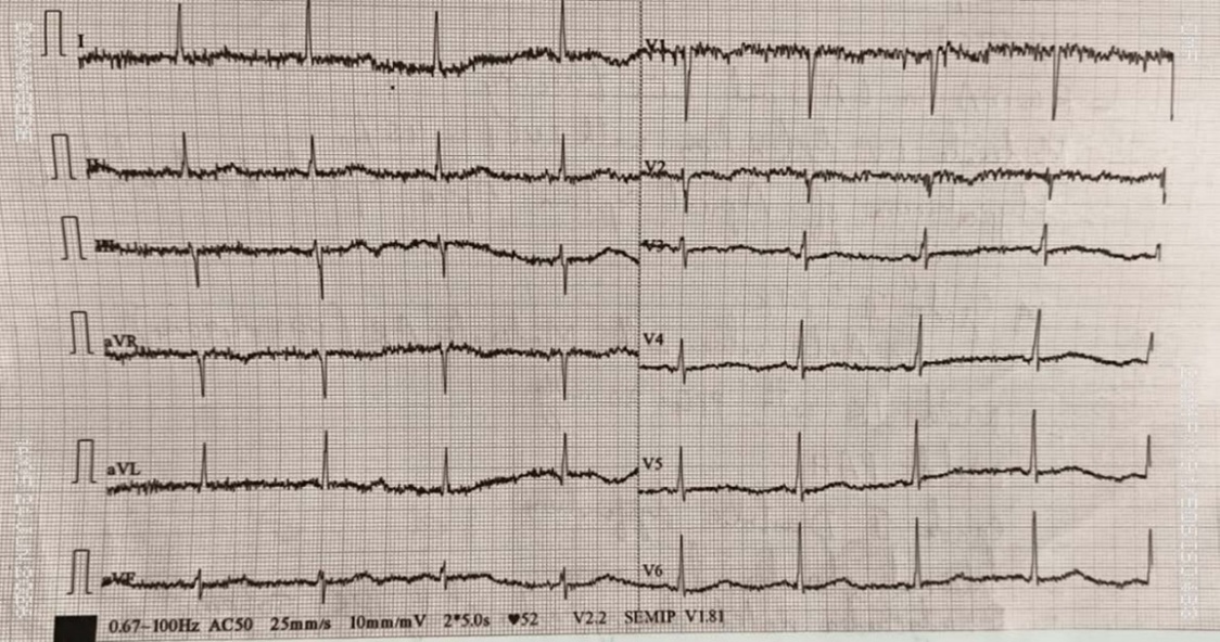
Initial ECG showed sinus bradycardia with PR prolongation, followed by atrial fibrillation with rapid ventricular response and T-wave inversions. Serial electrocardiograms demonstrated gradual resolution of the arrhythmia, with restoration of normal sinus rhythm within 24 hours of intensive care management (Figure 1).
3.7 Echocardiographic Evaluation
Initial Echocardiography
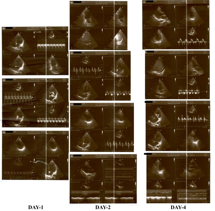
Urgent transthoracic echocardiography was performed due to persistent hemodynamic instability and a newly developed arrhythmia. The study demonstrated normal cardiac chamber dimensions, mild mitral regurgitation, moderate tricuspid regurgitation, and an estimated pulmonary artery systolic pressure of 40 mmHg. Severe global left ventricular hypokinesia was observed, resulting in marked left ventricular systolic dysfunction with an ejection fraction (LVEF) of 35%.
Repeat Echocardiography (48 Hours)
Repeat echocardiography performed 48 h later showed substantial improvement in cardiac function. Only mild residual global left ventricular hypokinesia was observed, with trivial mitral and tricuspid regurgitation. The left ventricular ejection fraction improved to 50%, and minimal bilateral pleural effusion was noted.
Follow-Up Echocardiography (Day 4)
A follow-up echocardiogram obtained on day four demonstrated complete recovery of left ventricular systolic function with normalization of chamber contractility and an LVEF of 55%. Cardiology consultation was obtained during hospitalization, and acute coronary syndrome was ruled out based on clinical assessment, serial ECG findings, negative cardiac biomarkers, and rapid reversibility of ventricular dysfunction (Figure 2).
3.8 Chest Imaging
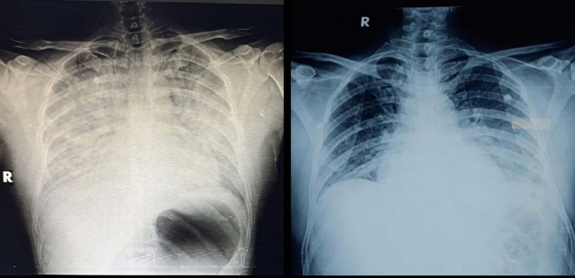
Initial chest radiography showed bilateral pulmonary infiltrates consistent with acute cardiogenic pulmonary edema in the setting of severe left ventricular dysfunction and elevated NT-proBNP. Serial chest radiographs demonstrated progressive resolution following ventilatory support, fluid optimization, and recovery of cardiac function, with complete clearing of pulmonary opacities on follow-up imaging (Figure 3).
3.9 Final Diagnosis
The patient was diagnosed with severe presumed cobra envenomation causing rapidly progressive neuroparalysis, respiratory failure, transient severe left ventricular systolic dysfunction (ejection fraction of 35%), atrial fibrillation with rapid ventricular response, and cardiogenic pulmonary edema. Rapid normalization of cardiac function and resolution of arrhythmia on follow-up confirmed reversible myocardial involvement associated with neurotoxic snake envenomation.
3.10 Therapeutic Interventions
Antivenom Therapy
Given the history of presumed cobra envenomation and the presence of rapidly progressive neurotoxic manifestations, prompt anti-snake venom (ASV) therapy was initiated. Polyvalent ASV manufactured by Bharat Serums and Vaccines Ltd. was administered after premedication with intravenous pheniramine (Avil) and hydrocortisone to minimize the risk of hypersensitivity reactions. An initial dose of 10 vials of ASV diluted in 250 mL of normal saline was infused over 1 h. Owing to the severity of envenomation and ongoing clinical concerns, five additional vials were administered, bringing the total to 15 during hospitalization.
Management of Neurotoxic Envenomation
Neurological examination on admission demonstrated severe neurotoxic envenomation with bilateral ptosis, impaired eyelid elevation, bulbar weakness, and progressive respiratory muscle paralysis, indicating impending respiratory failure. Owing to deteriorating consciousness, severe hypoxemia, and compromised airway protection, urgent endotracheal intubation and mechanical ventilation were initiated. Initial resuscitation included atropine (0.6 mg) and adrenaline (1 mg). Anticholinesterase therapy with neostigmine (2.5 mg) and glycopyrrolate (0.4 mg) was administered to improve neuromuscular transmission and counteract neurotoxic venom effects. These interventions, together with anti-snake venom therapy and intensive care support, resulted in progressive neurological recovery.
Respiratory Support
Ventilatory support was provided using the volume-controlled ventilation (VCV) mode with a tidal volume of 410 mL, respiratory rate of 28 breaths/min, positive end-expiratory pressure (PEEP) of 12 cmH₂O, and fraction of inspired oxygen (FiO₂) of 70%. The patient remained on invasive mechanical ventilation for 24 h, following which he was successfully extubated and transitioned to non-invasive ventilation (NIV). Intermittent NIV support was continued for an additional 48 h until complete respiratory stabilization was achieved.
Hemodynamic Support
The bite site showed no significant local swelling, necrosis, bleeding, or cellulitis, consistent with predominantly neurotoxic envenomation. The patient developed persistent hypotension and fluctuating heart rates, suggesting autonomic and cardiovascular involvement. Hemodynamic support with noradrenaline (up to 10 mcg/min) and dobutamine (up to 5 mcg/kg/min) was initiated and gradually discontinued following recovery of cardiac function and hemodynamic stability.
Adjunctive Therapies
Supportive ICU care included piperacillin–tazobactam, intravenous crystalloids, magnesium supplementation, enoxaparin, proton pump inhibitors, paracetamol, endotracheal suctioning, and continuous monitoring. These measures, combined with timely anti-snake venom therapy, ventilatory support, and hemodynamic management, contributed to progressive neurological, respiratory, and cardiovascular recovery.
3.11 Outcome and Follow-Up
The patient recovered rapidly following prompt anti-snake venom therapy and intensive care. Neurological deficits resolved progressively, permitting extubation after 24 hours and transition to non-invasive ventilation. Hemodynamic stability improved, allowing discontinuation of vasopressor and inotropic support. Serial electrocardiograms restored normal sinus rhythm, while echocardiography demonstrated recovery of left ventricular ejection fraction from 35% to 55% by day four. He was discharged after four days with complete neurological and cardiac recovery and remained asymptomatic during follow-up.
Timeline of Events
Table 1 summarizes the patient's clinical course, from the presumed cobra bite and progressive neurotoxic manifestations to respiratory failure, cardiovascular complications, anti-snake venom therapy, intensive care management, and complete neurological and cardiac recovery.
| Time Point | Clinical Event |
| 09:00 AM | Presumed cobra bite over the right lateral malleolar region |
| 10:00 AM | Initial first aid at the Primary Health Centre |
| 10:30 AM | Evaluation at Government Hospital, Davangere |
| 11:00 AM | Referred to a tertiary care center due to worsening neurological status and respiratory distress |
| 12:00 PM | Polyvalent antisnake venom (10 vials) initiated |
| Admission | GCS E1V1M1, severe hypoxemia, bradycardia, ptosis, bulbar weakness; intubation and mechanical ventilation initiated |
| First 24 hours | Development of atrial fibrillation and severe LV dysfunction (EF 35%); vasopressor and inotropic support started |
| 24 hours | Extubated and transitioned to non-invasive ventilation |
| 48 hours | Repeat echocardiography showed improvement of LVEF to 50% |
| Day 2 | Hemodynamic stabilization; vasopressors discontinued |
| Day 3 | Shifted from ICU to HDU |
| Day 4 | Echocardiography showed normalization of LVEF to 55%; discharged home |
| Follow-up | Asymptomatic with normal cardiac and neurological status |
3.12 Patient Perspective
At follow-up, the patient reported complete recovery without residual neurological, respiratory, or cardiovascular symptoms. He resumed normal daily activities independently and remained asymptomatic, with no recurrence of arrhythmias or functional limitations. The patient expressed satisfaction with the timely medical care and intensive care support that contributed to his recovery.
4. Discussion
4.1 Neurotoxic Effects of Cobra Envenomation
Cobra envenomation causes neuromuscular paralysis through pre- and postsynaptic neurotoxins that impair neuromuscular transmission. Patients typically present with ptosis, ophthalmoplegia, dysarthria, dysphagia, and progressive respiratory muscle paralysis [6]. Our patient developed bilateral ptosis, bulbar weakness, and respiratory failure requiring mechanical ventilation. Prompt airway protection and ventilatory support remain essential to reduce mortality [2].
4.2 Cardiac Manifestations of Snakebite
Although respiratory paralysis predominates, cardiovascular complications may occur through direct cardiotoxicity, inflammation, coronary vasospasm, autonomic dysfunction, and catecholamine-mediated myocardial injury. Experimental studies have demonstrated that certain components of snake venom can directly affect cardiac myocytes, resulting in impaired contractility and transient ventricular dysfunction [7]. Autonomic dysfunction may contribute to cardiovascular complications, as evidenced by bradycardia, hypotension, and atrial fibrillation in our patient [8]. Catecholamine surge and venom-induced myocardial injury may cause reversible myocardial stunning or myocarditis-like syndromes [9]. Elevated NT-proBNP with negative troponin supported transient myocardial dysfunction without myocardial infarction.
4.3 Reversible Cardiomyopathy
The patient developed severe but rapidly reversible left ventricular systolic dysfunction, with ejection fraction improving from 35% to 50% within 48 hours and to 55% by day four. This rapid recovery suggests transient myocardial stunning rather than structural cardiomyopathy or ischemic heart disease. The reversible left ventricular dysfunction may represent Takotsubo-like cardiomyopathy, toxic myocarditis, or stress-induced myocardial stunning following severe envenomation [10]. However, the rapid recovery of ventricular function with negative troponin levels favors transient myocardial stunning rather than irreversible myocardial necrosis or structural cardiomyopathy [11].
4.4 Pulmonary Edema and Respiratory Complications
The patient developed bilateral homogeneous pulmonary infiltrates during the acute phase of the illness. In conjunction with severe left ventricular dysfunction, elevated NT-proBNP levels, and rapid radiological resolution following cardiovascular stabilization, these findings were most consistent with acute cardiogenic pulmonary edema. Acute left ventricular failure can elevate pulmonary capillary hydrostatic pressure, leading to alveolar fluid accumulation and impaired gas exchange. Although ARDS, aspiration, and neurogenic pulmonary edema were considered, rapid radiographic improvement with recovery of cardiac function strongly favored cardiogenic pulmonary edema as the underlying mechanism [12].
4.5 Importance of Early Echocardiography
Early echocardiography is essential in severe neurotoxic snakebite with hemodynamic instability, as cardiac complications may be overlooked. In our patient, it identified severe left ventricular dysfunction, enabling timely inotropic therapy. Serial echocardiography confirmed rapid recovery and guided management, highlighting the importance of early recognition of occult cardiac involvement [8].
4.6 Comparison with Previously Reported Cases
Cardiac dysfunction following neurotoxic snakebites is uncommon but has been increasingly reported in the literature. Sunil et al. described a spectrum of cardiovascular abnormalities following snakebite, including arrhythmias, conduction disturbances, and myocardial dysfunction [13]. Dissanayake et al. reported reversible cardiomyopathy associated with snake envenomation, with echocardiographic recovery occurring within days to weeks following supportive care [14]. Similar cases of transient left ventricular dysfunction and pulmonary edema have also been described following cobra and krait envenomation, suggesting that reversible myocardial stunning may be an underrecognized manifestation of severe neurotoxic snakebite [15]. Our case highlights reversible myocardial involvement in severe neurotoxic snakebite, emphasizing early diagnosis, intensive supportive care, and vigilant cardiac monitoring.
5. Conclusion
Neurotoxic snake envenomation may rarely cause reversible cardiovascular complications, including atrial fibrillation, pulmonary edema, and transient left ventricular systolic dysfunction. This case emphasizes the importance of early cardiac evaluation with electrocardiography and echocardiography, alongside prompt anti-snake venom therapy, airway protection, ventilatory support, and intensive care. Timely recognition and supportive management can result in complete neurological and cardiac recovery.
6. Declarations
Ethics Statement
Institutional Ethics Committee approval was not required for the publication of this single anonymized case report in accordance with the institutional policy. Written informed consent for publication of the clinical details and accompanying images was obtained from the patient.
Conflict of Interest
The authors declare that they have no conflicts of interest related to this work.
Funding
The authors received no specific funding for this work from any funding agency in the public, commercial, or not-for-profit sectors.
Acknowledgments
The authors would like to express their sincere gratitude to the Intensive Care Unit, Cardiology, Emergency Medicine, and Nursing teams for their dedicated efforts in the diagnosis, management, and the patient's successful recovery. We are particularly grateful to the patient and his family for their cooperation and for providing informed consent for the publication of this case report. Their willingness to share this clinical experience has contributed to a better understanding of the rare cardiovascular manifestations associated with neurotoxic snake envenomation. We also acknowledge the support of our institution in facilitating the patient's clinical care and follow-up visits.