Introduction
Ulcerative colitis is a chronic relapsing disease of the gastrointestinal tract having multifactorial etiology and complex pathogenesis. The assessment of disease activity is based on combination of clinical, colonoscopic and histopathological findings [1,8]. There are around twenty histological scoring systems in UC which have been described till now like Riley score, Chicago score, Gupta Index, Gramelich Index, Nancy Index, Robarts Index, ECAP Score etc [2,17]. Among these, the Original Geboes score (GS) has good reproducibility and modest colonoscopic agreement [5]. It is comprised of 7 grades which evaluate all aspects of mucosal injury. However, its utility is limited in daily routine practice. Aranzazu Jauregui-Amezaga et al.,[6], sought to streamline the assessment of histological activity in ulcerative colitis (UC) by introducing a Simplified Geboes Score (SGS) and evaluating its performance against the established. Their investigation focused on patients newly diagnosed with active UC, aiming to determine whether the SGS could offer a practical alternative to the OGS in clinical settings. The SGS was designed to include only variables directly linked to active inflammatory disease (including basal plasmacytosis), potentially simplifying the scoring process compared to the comprehensive OGS. What was finally devised was 5 grade scale that had its own respective sub-grades. The results of the study indicated that the assessments of histological activity based on both the OGS and the SGS were similar in patients with newly diagnosed active UC. Specifically, the correlation between endoscopic findings and histological scores did not exhibit significant discrepancies between the two scoring systems. Moreover, the inter-observer agreement for the SGS was moderate across all grades, suggesting reasonable consistency among different readers when using this simplified scoring method.
Materials and Methods
The aim of this study was to assess the comparability of OGS and SGS along with assessing the ease of application of SGS in place of OGS. This was an observational study conducted in the department of pathology, St. Stephen’s Hospital, Tis Hazari, Delhi and the cases were obtained from the department of Gastroenterology, St. Stephen’s Hospital, Delhi. A total of 87 patients were included in this study from January 2018 to August 2023.
Clinical parameters (Age, sex, clinical symptoms e.g bleeding PR, diarrhoea, abdominal pain etc) and colonoscopic findings (site of involvement, appearance of lesions and site of biopsy) were noted form the medical records.
All histologically diagnosed endoscopic biopsies of UC irrespective of age and gender were included in the study while Inadequate biopsies, Non-specific colitis and Non-specific ulcers of colon were excluded.
The endoscopic biopsies of the patients in whom IBD were suspected upon colonoscopy was obtained. No biopsies from normal appearing colon have been included. The gastroenterologist has performed colonoscopy of the patients. The biopsies were taken and put in properly labelled containers filled with 10% formalin and sent for histopathological examination. The biopsies were processed routinely, and paraffin embedded sections stained with Hematoxylin & Eosin were made.
The stained sections were scored using 2 histologic assessment methods i.e. OGS and SGS. The histologic score used for comparison with the endoscopic score for every biopsy represented the most severely inflamed area of that patient.
The OGS contains 7 different parameters, ranging in score from 0.0 (Normal mucosa) to 5.4 (ulceration and / or granulation) while the SGS ranges from 0.0 (Normal mucosa) to 4.4 (ulceration and /or granulation tissue) (Table I).
| Geboes Score | Grade | Simplified Geboes Score (SGS) |
| Architectural changes | Grade 0 | No inflammatory activity |
| Chronic inflammatory infiltrate | Grade 1 | Basal plasmacytosis |
| Eosinophils in lamina propria | Grade 2A | Eosinophils in lamina propria |
| Neutrophils in lamina propria | Grade 2B | Neutrophils in lamina propria |
| Neutrophils in epithelium | Grade 3 | Neutrophils in epithelium |
| Crypt destruction | Grade 4 | Epithelial injury (in crypt and surface epithelium) |
| Erosion and ulcerations | Grade 5 |
To allow for good comparison between the two histologic scores, the original geboes score was recoded and compiled, to correspond
to the subcategories of the simplified geboes score (Table II).
| Simplified Geboes Score | Geboes Score |
| GRADE (0): 0.0- No abnormality. | GRADE (0): 0.0- No abnormality |
| 0.1- Presence of architectural changes | 0.1- Mild abnormality |
| 0.2- Presence of architectural changes and chronic mononuclear cell infiltrate | 0.2- Mild/ moderate diffuse/multifocal abnormality 0.3- Severe diffuse/multifocal abnormality |
| GRADE (1): 1.0- No increase in Basal plasma cells | GRADE (1): 1.0- No increase in chronic inflammatory infiltrate |
| 1.1- Mild increase in Basal plasma cells | 1.1- Mild but unequivocal increase in chronic inflammatory infiltrate |
| 1.2- Marked increase in Basal plasma cells | 1.2- Moderate increase in chronic inflammatory infiltrate 1.3- Marked increase in chronic inflammatory infiltrate |
| GRADE (2A): 2A.0- No increase in eosinophils in lamina propria | GRADE (2A): 2A.0- No increase in eosinophils in lamina propria |
| 2A.1- Mild increase in eosinophils in lamina propria | 2A.1- Mild but unequivocal increase in eosinophils in lamina propria |
| 2A.2- Marked increase in eosinophils in lamina propria | 2A.2- Moderate increase in eosinophils in lamina propria 2A.3- Marked increase in eosinophils in lamina propria |
| GRADE (2B): 2B.0- No increase in neutrophils in lamina propria | GRADE (2B): 2B.0- No increase in neutrophils in lamina propria |
| 2B.1- Mild increase in neutrophils in lamina propria | 2B.1- Mild but unequivocal increase in neutrophils in lamina propria |
| 2B.2- Marked increase in neutrophils in lamina propria | 2B.2- Moderate increase in neutrophils in lamina propria 2B.3- Marked increase in neutrophils in lamina propria |
| GRADE (3): 3.0- No neutrophils in the epithelium | GRADE (3): 3.0- No neutrophils in the epithelium |
| 3.1- <50% of crypts involved | 3.1- <5% of crypts involved 3.2- <50% of crypts involved |
| 3.2- >50% of crypts involved | 3.3- >50% of crypts involved |
| GRADE (4): 4.0- No epithelial injury (in crypt and surface epithelium) | GRADE (4): 4.0- No crypt destruction |
| 4.1- Marked attenuation | 4.1- Probable: Local excess of neutrophils in part of the crypts 4.2- Probable: Marked attenuation |
| 4.2- Probable crypt destruction: probable erosion | 4.3- Unequivocal crypt destruction |
| 4.3- Unequivocal crypt destruction: unequivocal erosion | GRADE (5): 5.0- No ulceration, erosion, granulation tissue 5.1- Recovering epithelium with adjacent inflammation 5.2- Probable erosion: focally stripped 5.3 unequivocal erosion |
| 4.4- Ulcer/granulation tissue | 5.4- Ulcer/granulation tissue |
Data was coded and recorded in MS Excel spreadsheet program. SPSS v23 (IBM corp.) was used for data analysis. Descriptive statistics were elaborated in the form of means/standard deviations and medians for continuous variables, frequencies and percentages for categorical variables. Data was presented in a graphical manner wherever appropriate for data visualization using histograms/column charts for continuous data and bar charts/pie charts for categorical data. Chi- squared test was used for group comparisons for categorical data. In case the expected frequency in the contingency tables was found to be <5% for >20 of the cells, Fisher’s Exact test was used instead. Statistical significance will be kept at p<0.05.
Results
The age of the patients was in the range 16 to 78 years with mean age being 40.97 years. Maximum patients were seen in the age group of 21-30 and 31-40 years. Out of 87 patients, 67.8% of patients were male and 32.2% were female, thereby indicating male predominance. In our study, bleeding per rectum (36%) was the most frequent complaint in both genders followed by diarrhoea with blood, abdominal pain, diarrhea, increase in frequency of stools among others. Of the 87 cases, 50.5% biopsies from the rectum while only 2.3% and 5.7% cases were from the terminal ileum and caecum, respectively. On colonoscopy, 52.9% of cases showed loss of vascular pattern followed by superficial and diffuse ulceration.
The distribution of OGS showed the majority within Grade 5, accounting for 60.9% of cases collectively (5.1: 18.4 %, 5.2: 1.1%, 5.3: 1.1%, 5.4: 40.2%). Grade 4 accounted for 21.8% of cases (4.1: 20.7%, 4.3: 1.1%), while Grade 3 comprised 14.9% of cases (3.1: 10.3%, 3.2: 4.6%). Grade 2B was the least represented, constituting 2.2% of participants (2B.1: 1.1%, 2B.2: 1.1%).
Similarly, the distribution of SGS showed the majority within Grade 4, accounting for 82.7% of cases collectively (4.1: 20.7%, 4.2: 1.1%, 4.3: 20.7%, 4.4: 40.2%). Grade 3 accounted for 14.9% of participants (3.1: 14.9%), while Grade 2B was the least represented, constituting 2.2% of participants (2B.1: 1.1%, 2B.2: 1.1%).
Chi-squared test was used to explore the association between OGS and SGS. There was a significant difference between the various groups in terms of distribution of SGS (χ2 = 522.000, p = <0.001). The strength of association between the two variables (Cramer's V) was 1 i.e High Association (Table III).
| SGS | OGS | Chi-Squared Test | |||||||||||
| `2B.1 | `2B.2 | `3.1 | `3.2 | `4.1 | `4.3 | `5.1 | `5.2 | `5.3 | `5.4 | Total | χ2 | P Value | |
| `2B.1 | 1 (100.0%) | 0 (0.0%) | 0 (0.0%) | 0 (0.0%) | 0 (0.0%) | 0 (0.0%) | 0 (0.0%) | 0 (0.0%) | 0 (0.0%) | 0 (0.0%) | 1 (1.1%) | 522.000 | <0.001 |
| `2B.2 | 0 (0.0%) | 1 (100.0%) | 0 (0.0%) | 0 (0.0%) | 0 (0.0%) | 0 (0.0%) | 0 (0.0%) | 0 (0.0%) | 0 (0.0%) | 0 (0.0%) | 1 (1.1%) | ||
| `3.1 | 0 (0.0%) | 0 (0.0%) | 9 (100.0%) | 4 (100.0%) | 0 (0.0%) | 0 (0.0%) | 0 (0.0%) | 0 (0.0%) | 0 (0.0%) | 0 (0.0%) | 13 (14.9%) | ||
| `4.1 | 0 (0.0%) | 0 (0.0%) | 0 (0.0%) | 0 (0.0%) | 18 (100.0%) | 0 (0.0%) | 0 (0.0%) | 0 (0.0%) | 0 (0.0%) | 0 (0.0%) | 18 (20.7%) | ||
| `4.2 | 0 (0.0%) | 0 (0.0%) | 0 (0.0%) | 0 (0.0%) | 0 (0.0%) | 1 (100.0%) | 0 (0.0%) | 0 (0.0%) | 0 (0.0%) | 0 (0.0%) | 1 (1.1%) | ||
| `4.3 | 0 (0.0%) | 0 (0.0%) | 0 (0.0%) | 0 (0.0%) | 0 (0.0%) | 0 (0.0%) | 16 (100.0%) | 1 (100.0%) | 1 (100.0%) | 0 (0.0%) | 18 (20.7%) | ||
| `4.4 | 0 (0.0%) | 0 (0.0%) | 0 (0.0%) | 0 (0.0%) | 0 (0.0%) | 0 (0.0%) | 0 (0.0%) | 0 (0.0%) | 0 (0.0%) | 35 (100.0%) | 35 (40.2%) | ||
| Total | 1 (100.0%) | 1 (100.0%) | 9 (100.0%) | 4 (100.0%) | 18 (100.0%) | 1 (100.0%) | 16 (100.0%) | 1 (100.0%) | 1 (100.0%) | 35 (100.0%) | 87 (100.0%) |
Discussion
Evaluation of Individual Histological Parameters
Basal Plasmacytosis
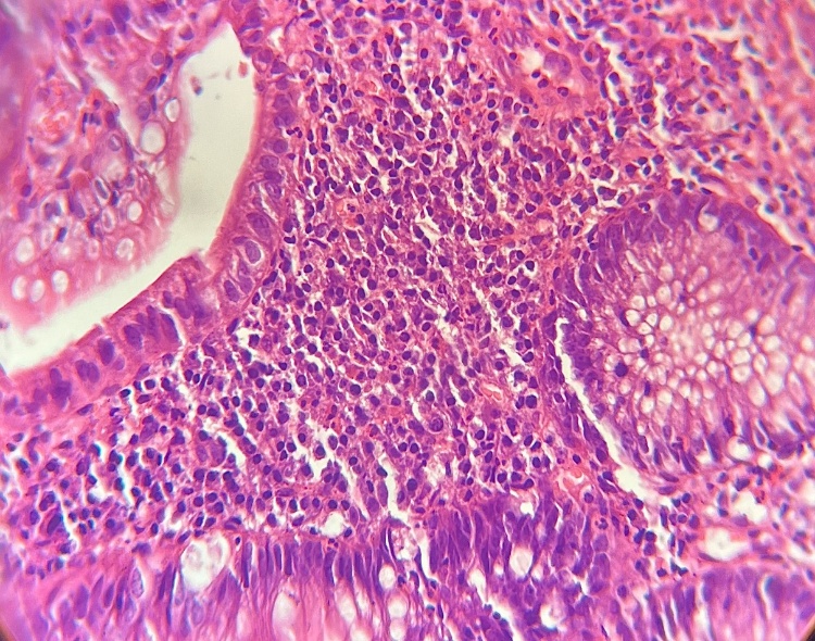
Basal plasmacytosis [19] (Grade I of SGS) was observed in the biopsies of all the patients. In 32 cases, the basal plasmacytosis was minimal and in 55 it was marked. The presence of BP also showed an association with active microscopic disease that was indicated by SGS of 3.1 and higher (presence of neutrophils in the crypts). In the study by V. Villanacci et al.,[9], BP was present in at least one colonic segment in all IBD segments and sparsely found in other forms of colitis and in controls. It was found that the probability of IBD diagnosis markedly increases with the number of segments positive for the presence of BP. The earliest diagnostic feature with the highest predictive value for the diagnosis of UC was the presence of basal plasmacytosis [19].
Neutrophils and Epithelial Injury
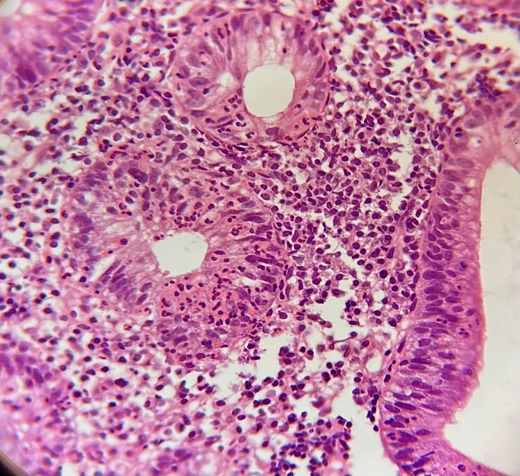
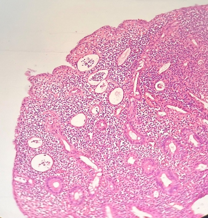
Microscopic activity is based on the presence of neutrophils or defined as damage of the surface and crypt epithelium typically in conjunction with neutrophils [22]. Neutrophils in lamina propria exclusively were observed in 2 cases i.e. grade 2. Neutrophils in the epithelium were observed in 13 cases and the rest of the cases showed epithelial injury in the form of crypt abscess, crypt destruction, erosion, ulceration and the presence of regenerative changes (Figure 3). Many studies with leucocyte scanning have supported the use of neutrophils as an indicator of disease activity [15]. In a study by Pai et al.,[20], relapsers were more likely to have neutrophillic inflammation on biopsy, when compared with non relapsers of UC.
Comparison of OGS AND SGS
The two scores were comparable and represented the histopathological status of the biopsies. However, in SGS, in the absence of ulceration/erosion on microscopy, one cannot comment on the regenerative change. Regenerating epithelium is not a parameter in SGS. So, if the biopsy shows regenerative epitheium and no ulceration/erosion, SGS is at a falsely lower grade of disease i.e 4.0 or 4.1 instead of 4.3 (Figure 3).
Conclusion
The two scores, GS and SGS were comparable and represented the histopathological status of the biopsies.
As SGS includes basal plasmacytosis (early indicator of active microscopic disease) and reduced number of grading criteria, it can be used to assess microscopic disease of UC in daily routine practice.
Declarations
Ethical Clearance
Approved by the Institutional Ethics Committee of St. Stephen’s Hospital, reference no. SSHEC/May-22/018.
Conflict of interest
The authors declare no conflict of interest
Funding/ financial support
This study received no funding/financial support.
Contributors
Dr. Tanvi Gokhale, Dr. Molly Joseph, Dr. Richa Jindal, Dr. Kuldeep Kaur (St. Stephen’s Hospital, Delhi)
Acknowledgements
The authors acknowledge the support of the Department of Pathology and the cooperation of the clinical team in the successful completion of this study.